VARIATIONS/UNUSUAL SITUATIONS
A disc excision (with straight or angled edges) may be performed either for biopsy purposes or for biopsy and definitive treatment. This option
is chosen when healing by second intention is preferred or when it is desirable to remove the tumor first and then repair the dog-ears during the closure. Second intention healing may be advantageous when the risk of adverse reactions with primary closure is high. For example, excisions of even small tumors on the anterior lower leg that are closed primarily can produce wounds with tension and wounds that are more likely than those at other sites to dehisce, become infected, or be painful. This is especially true if undermining is inadequate and weight-bearing and limb dependency is not minimized for the first 5–7 postoperative days.
Another option for wounds with too much tension on the anterior lower leg is the placement of a skin graft, but postoperative care requires leg elevation and the absolute avoidance of weight-bearing. Alternatively, the wound can be closed, either partially or completely, with a pursestring suture. The latter is a running suture that is placed around the entire perimeter of the disc. After its placement, tension is applied to the ends of the suture until maximum constriction is obtained and then the ends are knotted. Advantages of the pursestring suture are speed, distribution of tension circumferentially, and amelioration of the need for the removal of excess normal skin in order to complete a fusiform excision. Since the time for complete re-epithelialization of a wound undergoing second intention healing is related to the diameter of the largest circle that can be drawn within the wound, even closure of a wound by 30% can result in a significant reduction in healing time. This smaller wound can also be grafted if desired.
When wounds heal by second intention, postoperative care is less burdensome, as the patient may resume normal activities almost immediately while dressing and bandaging the wound daily. However, compared to a linear excision, re-epithelialization requires a significantly longer period of time; the latter varies directly with the size of the wound and can range from 2 weeks to several months on the distal lower extremities. The patient also needs to consider the resultant scar, i.e. a depressed circular scar versus a graft versus a linear scar.
Removal of melanocytic nevi on the upper trunk and shoulders via disc excision or saucerization (angled edges) results in a smaller, often more cosmetically acceptable scar than does a fusiform excision. Also, the excision of tumors in sites such as the ear may be amenable to disc excision followed by second intention healing.
Performing a disc excision and then repairing the dog-ears during the closure allows the surgeon to pay particular attention to the issue of appropriate curative margins without dealing simultaneously with the cosmetic and functional issues of the closure. Once the tumor has been removed and the area widely undermined, the line of closure can be determined, that is, the closure with the least amount of tension that fits into the dominant lines of the site. The central portion of the
wound is apposed by subcutaneous and epidermal sutures or by a large simple interrupted temporary suture and then the dog-ears are repaired. Since the cosmetic and functional issues are addressed after excision of the tumor and undermining, the resulting scar can be optimized.
The shape of the excision can also be modified via a procedure known as an S-plasty. Most useful on convex surfaces, this variation increases the total length of the scar while keeping the linear distance between the two apices constant. In a simple fusiform excision, the tension is located primarily in the center of the wound and is exerted along a single vector that is perpendicular to the axis of the wound. On the other hand, an S-plasty redirects and redistributes tension into multiple vectors along the wound, thereby minimizing tension in the center of the wound; in addition, the central tension vector is perpendicular to the primary tension vectors at the apices of the wound. Because much of the tension is displaced from the area of maximal convexity, the likelihood of a central depression or dehiscence is reduced.
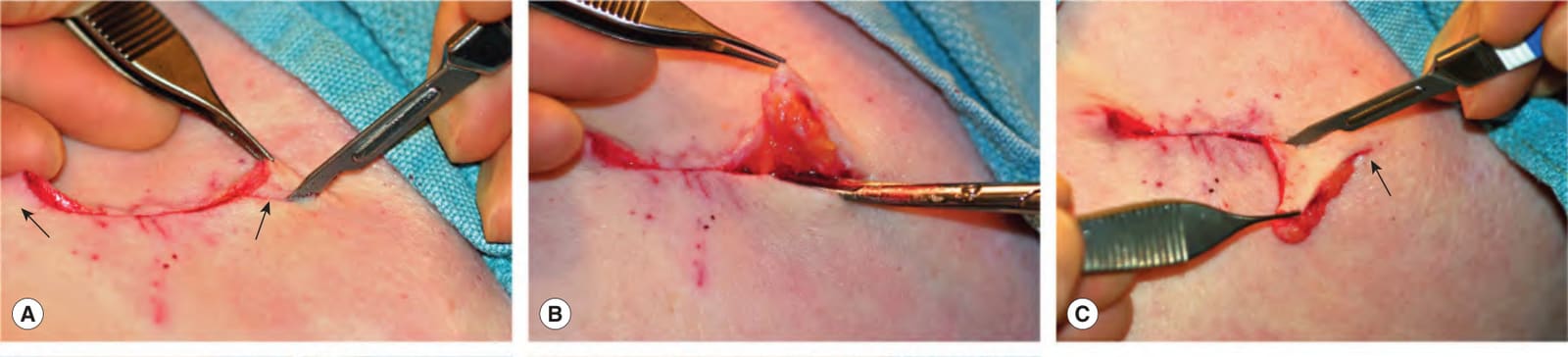
Fig. 146.16 Repair of standing cones by straight excision.A Standing cones are protrusions that form at each end during closure of a disc-shaped defect (arrows). The first limb of the standing cone is incised to the subcutaneous compartment by scissors or a blade. B The wound is undermined beneath the entire standing cone in the same plane as the rest of the defect. C The undermined standing cone is draped over the incision; the arrow indicates the apex which lies flat against the underlying skin surface. Scissors or a blade is used to complete the excision of the second limb of the standing cone. D The redundant tissue removed is in the shape of a triangle, often called a Burow’s triangle. E Subcutaneous and epidermal sutures are placed to complete the closure.
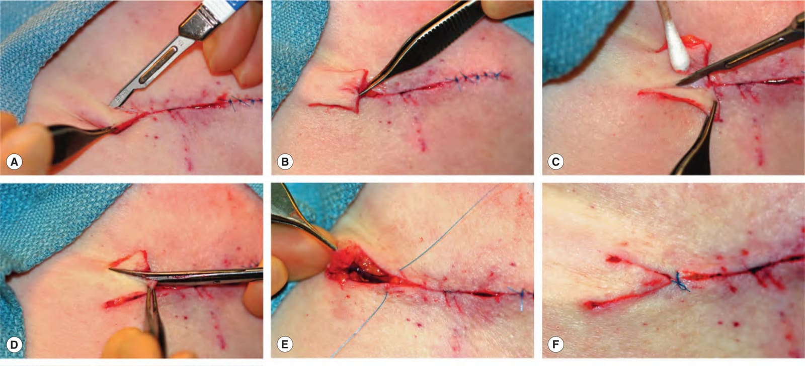
Fig. 146.17 Repair of standing cones by M-plasty.A Each limb of the standing cone is cut at a 30–45° angle from the end of the wound to about halfway toward the apex of the redundancy. B After both limbs are cut and the wound is fully undermined beneath the standing cone, the redundant tissue can be draped over the incision. At this point, an M is visible. C A Burow’s triangle is removed from half of the draped tissue. D A second Burow’s triangle is excised from the opposite side of the draped tissue. E A three-corner stitch (half-buried horizontal suture) is used to pull the point of the M-plasty into appropriate position. The needle enters the epidermis proximal to the expected position of the point, passes into the dermis, crosses the wound and enters the dermis of the tip. It then passes horizontally through the dermis of the tip, crosses the wound again, enters the opposite dermis and passes to the epidermal surface. F The knot of the corner stitch lies across the wound proximal to the position of the point of the M-plasty. G The closure can then be completed and the sutured wound lies flat.
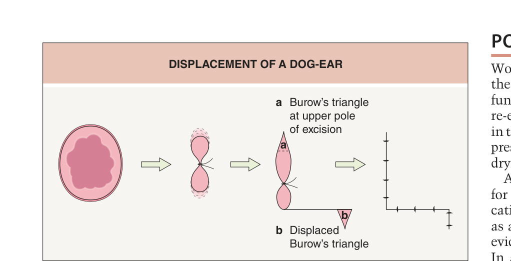
Fig. 146.18 Displacement of a dog-ear.