變化型/不尋常情況 (VARIATIONS/UNUSUAL SITUATIONS)
圓盤式切除(disc excision,邊緣可為垂直或斜角)可用於切片目的,或同時作為切片與確定性治療。此選項
適用於偏好二期癒合(healing by second intention)時,或希望先移除腫瘤、然後在閉合過程中再修補狗耳(dog-ears)時。當一期閉合(primary closure)發生不良反應的風險高時,二期癒合可能較有利。舉例來說,即使是小腫瘤,若在小腿前側切除後以一期閉合,也可能產生有張力的傷口,且相較於其他部位的傷口,更容易發生傷口裂開、感染或疼痛。若剝離不足,且術後最初 5–7 天未將負重與肢體下垂降至最低,情況更是如此。
小腿前側張力過大的傷口,另一個選項是進行植皮,但術後照護需要抬高腿部並絕對避免負重。或者,可用荷包縫合(pursestring suture)將傷口部分或完全閉合。後者是繞著圓盤整個周緣所置放的連續縫線。置放後,對縫線兩端施加張力直到達成最大收縮,然後將兩端打結。荷包縫合的優點是快速、將張力呈環周分佈,並免除為完成梭形切除而須移除多餘正常皮膚的必要。由於以二期癒合的傷口達成完全再上皮化所需的時間,與該傷口內可畫出的最大圓的直徑有關,即使將傷口閉合 30% 也能顯著縮短癒合時間。若有需要,此較小的傷口亦可植皮。
當傷口以二期癒合時,術後照護的負擔較輕,因為病人幾乎可立即恢復正常活動,同時每日為傷口換藥包紮。然而,相較於線性切除,再上皮化所需的時間顯著較長;後者與傷口大小直接相關,在下肢遠端可從 2 週到數個月不等。病人也需要考量最終形成的疤痕,即凹陷的圓形疤痕、植皮,或線性疤痕之間的取捨。
以圓盤式切除或碟形切除(saucerization,斜角邊緣)移除上軀幹與肩部的黑色素細胞痣(melanocytic nevi),所形成的疤痕比梭形切除更小,通常在美觀上也更可接受。此外,如耳部等部位的腫瘤切除,可能適合以圓盤式切除後接二期癒合。
先進行圓盤式切除、然後在閉合過程中修補狗耳,可讓外科醫師特別專注於適當的治療性邊界問題,而不必同時處理閉合的美觀與功能議題。一旦腫瘤已被移除且該區域已廣泛剝離,即可決定閉合線,也就是張力最小且符合該部位主要皮紋線的閉合方式。傷口的
中央部分以皮下與表皮縫線、或以一針大的單純間斷暫時縫線對合,然後再修補狗耳。由於美觀與功能議題是在腫瘤切除與剝離之後才處理,所形成的疤痕可達到最佳化。
切除的形狀也可透過一種稱為 S-plasty 的術式加以修改。此變化型在凸面上最為有用,它增加疤痕的總長度,同時保持兩個頂點之間的直線距離不變。在單純的梭形切除中,張力主要位於傷口中央,並沿著單一與傷口軸垂直的向量作用。相對地,S-plasty 將張力重新導向並沿傷口重新分佈為多個向量,從而使傷口中央的張力最小化;此外,中央的張力向量與傷口頂點處的主要張力向量互相垂直。由於大部分張力自最大凸起處被移轉開來,中央凹陷或傷口裂開的可能性因而降低。
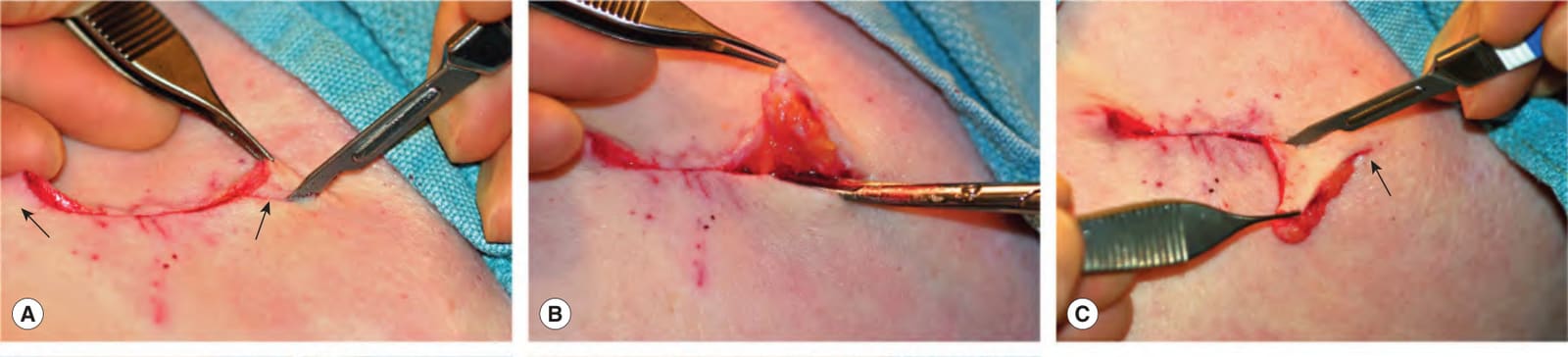
Fig. 146.16 Repair of standing cones by straight excision.A Standing cones are protrusions that form at each end during closure of a disc-shaped defect (arrows). The first limb of the standing cone is incised to the subcutaneous compartment by scissors or a blade. B The wound is undermined beneath the entire standing cone in the same plane as the rest of the defect. C The undermined standing cone is draped over the incision; the arrow indicates the apex which lies flat against the underlying skin surface. Scissors or a blade is used to complete the excision of the second limb of the standing cone. D The redundant tissue removed is in the shape of a triangle, often called a Burow’s triangle. E Subcutaneous and epidermal sutures are placed to complete the closure.
圖 146.16:以直線切除修補立體錐(standing cones)。A 立體錐是在閉合圓盤形缺損時於兩端形成的隆起(箭頭)。以剪刀或刀片將立體錐的第一臂切開至皮下腔室。B 在整個立體錐下方、與缺損其餘部分同一平面進行剝離。C 將剝離後的立體錐覆蓋於切口之上;箭頭指出平貼於下方皮膚表面的頂點。以剪刀或刀片完成立體錐第二臂的切除。D 所移除的多餘組織呈三角形,常稱為 Burow 三角(Burow’s triangle)。E 置放皮下與表皮縫線以完成閉合。
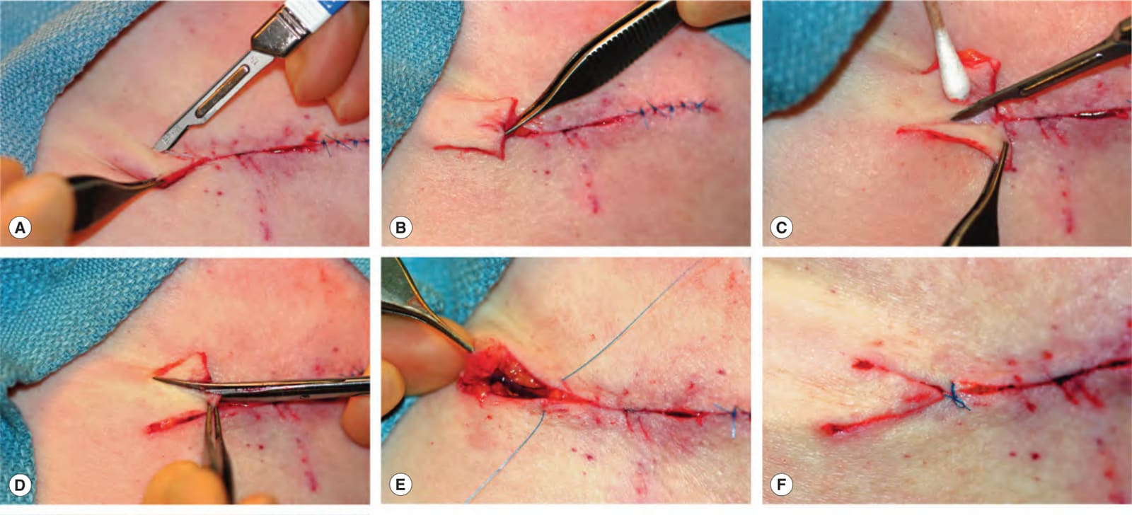
Fig. 146.17 Repair of standing cones by M-plasty.A Each limb of the standing cone is cut at a 30–45° angle from the end of the wound to about halfway toward the apex of the redundancy. B After both limbs are cut and the wound is fully undermined beneath the standing cone, the redundant tissue can be draped over the incision. At this point, an M is visible. C A Burow’s triangle is removed from half of the draped tissue. D A second Burow’s triangle is excised from the opposite side of the draped tissue. E A three-corner stitch (half-buried horizontal suture) is used to pull the point of the M-plasty into appropriate position. The needle enters the epidermis proximal to the expected position of the point, passes into the dermis, crosses the wound and enters the dermis of the tip. It then passes horizontally through the dermis of the tip, crosses the wound again, enters the opposite dermis and passes to the epidermal surface. F The knot of the corner stitch lies across the wound proximal to the position of the point of the M-plasty. G The closure can then be completed and the sutured wound lies flat.
圖 146.17:以 M-plasty 修補立體錐。A 立體錐的每一臂自傷口末端以 30–45° 角切開,切至朝多餘組織頂點約一半的位置。B 兩臂都切開且立體錐下方的傷口已充分剝離後,多餘組織即可覆蓋於切口之上。此時可看見一個 M 形。C 自覆蓋組織的一半切除一個 Burow 三角。D 自覆蓋組織的對側切除第二個 Burow 三角。E 以三角縫合(three-corner stitch,半埋入式水平縫線)將 M-plasty 的尖端拉至適當位置。針自尖端預期位置的近端進入表皮,進入真皮,越過傷口並進入尖端的真皮。接著水平穿過尖端的真皮,再次越過傷口,進入對側真皮並穿至表皮表面。F 角縫線的結橫跨傷口,位於 M-plasty 尖端位置的近端。G 接著即可完成閉合,縫合後的傷口呈平坦。
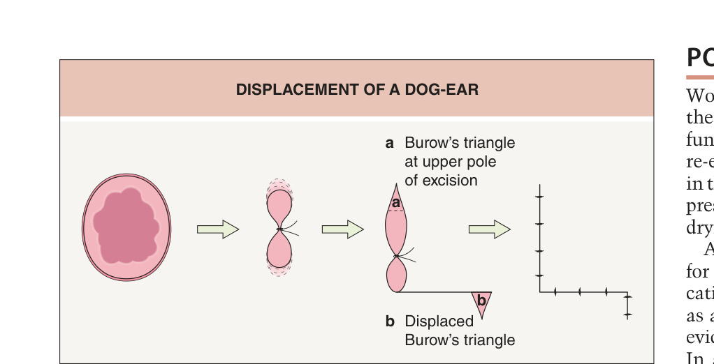
Fig. 146.18 Displacement of a dog-ear.
圖 146.18:狗耳的位移。