BACKGROUND
At the patient’s side, it is always helpful to have an on-demand recall of flap options. With experience, this is automatic, but those with less experience may find the mnemonic STARTS helpful (Table 147.1). It is incumbent upon the surgeon to synthesize the many factors that may affect the choice of the most appropriate flap for a defect. The characteristics of the wound, qualities of the adjacent skin, the functional aspects of the affected anatomic site, the pertinent aesthetic considerations, the anatomic borders in proximity to the defect, and the age and expectations of the patient all contribute to deciding the most appropriate reconstruction.
The most important goal in reconstruction is functional aesthetics. This means that the reconstruction should first and foremost maintain all vital functions and accomplish this aesthetically. Not all aesthetic reconstructions are functional, but all functional reconstructions can be aesthetic.
Fundamentals of Flap Design and Suturing Technique
Technical precision, including suturing technique, may be the most important variable in achieving an outstanding result. The first important step is to prepare the wound edges of both the defect and the flap. All incisions should be made at a 90° angle to the skin surface such that the wall of the defect and the edge of the flap are square. Secondly, the wound base should be made a uniform depth. Whether reconstructing with an elliptical side-to-side closure or with any of the more intricate flaps, the contour of the wound base will persist and may affect the long-term appearance of the wound. Likewise, the thickness of the flap should be uniform. Although the optimal plane of flap dissection is somewhere between the upper mid subcutaneous and deep subcutaneous tissue, the ideal thickness of a flap varies and must be determined on a case-by-case basis. Flaps constructed too thinly can develop vascular compromise or may lead to an unnatural appearance. Conversely, a very thick flap may be bulky, have decreased mobility or be more difficult to manipulate while suturing, and it may unintentionally damage underlying neurovascular structures or muscles. Hair-bearing skin mandates attention to the level of the hair follicles.
Widespread undermining of the subcutaneous tissue adjacent to both the defect and the donor site defect is important for creating a broad “plate-like scar” (Fig. 147.1). This results in an even distribution of scar contraction over a greater area and diffuses the effects of contraction such that it is not focused on the flap itself or the incision lines of the flap (Fig. 147.2). Undermining should be uniform and complete, including the areas of low tissue tension such as the tips of an ellipse or the pivot points of flaps.
Next, meticulous pinpoint hemostasis with complete visualization of all undermined skin with the help of an assistant is critical. Excessive postoperative bleeding within a sutured wound often results in an inferior outcome. It should be emphasized, however, that extensive tissue injury caused by electrosurgical hemostasis should be avoided as fervently as postoperative bleeding, since this can contribute equally to unwanted results. With a well-prepared wound and flap that are characterized by uniformity in thickness, sharply squared edges, well-loosened tissue margins, and excellent hemostasis, the wound is ready for suturing.
An adequately undermined flap may still lead to distortion of delicate structures if the flap is large, heavy, or moves under considerable tension. A flap suspension suture may be necessary to avoid distortion or displacement of delicate tissue. These sutures are placed in fixed or rigid structures underlying the flap. Periosteal sutures represent a common anchoring technique and they are placed from the periosteum through the undersurface of the flap into the deep dermis. When this suture is placed in the undersurface of the flap, it should correspond topographically to its desired location relative to the periosteal suture. More than one suspension suture may be needed and should be placed in a direction parallel to the flap blood supply to avoid vascular compromise of the flap.
Whether it is a side-to-side closure or a flap, eversion of the skin edges (along with undermining) is also essential (see Fig. 147.1). Given their longer period of effectiveness, as compared to epidermal sutures, the author prefers the use of subcutaneous (“buried”) vertical mattress sutures to achieve eversion (see Ch. 146).
Flap Movement Characteristics
It is helpful to broadly characterize flap types according to their mechanism of skin movement and rearrangement. By understanding the dynamics of each flap, it is easier to select one that will provide functionally aesthetic results. Upon consideration of the unique characteristics of the defect, the donor skin, and the functional and aesthetic issues of reconstruction in the context of the mechanics of flap tissue movement, a rational decision regarding the method of closure can be made. The components that flaps share include the flap body, the pedicle or vascular base, primary and secondary defects, and, as an integral part of flap dynamics, the primary and secondary movements (Table 147.2, Fig. 147.3).
Most tissue movement results in some degree of tension. This tension creates vectors of force that tend to return moving skin back to its original position. The primary movement of a flap creates a primary tension vector, while the secondary movement (during closure of the donor site defect) creates a secondary tension vector. Consideration of these forces and the effects of these forces on adjacent tissue and even the blood supply of the flap is the primary factor in selecting one flap over another. The most perfectly executed flap that, via tension vectors, results in the distortion of a free margin (e.g. a lip or eyelid) is of little value to the patient or the goal of aesthetic restoration. Therefore, the initial and most important skill required for the proper execution of flaps is the ability to anticipate the direction and amount of tension created by a particular flap and to predict what effects this tension will have on adjacent structures (see Figs. 147.2 & 147.3). Secondarily, judicious use of tension sutures is helpful.
There are four broad categories of flaps based upon how the tissue is rearranged to achieve closure (Table 147.3). In the first category, one or both Burow’s triangles that would be removed if the wound were closed by a simple side-to-side closure are displaced. The second includes the flaps that reconfigure defects, while the third includes flaps that reorient tissue from adjacent pools of laxity into the defect. Finally, there is a category of flaps that enables defect closure by tissue importation.
Burow’s triangle displacement flaps
Flaps that displace Burow’s triangles to a “convenient” location distant from the defect include the single tangent advancement flap (Burow’s flap), the bilateral single tangent advancement flap (A-to-T flap), the double tangent advancement flap (U-flap), the bilateral double tangent advancement flap (H-flap), and rotation flaps (e.g. the dorsal nasal or Rieger flap, Mustarde flap, helical advancement flap) (Fig. 147.4).
These flaps displace Burow’s triangles along straight or curved incision(s) tangential to the wound.
The key features of these flaps are: (1) they displace Burow’s triangles to a distant site; (2) there is limited reorientation of tissue; and (3) their movement is dependent upon the intrinsic elasticity of the skin. Just as a simple side-to-side closure performed in stiff, inelastic skin would result in a tense closure, so would a Burow’s triangle displacementtype flap. When the surrounding skin is elastic and “loose”, the wound edges can be closed under minimal tension in both a side-to-side closure and with these flaps. Thus, the only advantage that these flaps confer over a side-to-side closure in tight skin is that a larger area of skin is typically undermined, which releases the flap from the tethering effect of the subcutaneous tissue. This allows for a modest decrease in wound closure tension. The primary utility of these flaps is in defects where, for functional or aesthetic reasons, it is inconvenient to remove a Burow’s triangle immediately adjacent to the defect. An example of this is a defect above or below the eye in which displacement of the Burow’s triangle is useful to avoid removing a triangle from the eyebrow or eyelid (Fig. 147.5).
Defect reconfiguration flaps
The pedicled V-to-Y advancement flap is unique in that it is an advancement of tissue with its vascular base as a pedicle of subcutaneous tissue and muscle oriented in a plane perpendicular to the skin and at 90° to the more traditional advancement or rotation flaps. In this case, the tissue redundancy created at the pivot point of the flap is buried in the subcutaneous plane at the advancing edge of the flap (see below). Fundamentally, the pedicled V-to-Y flap reconfigures the typical roundish shape of the wound to a sharp, angled, geometric shape that is easily closed without standing cones or distortion of surrounding structures (Fig. 147.6). Movement of this flap is entirely dependent on the elasticity of the tissue that comprises the vascular pedicle of the flap.
Tissue reorientation flaps
Tissue reorientation flaps are characterized by the lifting of skin from an adjacent donor site and reorienting it through the transposition of the flap over a peninsula of skin between the donor site and the defect. There is a pivot point at the base of this flap (see Fig. 147.3). The tissue movement depends on the presence of laxity in adjacent tissue. Although intrinsic elasticity of the flap skin may facilitate the ease of execution, without ample laxity in the donor site, the transposition cannot be performed. Some of the flaps that fall into this category include the rhombic transposition flap (see Fig. 147.2), the bilobed transposition flap, and the nasolabial transposition flap (see Table 147.3).
To better understand the reorientation of tissue that occurs in these flaps, a review of the most basic tissue reorientation flap, the Z-plasty, is helpful. Z-plasties transpose two angular flaps, placing them in a complementary fashion into the defects of the other flap (Fig. 147.7). This reorientation of tissue ultimately results in the lengthening of the skin in the direction of the middle arm of the Z-plasty (Fig. 147.8). There is a complementary decrease in the length of skin perpendicular to this same arm. The amount of lengthening is proportional to the angles of the flap(s). Put another way, tissue gain in the direction of the middle arm is proportional to the width of the base of the flaps. This is effectively the mechanism of tissue reorientation in transposition flaps. Upon careful examination, Z-plasties can be found within the design of transposition flaps (Fig. 147.9). Therefore, just as with Z-plasties, transposition flaps work because the reorientation of the tissue results in lengthening of tissue in the direction of the skin deficit at the expense of the laxity in the donor site.
It is helpful to understand the utility of these principles of reorientation – an example of this would be a defect on the nasal tip (Fig. 147.10). The skin is typically very tight in this area, making simple sideto-side closure in the vertical direction difficult. Side-to-side closure in the horizontal direction is impractical because of the distortion that would occur to the free margin of the ala. When one looks for donor sites, there is typically a substantial amount of laxity on the mid nasal dorsum and nasal sidewall. A bilobed flap allows utilization of this laxity through tissue reorientation using two lobes. The underlying mechanism of tissue reorientation is a series of Z-plasties inherent in this flap. Since tissue gain in Z-plasties is in the direction of the middle arm of the Z-plasty, a substantial reorientation and lengthening of tissue in the desirable direction for repair of the defect occurs without distortion. In contrast to the Burow’s triangle displacement flap, this flap does not rely upon intrinsic elasticity of the flap skin. This is key to avoiding distortion of the alar rim.
Tissue importation flaps
Large defects, or defects in areas where tissue laxity is lacking, may be reconstructed by importing vascularized skin from a site that is not adjacent to the defect (see Table 147.3). Tissue importation flaps are typically derived from a distant donor site with ample tissue laxity and, most importantly, an excellent blood supply. This blood supply may be based on a particular artery (e.g. paramedian forehead flap based on the supratrochlear artery) or by a rich, random-pattern blood supply (e.g. the retroauricular pedicle flap).
These flaps are typically two-staged flaps requiring a period of vascular ingrowth from the wound bed of the defect. This period ranges from 2 to 6 weeks, with 3 weeks being the most commonly reported time point for transection of the pedicle. In the head and neck region, defect sites where tissue importation flaps are commonly employed include the nose, ear, eyelid, and lip.
There are also single-staged tissue importation flaps. These are tunneled flaps in which skin from a non-adjacent site with a subcutaneous and muscular pedicle is passed through a “tunnel” to the defect site. The pedicle lies within the tunnel providing blood supply to the flap.
The appropriate selection and execution of these four broad flap categories are discussed in more detail below.
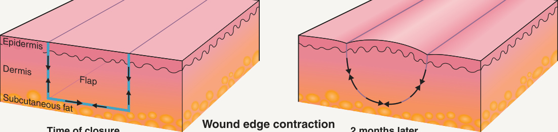
Fig. 147.1 Scar contraction.A Sutured wound without wound edge eversion and without undermining of the surrounding tissue. B The results of scar contraction, leading to inversion of the scars and trapdoor deformity of the flap. C A wound sutured with eversion and wide undermining. D The canceling effect of scar contraction and wound eversion as well as the diffusion of scar contraction in the deep, undermined wound plane (“plate-like” scar).
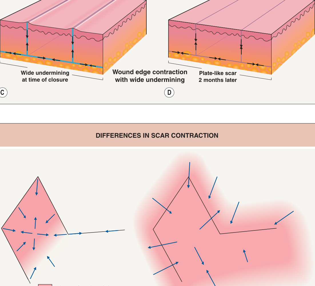
Fig. 147.2 Differences in scar contraction. Depiction of differences between wounds that are undermined and those that are not undermined prior to suture placement. A The contraction forces are focused on the margins of the flap in a wound that was not widely undermined. B Broad diffusion of contractile forces that are not concentrated at the flap margins, making trapdoor deformity (“pincushioning”) less likely in the widely undermined flap.
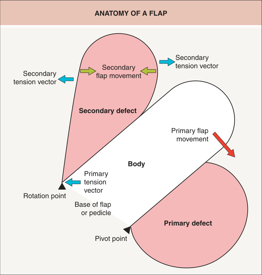
Fig. 147.3 Anatomy of a flap. Definitions of the components of a flap are outlined in Table 147.2.
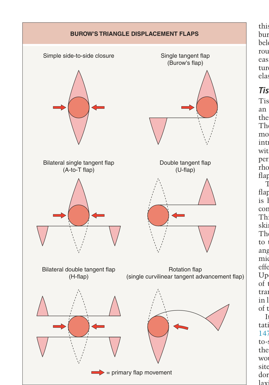
Fig. 147.4 Burow’s triangle displacement flaps.
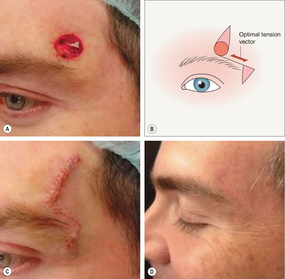
Fig. 147.5 Single tangent advancement flap
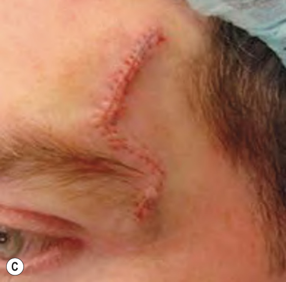
Fig. 147.6 The pedicled V-to-Y flap. Reconfiguration of the defect (pink shaded area) in A to the defect in B. The shaded secondary defect in B is comprised entirely of acute angles which are easily closed primarily (C). Also see Fig. 147.20.
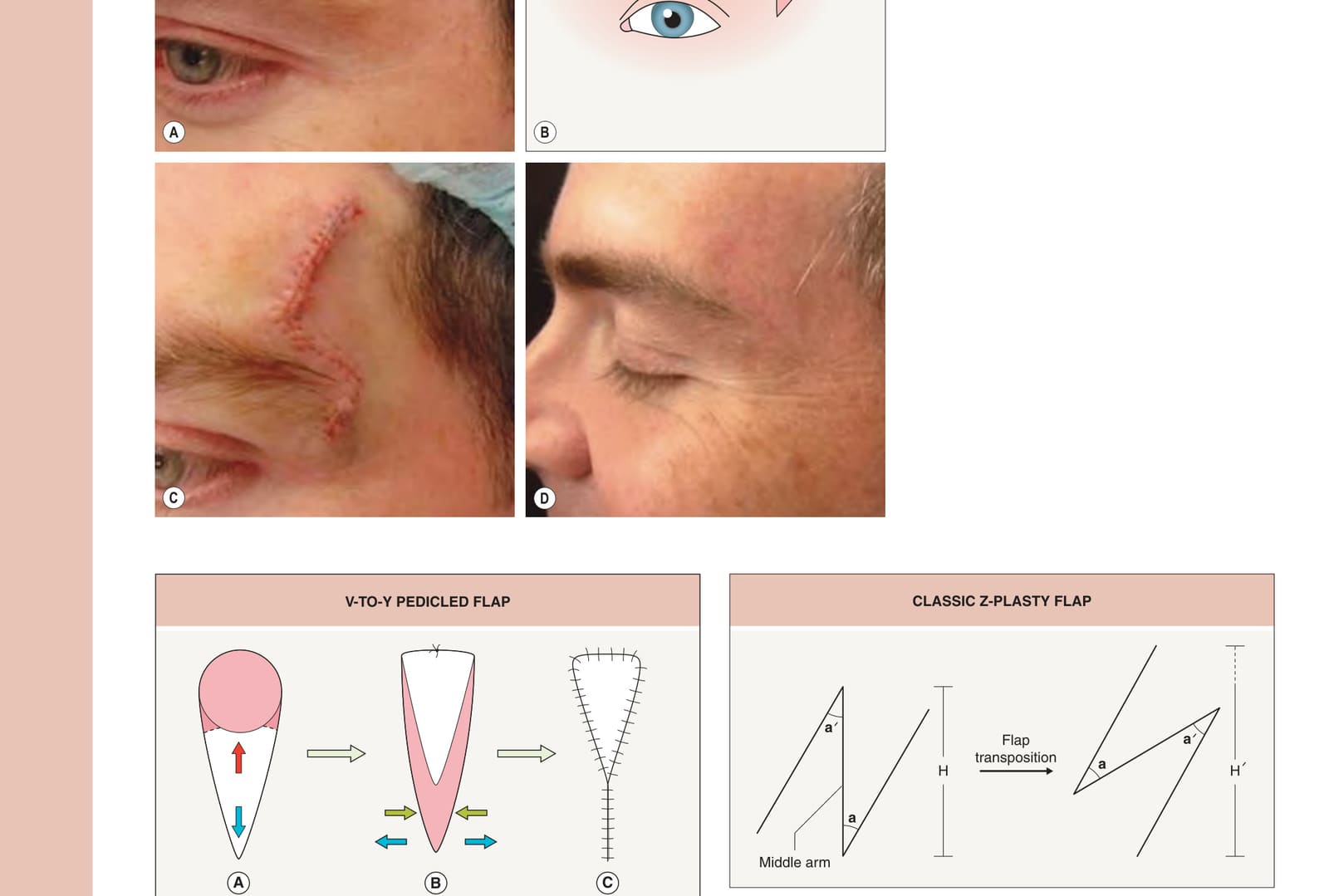
Fig. 147.7 Classic Z-plasty flap. The directional tissue gain (dotted line) is depicted (H to H′) after flap transposition in the direction of the middle arm of the Z-plasty.
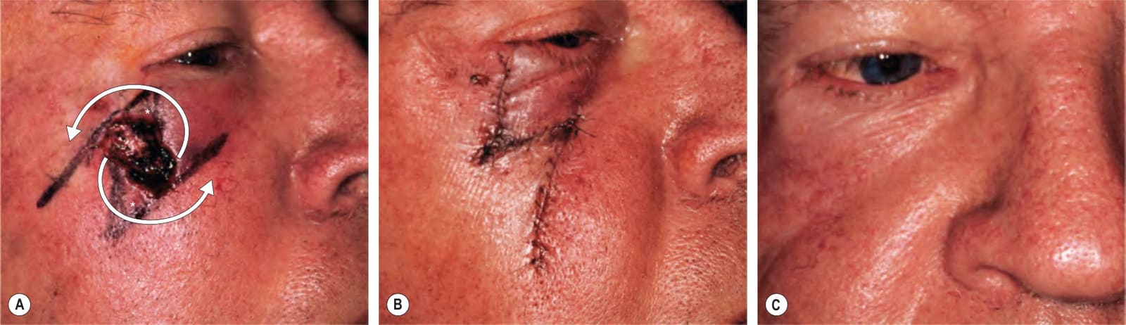
Fig. 147.8 Classic Z-plasty flap.A Defect close to the eyelid margin. The middle arm of this Z-plasty flap is composed of the defect plus the Burow’s triangles excised above and below it to facilitate the transposition of the flaps (*). The medial flap is transposed over the lateral flap (arrows). B The wound is closed via a Z-plasty closure with the transposition of flaps at the upper medial and lower lateral aspects of the wound. This is done to lengthen tissue in the direction of the palpebral margin in order to prevent ectropion as demonstrated at the 4-month postoperative visit (C).
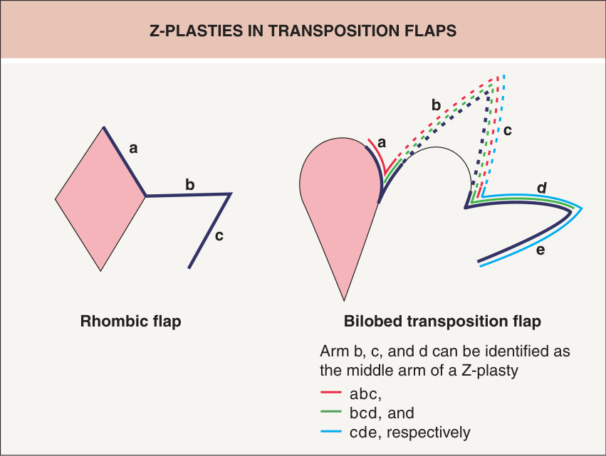
Fig. 147.9 Z-plasties in transposition flaps. Z-plasties are highlighted within the designs of both the rhombic and the bilobed transposition flap. Note in the rhombic flap the middle arm of the Z-plasty is line (b) and is the direction of anticipated tissue lengthening. In the bilobed transposition flap diagram, there is a series of three Z-plasties all contributing to tissue gain in the desired direction of the defect.
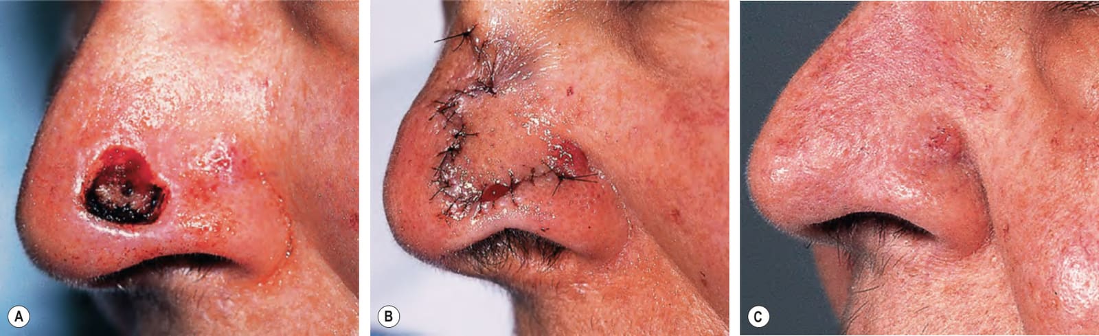
Fig. 147.10 Bilobed flap. A Defect on the nasal tip adjacent to the free margin of the ala. Bilobed flap reconstruction with minimal distortion of the alar rim (B) and long-term result (C).
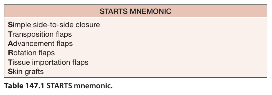
Table 147.1 STARTS mnemonic.
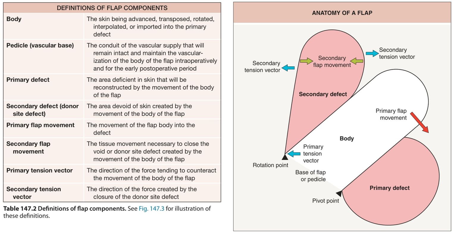
Table 147.2 Definitions of flap components. See Fig. 147.3 for illustration of these definitions.
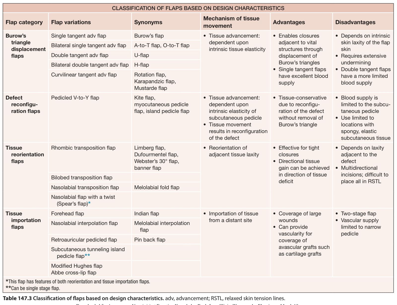
Table 147.3 Classification of flaps based on design characteristics. adv, advancement; RSTL, relaxed skin tension lines.