WOUND HEALING CONSIDERATIONS
Wound healing after skin grafting proceeds through a unique series of events. The first 24-hour period following graft placement is termed
the stage of plasmatic imbibition, during which fibrin glue attaches the graft to its recipient bed, allowing it to take up the underlying wound exudate and to become edematous, gaining up to 40% in weight. The graft thereby remains hydrated and obtains a supply of nutrients, which maintains graft vessel patency until revascularization begins. The fibrin beneath the graft is subsequently replaced by granulation tissue, which attaches the graft permanently to its bed.
With proper apposition of the graft to its bed, revascularization may proceed. Anastomoses begin to form within 48–72 hours of grafting between the recipient bed and pre-existing vessels in the dermis of the graft, a process known as inosculation. Vascular proliferation occurs next, with sprouting and budding of vessels in the graft and its bed. Even relatively avascular tissue may be grafted, as long as the avascular area is small and surrounded by a rich vascular supply. Through a process known as the bridging phenomenon, vascular connections arising from the vascularized part of the recipient bed allow blood flow to occur through pre-existing graft vasculature, so that nutrients reach the part of the graft overlying the avascular area. Full circulation is restored to the graft within 4–7 days.
Restoration of the lymphatic circulation parallels restoration of the blood supply over the first week. Epidermal proliferation occurs between the 4th and 8th day post-transplant and persists for several weeks. Degeneration of sebaceous and eccrine glands may occur initially, but subsequent glandular regeneration may allow partial function to be maintained. Graft reinnervation and return of sensory nerve function may begin as early as 2–4 weeks after grafting, although patients do not usually regain full sensation for many months.
If extension of the ischemic period occurs, decreased graft survival may result (Table 148.3). Hematoma or seroma formation, infection, or mechanical shear forces may disrupt the fragile vascular connections between the graft and its bed. These complications tend to affect FTSGs (which have a greater volume to nourish and revitalize) more than STSGs.
Even after the ischemic period is past, other factors may decrease the vascular supply nourishing the undersurface of the graft. The most important of these is cigarette smoking or use of other nicotinecontaining products, but diabetes mellitus, protein deprivation, and severe trace element or vitamin deficiencies may also increase the risk of graft failure. Certain systemic medications, such as corticosteroids, chemotherapeutic agents, other immunosuppressive drugs and anticoagulants, may interfere with wound healing as well.
Other causes of graft failure include: insufficient vascularity due to necrotic debris within the recipient bed, hematoma, seroma, an avascular wound bed, previous radiotherapy, infection, excessive graft tension, mechanical shearing forces, and improper postoperative care (see Table 148.3). The most common infectious agents associated with graft failure include coagulase-positive staphylococci, β-hemolytic streptococci, and Pseudomonas spp. Pseudomonal infections are particularly common in auricular grafts. For all of these reasons, a thorough preoperative evaluation, meticulous intraoperative technique, and good postoperative care are essential to maximize graft survival.
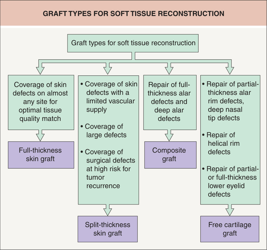
Fig. 148.1 Graft types for soft tissue reconstruction.
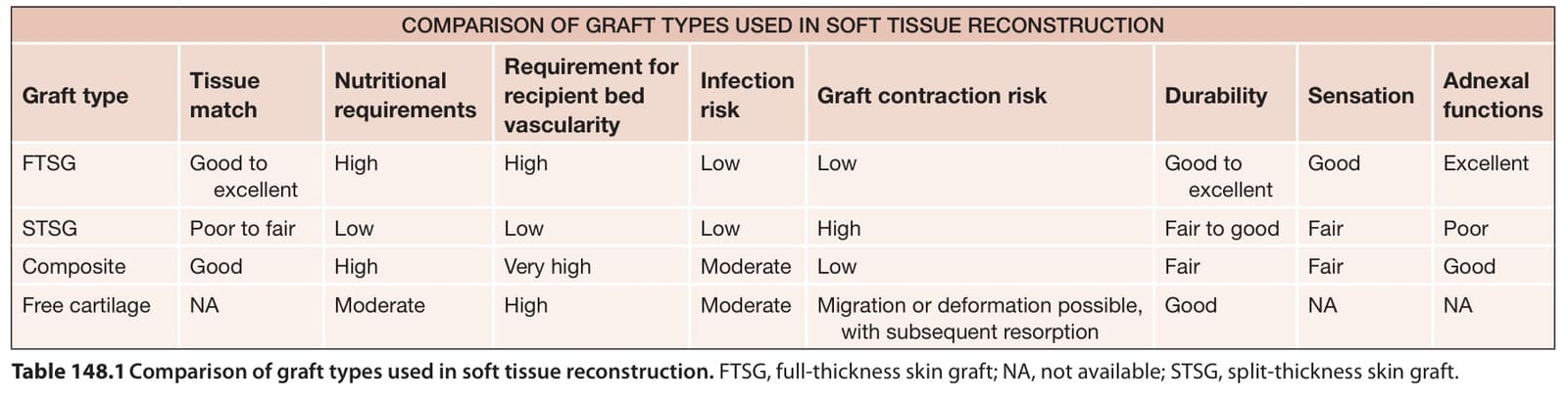
Table 148.1 Comparison of graft types used in soft tissue reconstruction. FTSG, full-thickness skin graft; NA, not available; STSG, split-thickness skin graft.
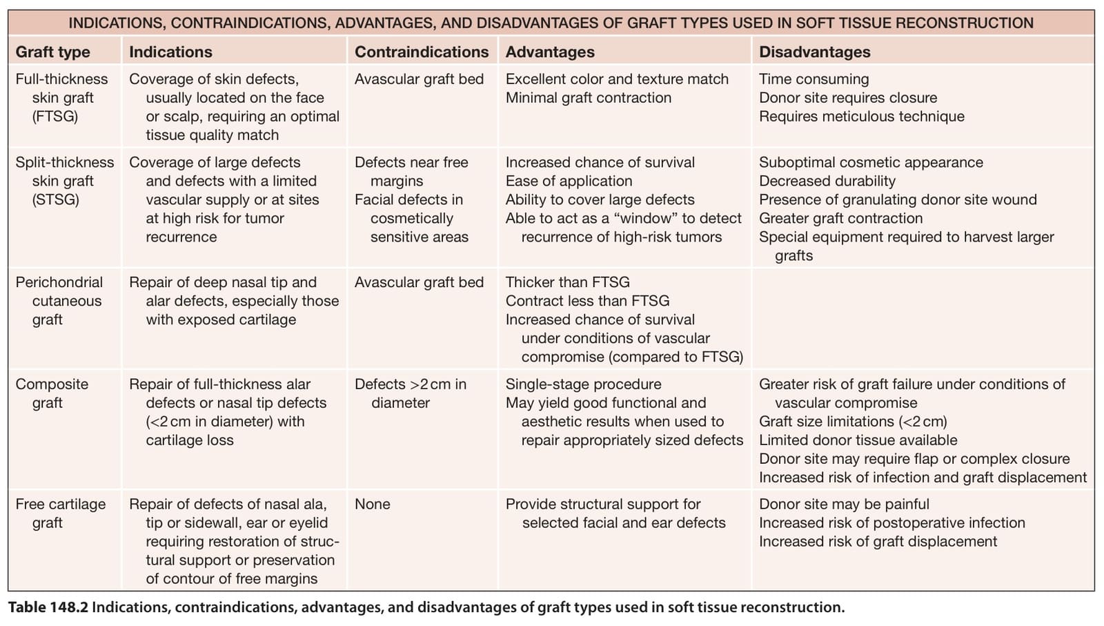
Table 148.2 Indications, contraindications, advantages, and disadvantages of graft types used in soft tissue reconstruction.
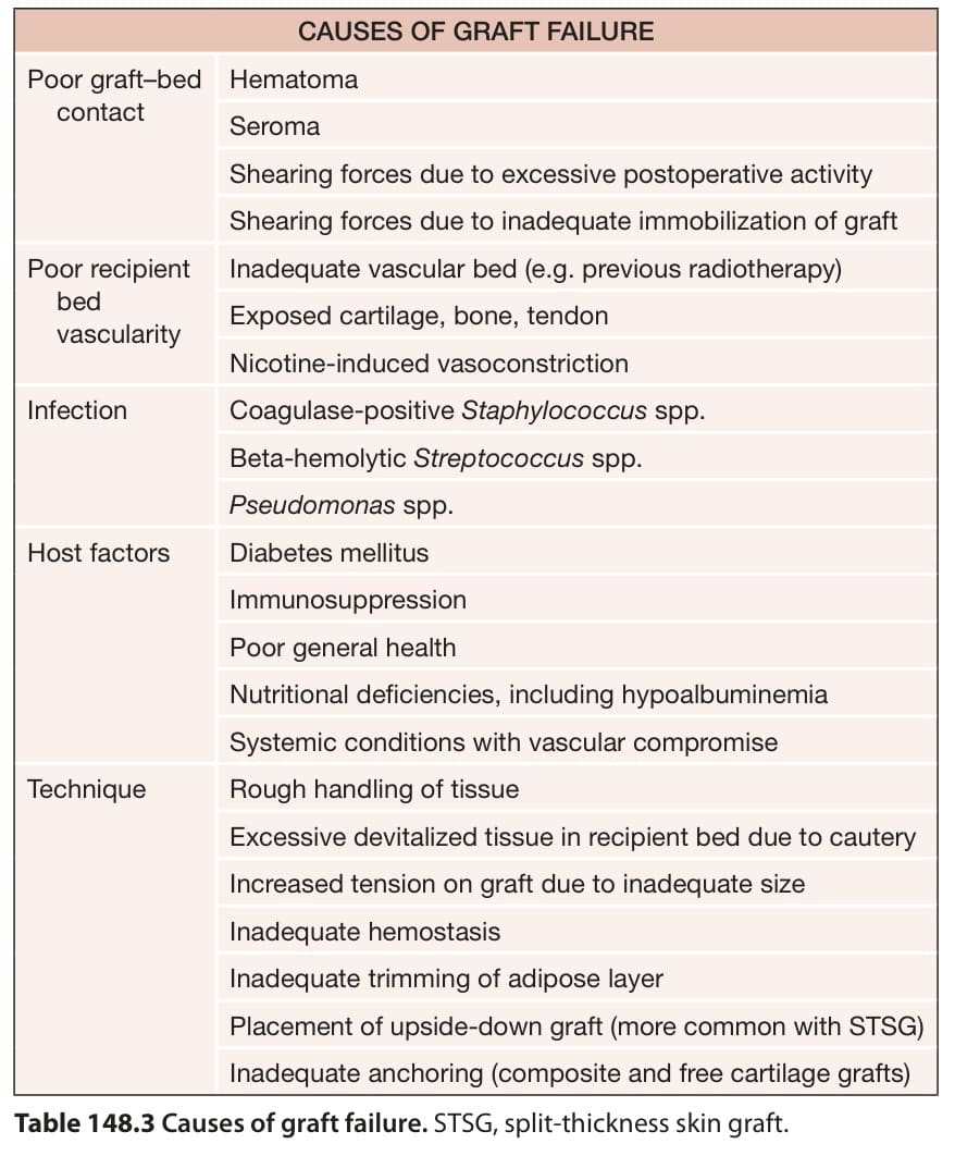
Table 148.3 Causes of graft failure. STSG, split-thickness skin graft.