FULL-THICKNESS SKIN GRAFTS
Indications/Contraindications
FTSGs are most commonly used to repair facial defects resulting from skin cancer removal. They may be used to repair defects at virtually any site, as long as the recipient bed has a sufficiently rich vascular supply to promote capillary regrowth and fibroblasts to supply collagen for graft adherence. Small avascular areas may be grafted due to the bridging phenomenon. Larger avascular areas, including patches of exposed bone, cartilage, tendon or nerve devoid of periosteum, perichondrium, peritenon or perineurium, respectively, are unable to support FTSGs. For this reason, FTSGs should not be placed over larger areas of avascular tissue.
Under the proper circumstances, FTSGs can provide excellent color, texture, and thickness match for facial defects because they include the full thickness of epidermis and dermis. Wound contraction is minimized since the full thickness of dermis is present, and adnexal structures remain intact. FTSGs may be especially useful for the cosmetically and functionally acceptable repair of defects of the nasal tip, dorsum, ala and lateral sidewall, as well as the lower eyelid and ear (Fig. 148.2).
Preoperative History and Donor Site Considerations
A thorough preoperative evaluation that includes questions regarding bleeding tendencies, alcohol use, use of anticoagulant medications (including aspirin and nonsteroidal anti-inflammatory drugs), and a history of hypertension can help assess the risk of excessive bleeding and clot formation beneath the graft. Diabetes mellitus, nutritional deficiencies, cigarette smoking, or the use of other nicotine-containing
products may increase the risk of graft failure, and any history of these should be identified preoperatively.
Selection of a donor site for full-thickness skin grafting depends upon the color, texture, thickness, and sebaceous qualities of the skin
surrounding the defect (Tables 148.4 and 148.5). Most FTSGs are taken from sun-exposed areas above the shoulders whose color, vascular pattern, texture, and distribution of adnexal structures best match the skin surrounding facial defects. The thinnest grafts are usually harvested from the upper eyelid or the postauricular sulcus. Medium-thickness grafts are often harvested from the preauricular and cervical regions, while thicker grafts may be taken from the supraclavicular or clavicular region, conchal bowl, or nasolabial fold. Donor skin thickness, irrespective of site, will vary from one patient to another. It is therefore important to examine all sites carefully to find the best possible tissue match. This approach will ensure optimal donor site selection for each individual patient and surgical defect.
A regional approach may, at times, be used to obtain the best possible match for a given defect. Grafts taken from redundant upper eyelid skin may be used to repair lower eyelid defects, providing a good color and texture match in addition to a well-camouflaged donor site scar. Grafts used for lower eyelid defects should be oversized by 100%–200% to allow for contraction and to avoid ectropion. Due to its relatively large size and inconspicuous location, postauricular skin may also be useful as a donor site for eyelid and auricular defects. Because postauricular skin is relatively non-sun-exposed, grafts harvested from this region may not provide a good color or texture match for other facial defects.
Preauricular skin is more versatile and can be used to repair most nasal defects, since the thickness and degree of sun exposure of these areas tend to be comparable. Donor site scars in this region can be easily camouflaged, as in facelift surgery, to provide a cosmetically desirable result. Even bearded individuals have a 1–2 cm hairless zone in the preauricular region. Care must be taken not to harvest hairbearing skin in grafts taken from this area. Accidentally including mature follicular units may produce undesirable hair growth within the graft. Hair-bearing skin from the temporoparietal region may be used to repair eyebrow defects, where survival of the follicular units after transplantation may produce a superior cosmetic result. Skin from the nasolabial fold or the conchal bowl can sometimes be used to graft small nasal tip or ala defects with good tissue match. At times, sufficient skin laxity may be present, particularly on the nose or forehead, to allow for partial defect closure, utilizing the adjacent Burow’s triangle as a FTSG to repair the remainder of the defect, providing an excellent tissue match.
For larger defects requiring FTSGs of sun-damaged skin, such as the forehead and scalp vertex, the supraclavicular region or lateral neck can be used as donor sites. Again, care must be taken not to harvest
unwanted hair along with the graft. These donor sites are often more difficult to camouflage, especially in areas that might not always be covered by clothing, and must be carefully oriented. Although their color and texture match may be suboptimal, areas below the neck with thin, redundant skin, such as the upper inner arms, forearms and inguinal area, can also be considered as donor sites.
Description of Technique
Varying techniques for harvesting and placing FTSGs have been described. To perform a FTSG, a template of the defect is first made, using any flexible material, such as gauze, Telfa™ or aluminum foil, which can be bent to conform to the defect (Fig. 148.3A). After marking the periphery of the recipient site with a sterile marker, the template material is pressed against the defect, and the resulting outline of the inked margin serves as a guide to cut a perfect template. The template is applied to the donor site, and inking material is applied around it (Fig. 148.3B). The graft should be 3%–5% larger than the true template to allow for the natural contraction and shrinkage of the graft after it has been harvested. Often, just cutting around the outside of the inked margin accounts for this extra “safety” skin. For eyelid defects, grafts should be oversized significantly to minimize the risk of ectropion. Marking the donor site prior to local anesthesia prevents incorrect sizing due to tissue stretch from lidocaine infiltration. It is important to note that a donor site ellipse with a 3 : 1 ratio may not always be required, allowing for smaller donor defects.
After the donor site is marked, local anesthesia is injected into the donor and recipient sites. Epinephrine (adrenaline) may be used without compromising graft survival. If waterproof ink is not available for template marking, the outline of the template may be scored on the donor site with a needle. The donor site and recipient beds are then scrubbed with an antibacterial preparation such as povidone–iodine (especially around the eyes and ears) or chlorhexidine, excess scrub is removed, and sites are draped with sterile towels.
The donor site is excised with a scalpel to the level of the subcutaneous fat (Video 148.1). The graft is placed in a sterile bowl or Petri dish containing sterile normal saline, where it may remain for up to 1–2 hours. Grafts may be utilized up to 24 hours after harvesting if refrigerated or kept on ice. Before suturing the graft into place, defatting should occur. This is an essential step, since direct contact between the graft and its bed promotes connections between existing vessels as well as new vessel growth and nutritional support from the base of the defect. Adipose tissue adherent to the graft is poorly vascularized and is therefore not an effective medium for vessel growth between the graft and its bed.
When defatting is performed, the graft is placed dermal-side-up on the fingers or in the palm. All fat is trimmed from the underside using sharp scissors (Video 148.2). Fat is adequately removed when all of the yellow globular material is gone, leaving a white glistening dermal surface (Fig. 148.3C, Video 148.3). If indicated, part of the dermis can also be trimmed to allow for differences in recipient site depth, although adnexal structures may be removed in the process, potentially altering the cosmetic result. The graft is then placed dermis-down in its bed, and rotated and trimmed if necessary, to achieve a perfect fit (Fig. 148.3D, Videos 148.4 and 148.5). While one surgeon is trimming the graft, another may close the donor site, usually in a linear layered fashion with bilateral removal of standing cones (Fig. 148.3E). For conchal bowl donor sites, a 2- or 3-mm punch biopsy instrument may be used to remove full-thickness discs of cartilage, thereby facilitating second intention healing from the underlying dermis.
Contouring of donor skin in FTSGs may be challenging, particularly when repairing defects on the nasal tip, ala, lateral nasal sidewall, helical rim, and eyelid. Graft contouring often requires multiple trial placements within the recipient bed, as well as multiple trimmings, to obtain an optimal result. Removal of cartilage not needed for structural support from the recipient site may increase the chance of graft survival, particularly for auricular defects, without compromising the aesthetic appearance of the repair.
Securing the Graft
Perimeter sutures, basting sutures, support dressings, or a combination of any or all of these can be used to anchor the FTSG. Depending upon the graft’s size, four to eight interrupted 4–0 or 5–0 absorbable or non-absorbable sutures may be placed at opposite edges of the graft periphery (e.g. at 3, 6, 9 and 12 o’clock) to tack down its four quadrants. A simple running suture, such as 6–0 fast-absorbing chromic gut suture, may then be placed around the graft’s perimeter (Fig. 148.3F, Video 148.6). Great care is taken to achieve perfect epidermal wound approximation. The running suture is placed in almost epicuticular fashion, passing the needle first through the graft and then through the surrounding skin. Placing the suture slightly higher in the dermis on the graft side and slightly deeper in the dermis of the surrounding skin makes suture placement easier, prevents tenting of the graft edges, and maximizes graft–recipient bed contact.
Using tissue adhesives in full-thickness skin grafting has been investigated. In a pilot study, it was found that applying cyanoacrylate tissue adhesive to secure FTSGs to their recipient bed wound edges produced cosmetic results essentially identical to those achieved with suturing. Because these adhesives set virtually instantaneously, the graft must be perfectly trimmed and aligned before adhesive application, since fine adjustments in positioning the graft’s edges are not easily made. Tissue adhesives may represent a time-saving alternative for securing the periphery of certain grafts, particularly those in relatively immobile areas such as the temple, forehead, and distal nose.
Basting or quilting sutures, usually simple interrupted 6–0 fastabsorbing gut sutures, can occasionally be placed to secure the central portion of the graft. They can be useful in securing large grafts to provide extra support against movement, as well as grafts placed on a concave surface where tenting could possibly occur. All sutures should be snug, but not strangulating, so as not to compromise graft survival.
Immobilization of the graft over its bed can be maximized with pressure dressings. Classically, tie-over bolster dressings have been employed to immobilize grafts, using anchoring stents to ensure direct contact between the graft and its bed. These stents may consist of various materials including Xeroform™ gauze (bismuth tribromophenate-petrolatum-impregnated gauze), cotton balls, foam rubber, sponges, plastic beads, or disks. Although sutures or skin staples are usually employed to anchor the stent, adhesive wound closure tapes or Steri-Strips™ can also be applied to exert even pressure on the bolster. Adherence of the dressing to the graft, which tends to pull the graft from its bed at the time of dressing removal, is minimized by first applying petrolatum ointment, a non-adherent contact dressing, or Xeroform™ gauze to the graft site. Pressure or tie-over dressings help immobilize the newly placed graft during the critical period of revascularization, thereby preventing hematoma or seroma formation. While such dressings may not be necessary to secure very small FTSGs, a recent literature review found that graft take, cosmetic outcomes, and postoperative complications after use of either tie-over bolsters or quilting sutures were comparable.
The simplest bolster consists of Xeroform™ gauze alone, which is molded and placed to apply pressure to the graft. One end of each peripheral non-absorbable suture is cut long, to a length of approximately 3–6 cm, and its opposing suture is left uncut (Video 148.7). The suture ends can then be tied over the dressing two at a time (i.e. 12 o’clock to 6 o’clock and 3 o’clock to 9 o’clock) to secure the bolster (Fig. 148.3G, Video 148.8). Alternatives to tie-over sutures include the running bolster suture, staples, and the “Lilliputian” technique. A light dressing consisting of Telfa™ and Hypafix® may be placed over the graft. A pressure dressing should be placed over the donor site for 24 hours.
Postoperative Care
After the pressure dressings are removed, careful wound care should be performed once to twice daily. One method is to gently clean both the donor site and the area immediately surrounding the bolster with hydrogen peroxide to remove all crusts, followed by application of petrolatum ointment (Vaseline®). The bolster is not disturbed until dressing removal at 1 week, at which point the bolster and all tie-over sutures are removed. Steri-Strips™ may be applied to the donor site as needed after suture removal.
The ideal graft is light pink when the bolster is removed. Its color may range, however, from pink or red to darker blue or purple, depending on the extent of graft revascularization. Patients should be cautioned about these color changes beforehand. A bluish tinge may be a sign of ecchymosis rather than graft failure. At the 1-week time point, a white graft is a sign that the graft has not revascularized and will fail, requiring the underlying wound to heal secondarily. A black graft signals necrosis and is also undesirable. It is possible that the entire epidermal surface may become black and necrotic and then slough without adversely affecting the graft’s dermal portion, as re-epithelialization can occur from adnexal structures and the epithelial edges, with an acceptable cosmetic result. Therefore, eschars should not be debrided, since they can serve as natural dressings under which healing will progress. Patients should be counseled that the graft’s vascular supply remains fragile for weeks. For this reason, trauma, including direct shower water to the area, and excessive activity should be avoided for an additional 1 to 2 weeks. When there is a good color and texture match between the FTSG and the surrounding skin, the cosmetic result is excellent (Fig. 148.3H).
Variations/Unusual Situations
Purse-string suture
The purse-string suture is a subcuticular stitch placed around the periphery of a circular or oval surgical defect. This type of suture allows partial defect closure by advancing skin from the entire periphery of the wound and may help to cover areas of exposed cartilage or bone at the wound edges that could inhibit graft take. In addition, the graft required to cover the remainder of the defect is greatly reduced in size due to an ~50% decrease in the defect area. Because of this reduced defect size, the graft needed to complete the repair may be harvested from traditional preauricular, postauricular or supraclavicular donor sites, which provide the best color, texture, and thickness match for most sun-exposed areas. Furthermore, FTSG placement decreases the risk of significant wound contraction, resulting in better cosmetic and functional results than would be produced by split-thickness skin grafting or second intention healing.
Large scalp defects extending to bone can present a significant management challenge, which can be handled by placing a purse-string suture followed by a galeal hinge flap. After purse-string placement, the galea aponeurotica adjacent to the scalp defect is scored with a scalpel, and a partial-thickness galeal flap of sufficient size to cover all or a portion of the exposed periosteum or bone may be dissected free and transposed into place, using the same motion as turning a page of a book. This hinged galeal tissue serves to recreate the vascular bed required for successful graft placement and thus permits repair of deep and often difficult scalp defects in a single operative session.
Burow’s grafts
Defects of the nasal sidewall and dorsum, and sometimes of the forehead, lateral neck or other areas, may be repaired with Burow’s grafts. Because Burow’s grafts utilize skin adjacent to the defect, they tend to provide a cosmetically superior match compared to grafts harvested from distant locations. In general, these grafts are harvested from the skin superior to the defect if on the nose, and medial or lateral to the defect if on the forehead. The Burow’s triangle is excised (see Ch. 146) and the resulting donor defect is closed primarily, thereby partially decreasing the original defect size. The triangle is defatted, trimmed, and sutured into the defect. Operating time is decreased because a separate donor site is not required. Burow’s grafts tend to be limited in size. When insufficient laxity is present for partial donor site closure, or if the Burow’s triangle is not sufficiently large to cover the remaining defect, alternative donor sites should be sought.
Deep nasal defects
Deep postsurgical defects on the nose may, at times, pose a challenge for repair. Delaying FTSG placement for 12 to 14 days may increase the likelihood of graft survival over defects of the nasal tip and ala with denuded cartilage. Also, when tissue loss is deep in these areas, delaying skin grafting for 7 to 14 days may allow granulation tissue to fill in the defect such that a better contour may ultimately be achieved. Because the early granulating wound produces an excellent vascular bed, delayed grafting may be considered in patients with reduced wound bed vascularity, e.g. those who smoke or have had previous radiotherapy. At the time of delayed graft placement, the wound bed is debrided by rubbing with dry gauze and the wound edges refreshed by scraping them with a scalpel oriented perpendicular to the wound edges. Prophylactic antibiotics are often prescribed when placing delayed grafts.
If a depressed defect is anticipated, but immediate reconstruction is preferred, dermal grafts can be used as tissue filler prior to FTSG placement. Dermal grafts can effectively fill the defect, with little risk of resorption, and eliminate the need for a more complicated repair choice. The “drumhead” graft repair of deep nasal alar defects involves application of an overlying rigid plastic suspension coupled with an undersized graft, thereby preventing graft depression and nasal valve collapse; a cut portion of the periphery of the sterile plastic packaging from a suture packet can serve as the plastic suspension. It enables the graft to remain apposed to the recipient bed, suspended as it would be in its natural form if it were supported both by soft tissue and by nasal cartilage. A bolster can be placed overlying the graft.
Perichondrial cutaneous grafts (PCCGs) are actually composite grafts composed of full-thickness skin and subjacent perichondrium; PCCGs may be used as substitutes for FTSGs when reconstructing deep nasal tip and alar defects, particularly those with exposed cartilage. PCCGs are harvested from the conchal bowl, utilizing the subperichondrial plane as the plane of dissection. The cartilage is not removed, making this graft more analogous to a FTSG than a cartilage-containing composite graft. The advantages of PCCGs over FTSGs include the following: PCCGs are thicker, have a greater chance of survival under conditions of vascular compromise, contract less than FTSGs, and may achieve better skin texture match on the distal nose due to the sebaceous nature of conchal bowl skin.
Postoperative Complications
Complications of full-thickness skin grafting can be divided into short-term problems of graft failure and long-term functional and cosmetic problems. Short-term problems include infection, hematoma, seroma, and shearing of the graft over its bed. These problems are significant when they occur, but can usually be avoided. Infection after grafting of facial defects, in particular, is not often encountered, and oral antibiotics are not routinely given postoperatively. Nevertheless, it is important to be gentle while handling tissue intraoperatively and to minimize devitalized tissue created by electrocoagulation to minimize the risk of infection. Prophylactic oral antibiotics covering Staphylococcus and Streptococcus spp. may be helpful in selected patients, especially those with diabetes mellitus, immunosuppression, or a prolonged intraoperative time. Grafts on the ears, fingers, and legs may benefit from prophylactic antibiotics, as these sites can be prone to postoperative wound infections.
Hematoma and seroma formation can be avoided by meticulous intraoperative hemostasis, pressure dressings, and postoperative caution. After consultation with the patient’s internist, cardiologist, or primary care physician, patients may be instructed to avoid aspirin for 10 days before surgery, nonsteroidal anti-inflammatory drugs for 5 days before surgery, and alcohol for 2 days before and 2 days after surgery. In the past, patients taking antithrombotic medications were advised to discontinue them before surgery if their internist or cardiologist was in agreement. The current recommendations for management have, however, changed. While the risk of bleeding complications increases slightly if oral anticoagulants are not discontinued, most dermatologic surgeons do not routinely request that these medications be discontinued preoperatively, since the risk of morbidity from hemorrhagic complications is minimal compared to that of the thrombotic events potentially associated with their discontinuation. Patients are also advised not to engage in vigorous activity, heavy lifting, or bending for at least 2 weeks postoperatively. The latter measures help to inhibit graft movement and supplement the bolster’s effectiveness in minimizing shearing of the graft over its bed.
Long-term complications of FTSGs consist of cosmetic and functional problems. It is imperative to stress to the patient prior to graft placement that FTSGs usually take months to look natural. Preoperative counseling may help to alleviate fears concerning the graft’s appearance during the first weeks after bolster removal. Make-up can usually be applied 3–4 weeks after grafting. It is important to note that FTSGs are often depressed during their first 2–4 weeks. This depression will usually correct itself within 4–6 weeks.
Although careful donor site selection will minimize the color, texture, and contour deformities that can occur, patient and physician satisfaction with the cosmetic result may not be complete after healing has finished. Spot dermabrasion or laser resurfacing has traditionally been performed at 6 weeks to 6 months after grafting to correct differences in elevation between the graft and its surrounding skin, as well as to improve color and texture mismatch. While studies have focused on pulsed dye laser (PDL) treatment of postsurgical scars rather than FTSGs or STSGs, a meta-analysis found an average 72% rate of improvement or no recurrence of hypertrophic scars and keloids with PDL therapy. Graft hyperpigmentation can be addressed with a brief course of topical hydroquinone and/or tretinoin.
Functional complications of FTSGs occur primarily as a result of wound contraction. Grafts contract secondary to the centripetal movement of unopposed elastic fibers. A variable amount of shrinkage can therefore be expected, depending upon donor site thickness and elasticity. In one study, it was found that the area of FTSGs contracted by a mean of 38% by 16 weeks after placement, with grafts applied to the periorbital area and nose contracting more than those on the scalp and temples. Contraction usually increases as graft thickness decreases, and it is thought to occur in the fibrous layer under the graft, either in the bed itself or in the layer of scar tissue wedged between the graft and its bed. Complications due to graft contraction are usually minimal in FTSGs. If wound contraction does produce functional or cosmetic abnormalities, revisional surgery may be needed.
Future Directions
For defects with an inadequate vascular bed, full-thickness skin grafting may not be possible, necessitating repair with a less cosmetically elegant STSG. High-density, porous, polyethylene implants have therefore been designed which permit ingrowth of fibroneovascular tissue without changing the size or shape of the implant. When these prefabricated implants were placed in rabbits, it was found that they developed sufficient vascularity to sustain FTSGs. This technology holds promise for patients with deep nasal and ear defects, in whom an anatomically correct structure could be implanted, after which time would be allowed for fibrovascular tissue ingrowth, with full-thickness skin grafting performed later for a superior cosmetic result.
Progress is also being made on incorporating complex blood vessels into bioengineered skin equivalents, while 3D printing techniques have been investigated as a means of generating skin equivalents. Recently, a vascularized, perfusable skin graft was fabricated via 3D printing utilizing a bioink that contained human foreskin dermal fibroblasts, endothelial cells, and placental pericytes suspended in rat tail type I collagen to form a dermis. This was followed by 3D printing with a second bioink containing human foreskin keratinocytes to form an epidermis. In vitro, the keratinocytes replicated and matured to form a multilayered barrier while the endothelial cells and pericytes self-assembled into interconnected microvascular networks. With these advances in technology, it seems possible that, at some point, 3D printing of complex tissue-like constructs to use as skin grafts could become routine.
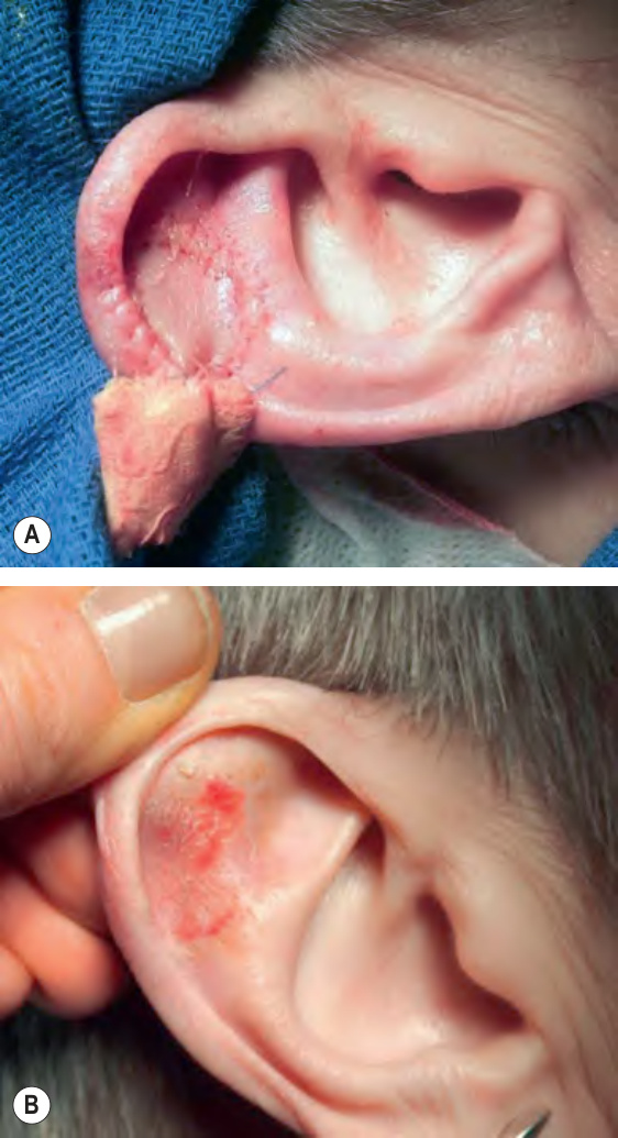
Fig. 148.2 Full-thickness skin graft.A Full-thickness skin graft taken from the preauricular region was used to repair this defect of the right upper anterior ear. B Eight-week postoperative result.
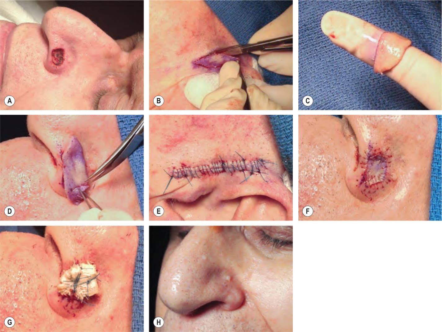
Fig. 148.3 Placement of a full-thickness skin graft.A Defect of the left nasal ala, measuring 1.8 × 1.2 cm, is present after removal of basal cell carcinoma via Mohs micrographic surgery. A preauricular donor site will provide the best match based upon color, sebaceous quality, degree of photodamage, and thickness. B With a sterile marker, a template has been made from the periphery of the recipient site and the template is then used to produce an inked margin at the donor site (center of wedge). Next, Burow’s triangles are outlined on either side of the template. Marking the donor site prior to injecting local anesthesia prevents incorrect sizing due to tissue stretch from lidocaine infiltration. The donor site is excised with a scalpel to the level of the subcutaneous fat. C The graft is carefully defatted with curved iris scissors so that only the white, glistening surface of the dermis remains. D The full-thickness skin graft is trimmed with curved iris scissors to ensure a perfect fit. E The preauricular donor site is closed primarily. F Full-thickness skin graft sewn into place with 6–0 fast-absorbing gut sutures. G Xeroform™ bolster sewn into place over the full-thickness skin graft with 5–0 polypropylene tie-over sutures. H Sixteen-week postoperative result. The graft shows good color and texture match with the surrounding skin.
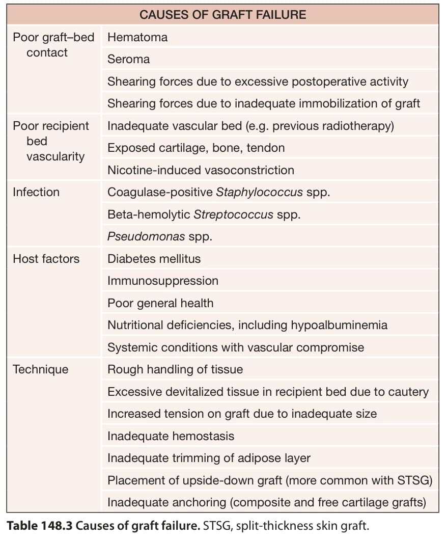
Table 148.3 Causes of graft failure. STSG, split-thickness skin graft.
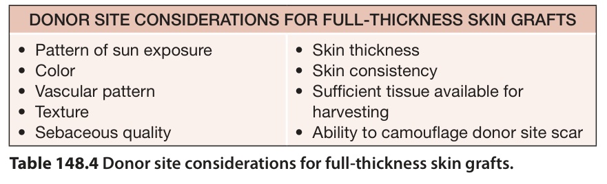
Table 148.4 Donor site considerations for full-thickness skin grafts.
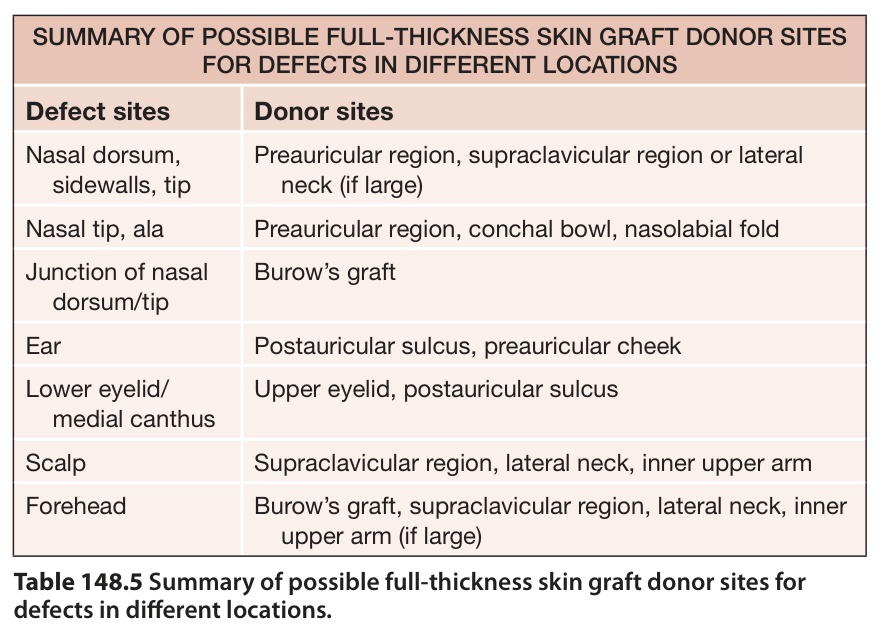
Table 148.5 Summary of possible full-thickness skin graft donor sites for defects in different locations.