SPLIT-THICKNESS SKIN GRAFTS
Split-thickness skin grafts (STSGs) consist of epidermis and a portion of the dermis. These grafts vary in thickness from ~0.005 to 0.030 inches (0.13–0.78 mm), and are classified as thin, medium, or thick, depending upon the amount of dermis included within the graft (Table 148.6).
Indications/Contraindications
STSGs contain less tissue requiring revascularization than FTSGs and are therefore more likely to survive when placed on almost any recipient bed, including those with a limited vascular supply. As a result, these grafts may be placed over periosteum, perichondrium, peritenon, and perineurium. STSGs are also used to cover large defects (Fig. 148.4A), particularly those that cannot be covered by a flap or would heal too slowly by second intention, as well as refractory venous leg ulcers. STSGs may be useful for covering surgical defects in sites at high risk for tumor recurrence, since recurrent tumor is usually visible when growing through split-thickness skin. If the tumor has not recurred after 1–2 years, the graft can be removed and definitive reconstruction performed.
Advantages of STSGs over FTSGs include their improved chance of survival under conditions of vascular compromise, their ease of application, their ability to cover large defects, and their ability to act as a “window” for recurrence of high-risk lesions. The principal disadvantages of STSGs include their suboptimal cosmetic appearance, the presence of a granulating donor site wound requiring postoperative care, greater contraction, and the special equipment required to harvest larger grafts. Furthermore, because of their relative thinness, STSGs may be less durable than FTSGs, necessitating regrafting or partial healing by second intention.
While thicker STSGs tend to be cosmetically superior to thinner ones, poor color and texture match with the surrounding skin often occurs after STSG placement. STSGs tend to be pale or white in color, hairless, and smooth, with impaired sweating due to the fact that adnexal structures are not removed in their entirety with the graft and do not survive. The contrast between the STSG and its surrounding skin can therefore produce a “tire-patch” appearance (Fig. 148.4B), which is more pronounced than that seen with FTSGs.
Preoperative History and Donor Site Considerations
Donor scar cosmesis should be taken into consideration when selecting a split-thickness donor site. The ease of postoperative care and the type of instrument used to harvest the graft may also help dictate donor site choice. Ideally, STSGs should be harvested from a site where a broad area of skin can be removed while still being concealed beneath clothing. The most common donor sites include the anterior, medial, and lateral portions of the upper thigh, the inner and outer aspects of the upper arm, and the inner aspect of the forearm. Lower back and abdominal skin can also be harvested. The anteromedial thigh is most frequently used as the donor site for STSGs, as harvesting and wound care are convenient, and wounds in this location do not interfere with ambulation. Donor site wounds on the buttocks tend to require assisted postoperative care, although their scars are ideally placed from a cosmetic standpoint. Power-driven dermatomes and large freehand knives require large flat donor surfaces, which may limit donor sites to the thighs, abdomen, and buttocks, while smaller grafts can be harvested freehand or with a power-driven dermatome.
Description of Grafting Techniques
A wide variety of techniques for harvesting and placing STSGs have been described. The instruments used to harvest STSGs can be classified into freehand and electric dermatomes. Freehand dermatomes include scalpel blades, double-edged razor blades, and knives such as the Weck blade. Although acceptable grafts can be obtained using these freehand devices, considerable technical expertise is required to harvest them.
A standard #15, 15c, or #10 blade can be an effective tool for harvesting small STSGs of medium thickness. After a template of
the defect is made, the donor site is marked, anesthetized with 1% or 2% lidocaine, or with a dilute lidocaine solution if the tumescent technique is to be employed, and scored lightly with the blade. The graft is then harvested by orienting the blade parallel to the skin and gently sweeping it just below the level of the epidermis, so that the blade is visible beneath the skin (Fig. 148.5). It is helpful to have an assistant apply traction to the donor site while the graft is harvested. Several blades may be required for harvesting, as blade sharpness diminishes quickly with multiple passes. This technique may be especially useful in harvesting small medium-thickness STSGs to repair auricular and postauricular defects.
Power-driven dermatomes became the standard method of harvesting larger STSGs after Brown developed the first such instrument in the 1940s. Electric dermatomes designed to harvest STSGs of varying thicknesses and widths, from several centimeters up to nearly 15 cm, are now commonly employed, and lithium ion battery-powered units are also available. Although STSGs can be obtained easily and reliably with any of these devices when properly used, graft quality is techniquedependent, and substantial irregularity in graft thickness and width may occur. The Zimmer dermatome, which was originally powered by compressed water-pumped nitrogen and subsequently modified into an electrically powered version, tends to harvest uniform grafts of predetermined width and thickness such that consistent graft quality tends to be less dependent on the operator’s technique. Multipurpose motor systems now exist which include a control box, foot switch, and autoclavable motor that can be used not only with dermatomes, but also with diamond fraises for dermabrasion, adjustable skin graft meshers, and other types of surgical handpieces, allowing a wide array of procedures to be performed using a single unit.
After the dermatome is prepared, the donor and recipient sites are anesthetized, prepped and draped in the usual sterile fashion. If chlorhexidine surgical scrub is used, a saline wash is employed to remove any excess scrub. The donor site is lubricated in advance with sterile mineral oil or another lubricant to ease travel of the dermatome over the skin. The handpiece is held on the donor site at an angle of 30–45°. A throttle
control is pressed to start the cut, and the unit is guided forward using light downward pressure to ensure that the cutting edge remains in continuous contact with the donor site. An assistant applies tension by pulling the skin away from the donor area to create a flat, even surface. As the dermatome glides over the donor skin, the graft emerges from the pocket of the dermatome, and is lifted away from the machine with tissue forceps or hemostats (Fig. 148.6A). Once a sufficiently large graft has been harvested, the dermatome is pulled away from the skin and the graft is placed in sterile saline or on sterile saline-soaked gauze (Fig. 148.6B). A modified harvesting technique using tumescent anesthesia and placement of a polyurethane membrane over the donor site has been reported to prevent graft shrinkage and dehydration and to facilitate graft fixation at the recipient site.
Securing the Graft
As in the case of FTSGs, STSGs should be secured so that infection, hematoma or seroma formation, and mechanical shearing forces can be prevented. Both the perimeter and the central portion of the graft must be secured for adequate nutritional support and to ensure graft survival. The edges of STSGs need not be as closely approximated to the surrounding wound edges as those of FTSGs, since overlapping skin will slough without affecting the ultimate cosmetic result. After the graft has been placed with the dermal side adherent to the recipient bed, its perimeter may be secured with sutures or staples. Centrally placed basting sutures may also be helpful in ensuring apposition of the graft to its bed. Once the graft has been secured and its bolster sewn into place, a non-adhesive dressing or pressure dressing may be applied as an additional precaution. Sutures or staples are removed after 7–10 days.
Donor Site Care
Harvesting STSGs creates a second wound, the donor defect, which often causes more postoperative discomfort than the grafted area itself.
This partial-thickness wound heals by second intention. While STSG donor sites were once treated with bulky occlusive dressings left in place for 10–14 days, the advent of transparent, vapor-permeable dressings such as Opsite® has improved both patient comfort and ease of care in the postoperative period (Fig. 148.6C). These dressings are advantageous because they allow the serosanguineous drainage that inevitably accumulates at the donor site to collect, keeping the wound moist and thereby shortening healing time. Because these dressings are transparent, the wound can easily be observed for complications during healing. In one review, these dressings were associated with the fastest healing rates, low infection rates, the least amount of pain, and lower cost when compared with other dressings.
After the skin around the donor site area is cleaned and dried, a thin coat of an adhesive such as Mastisol® is applied around the wound and allowed to dry. An Opsite® dressing is then placed over the wound. Paper tape is placed around the perimeter of the Opsite®, followed by a gauze dressing and an elastic bandage (e.g. Ace™ wrap).
Variations/Unusual Situations
Meshing the graft with scalpel slits may be performed to allow drainage of accumulated blood or serosanguineous material that could otherwise inhibit graft–bed contact. This technique may also be used to expand the surface area of STSGs. A meshing machine may be utilized to expand the graft’s surface area further by ratios ranging from 3 : 1 to 9 : 1. Meshing can facilitate coverage of a large recipient area with smaller donor grafts. Expanded meshed grafts placed experimentally on contaminated recipient beds have been found to exhibit increased take as compared to non-meshed donor skin.
Postoperative Care
During the first 24 hours after grafting, a large amount of serosanguineous fluid may accumulate beneath the donor site dressing. Patients need to be forewarned about this possibility so that they do not become alarmed. If necessary, the fluid can be drained with a needle and syringe, and an Opsite® patch applied. Alternatively, a new Opsite® dressing can be placed. The dressing can then be left in place until healing is complete. Depending on the STSG’s thickness, the donor site should fully re-epithelialize in 7–21 days. The scar usually evolves in color from pink to white over a period of months.
Complications
The complications of split-thickness skin grafting can be divided into early complications, which stem from failure of engraftment, and late complications. Failure of engraftment may result from hematoma or seroma formation, infection, or shearing forces. Late complications can be divided into cosmetic and functional problems (Fig. 148.7).
Color and texture mismatch of STSGs with the surrounding skin is predictable and expected. Grafts often remain erythematous for months to years after placement. More importantly, they may exhibit significant postoperative hyperpigmentation or hypopigmentation. Darker-skinned patients are especially prone to graft hyperpigmentation, despite observance of preventive measures. Patients should therefore minimize graft exposure to the sun without sunscreens for 6 months, and wear sunscreens consistently thereafter. The absence of adnexal structures predisposes to xerosis and a build-up of keratinous debris. The resultant scaling, pruritus, and dryness can be minimized with liberal emollient use.
Functional considerations are of paramount concern, since STSGs contract more than FTSGs and can create forces powerful enough to produce joint contracture if placed over or near joints. Contraction of facial grafts, especially near the nasal ala, the eyelid, the helical rim and the free margins of the vermilion border, may produce significant cosmetic deformities, including alar retraction, ectropion, helical rim distortion, and vermilion border distortion, respectively. Hypertrophic scarring of the graft and donor sites may also occur, which can be treated with corticosteroid-impregnated tape, intralesional corticosteroids, or pulsed dye laser. Graft fragility and breakdown can occur in areas of trauma, particularly in sites such as the lower leg, or in areas with little underlying soft tissue support, such as those directly overlying perichondrium or periosteum. These complications are not always avoidable, but forewarning patients may reduce unnecessary trauma to the area. Lastly, bullae can occur within graft sites, presumably related to decreased anchoring properties of the basement membrane zone (see Ch. 33).
Future Directions
In recent years, biosynthetic and tissue-engineered living skin replacements have been the focus of research in skin transplantation. Cultured human keratinocytes have been seeded onto type I collagen membranes petrolatum gauze to reconstitute the epidermis, enabling
A Hyperpigmentation of an entire STSG placed following excision of an in situ ungual melanoma; the sites of suture placement also developed hyperpigmentation. Increased scaling compared to the surrounding skin is commonly observed. B Mismatch of color, texture, and hair density in a skin graft of abdominal skin placed on the face following excision of a congenital melanocytic nevus. C Hypertrophic scarring of a STSG placed following partial excision of a large congenital melanocytic nevus on the arm one year prior; the skin overlying the elbow was not excised to prevent a joint contracture. D Growth of hair within a graft placed on the inner aspect of the foot following excision of a cutaneous melanoma. Courtesy Jean L. Bolognia, MD.
transfer of actively proliferating keratinocytes onto partial-thickness wounds. Human epidermal tissues mimicking the biochemical and morphologic properties of human skin have been established in vitro by growing human keratinocytes on a dermal fibroblast-containing collagen gel. Such skin-equivalent cultures are being used to reconstitute the epidermis in patients with full-thickness burn wounds and chronic ulcers (see Ch. 145).
Artificial dermis is now routinely used prior to full-thickness grafting, and an acellular allograft dermal matrix has been shown to provide a good bed for STSGs in patients with full- and partial-thickness burns. A tissue-engineered human skin equivalent, i.e. a bilayered product containing living human fibroblasts, keratinocytes and bovine collagen, is commercially available for the repair of surgical defects and coverage of lower leg ulcers that would previously have been repaired with STSGs. Dermal–epidermal composites using autologous keratinocytes and human allodermis may in the future be used for grafting full-thickness wounds in patients with burns, leg ulcers, or surgical wounds.
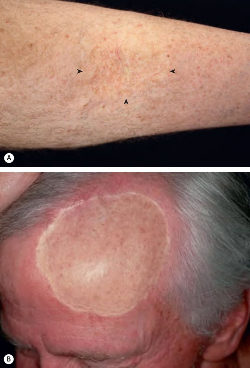
Fig. 148.4 Split-thickness skin grafts (STSGs).A A relatively thick STSG placed on the medial aspect of the lower extremity with excellent cosmetic results; arrowheads denote the edges of the graft. B STSG placed on the forehead and scalp several years prior, following excision of a cutaneous melanoma. Note the relative hypopigmentation, altered texture, and depression of the STSG compared to the normal surrounding skin. Courtesy Jean L. Bolognia, MD.
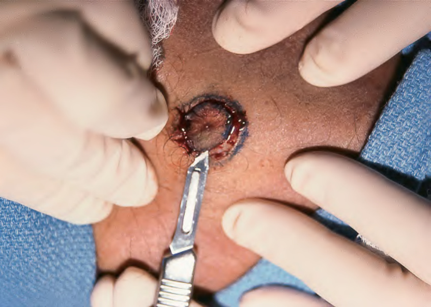
Fig. 148.5 Freehand harvesting of a small split-thickness skin graft from the upper outer arm. The blade is oriented parallel to the skin, and gently swept just below the level of the epidermis, so that it is just visible through the graft. An assistant applies traction to the donor site to facilitate harvesting.
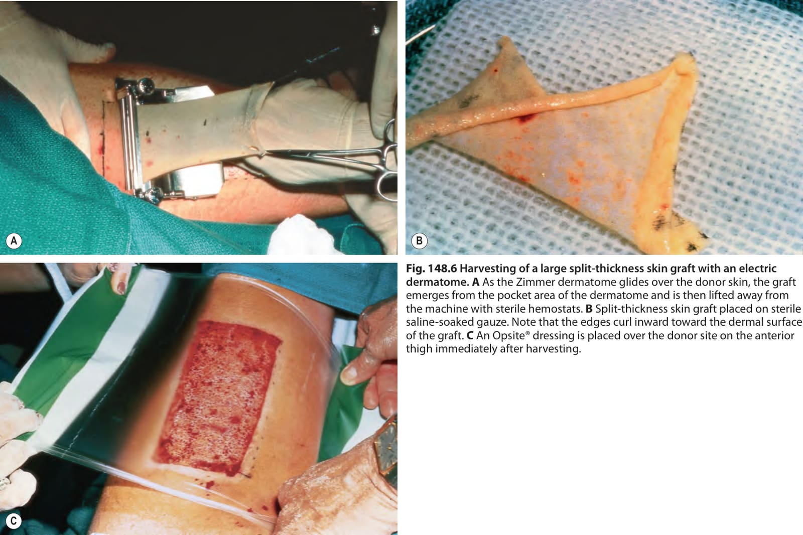
Fig. 148.6 Harvesting of a large split-thickness skin graft with an electric dermatome.A As the Zimmer dermatome glides over the donor skin, the graft emerges from the pocket area of the dermatome and is then lifted away from the machine with sterile hemostats. B Split-thickness skin graft placed on sterile saline-soaked gauze. Note that the edges curl inward toward the dermal surface of the graft. C An Opsite® dressing is placed over the donor site on the anterior thigh immediately after harvesting.
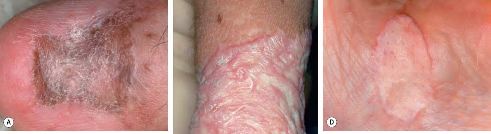
Fig. 148.7 Late complications of skin grafts.
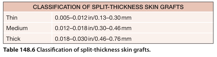
Table 148.6 Classification of split-thickness skin grafts.