COMPOSITE GRAFTS
Indications/Contraindications
Composite grafts are modified FTSGs, consisting of two or more tissue layers. In dermatologic surgery, these grafts are usually composed of skin and cartilage, although they may be composed of skin and fat or skin and perichondrium. Composite grafts are especially useful for repairing full-thickness alar rim defects, as well as nasal tip defects with cartilage loss. Full-thickness nasal mucosal defects can be repaired using composite grafts to provide mucosal lining and structural support with a nasolabial or forehead flap placed thereafter to reconstruct the overlying soft tissue defect (see Fig. 147.29). As discussed earlier, conchal bowl composite grafts or PCCGs, consisting of skin and perichondrium, may be used (with or without an underlying cartilage graft) for deeper nasal defects requiring skin coverage, especially defects with exposed cartilage.
Preoperative History and Considerations
Composite grafts require rapid revascularization to survive. Early re-establishment of circulation occurs via direct vessel anastomoses between the subdermal plexus of the graft and the subdermal plexus of the wound edge. Since composite grafts are dependent upon this bridging phenomenon for their survival, they are of necessity limited in size, with no point being more than 1 cm from a vascular source, as the risk of central necrosis increases significantly at graft diameters >2 cm3,47. Composite grafts for nasal ala and ear reconstruction are possible because of the rich vascular supply of the nose and ear, and because of the small surface areas generally involved. Composite grafts, like FTSGs, are threatened by excessive shearing forces of the graft over its bed, which prevent revascularization.
Composite grafts pass through four stages as they heal. After graft placement, the tissue blanches completely. By 6 hours, the graft becomes pale pink, signifying anastomosis of the graft vessels with those of the recipient site. At 12–24 hours, the graft appears dusky blue, reflecting venous congestion, and by 3–7 days, it should be pink, indicating graft survival.
Donor Site Considerations for Composite Grafts
The complexity of nasal ala anatomy makes reconstruction of fullthickness defects in this area difficult. There may be insufficient nasal skin to develop adequate local flaps, and nasolabial flaps, which provide reasonable defect coverage, generate other cosmetic deformities. Loss of alar tissue support also creates a functional deficit, as the alar skin is liable literally to “blow in the breeze” on inspiration and expiration. Composite grafts provide an excellent cosmetic and functional alternative for repair of full-thickness alar rim defects <2 cm in diameter. Grafts taken from the earlobe have been used successfully for this type of repair, although grafts taken from the cartilaginous portion of the ear are more frequently used.
Donor sites for harvesting auricular composite grafts include the helical crus, helical rim, and conchal bowl (see Ch. 142). Small alar defects with cartilage loss can be elegantly repaired using the helical crus as donor site, while more substantial defects may require repair using the helical rim or conchal bowl, since the crus may not provide sufficient inner lining for the graft. Donor defects involving the helical crus can be repaired with minimal scar formation, while wedge excisions are usually necessary to repair helical rim donor sites. Conchal bowl donor sites heal by second intention.
The advantages of auricular composite grafts in repairing fullthickness alar rim defects relate mainly to the presence of cartilage, which provides structural support and stability, with prevention of alar distortion during inspiration and at rest. Disadvantages include a higher risk of graft failure with an increased number of tissue layers, substantial graft size limitations, and limited donor tissue availability. Nevertheless, these grafts, when properly applied, can yield outstanding results. In one animal study, maintaining an intact skin-to-cartilage interface and an equivalent skin-to-cartilage ratio improved composite graft survival; however, when cartilage size exceeded skin size, or when skin and cartilage were separated before transfer, survival decreased, highlighting the importance of an intact skin-to-cartilage interface in optimizing graft survival.
Composite grafts used for full-thickness nasal mucosal repair are usually obtained from the triangular fossa, scapha, conchal cavum, cymba, or helical crus (see Fig. 142.13). The appropriate donor site is that which best matches the contour of the surgical defect. These donor sites are usually allowed to heal by second intention, with good aesthetic outcomes.
Description of Technique
Composite grafts used for alar repair are performed as follows (Fig. 148.8): the donor and recipient sites are anesthetized using local anesthesia, and prepped with an antiseptic solution. If the alar tissue is scarred and retracted, the area must be vigorously debrided to assure the best possible blood supply for the graft. The defect is then
measured and a template made as described previously. The donor site is marked and anesthetized, and the graft is harvested. The tissue is handled very gently and placed in sterile saline until ready for placement.
Due to the fragility of composite grafts, a tongue-in-groove technique is recommended to maximize graft stability and increase graft survival. Two cartilaginous wings are marked out and anesthetized on either side of the donor site prior to graft harvesting. After the graft is harvested, the skin overlying these two cartilaginous wings is removed, leaving the cartilage with its overlying perichondrium (Fig. 148.8B). These wings are then inserted into pockets prepared within the alar tissue of both sides of the defect (Fig. 148.8C) such that the graft interlocks with its recipient bed (Fig. 148.8D). Interlocking the graft with its bed may help to minimize shearing forces and provide a larger surface area for revascularization.
The graft is sutured into place in two layers. The undersurface of the graft, which replaces the inner lining of the nose, can be secured first using a 6–0 absorbable suture. The skin is then closed with a 6–0 non-absorbable suture, taking very small tissue bites to minimize vessel strangulation and to maximize the number of potential vessels available for reanastomosis. The needle should pass through the mucosal portion of the graft first, then through the outer epithelial edge of the graft, so that the knots are tied externally, and are not buried between the graft and its recipient bed. The cartilage does not need to be sutured, as it will heal on its own. A Vaseline® gauze or Xeroform™ dressing can then be placed in the nasal vestibule for support, and petrolatum ointment applied to the external suture line. A non-stick dressing is then applied to protect the graft from external injury.
Conchal bowl composite grafts used for reconstruction of deep alar defects are harvested in essentially the same way as composite grafts taken from the helical crus. Potential advantages of the conchal bowl technique include increased bulk, which may be superior in filling deep partial-thickness alar defects, and an excellent cosmetic match for the sebaceous skin of the nose.
Donor Site Closure48
Helical crus defects can often be closed in a side-to-side fashion. A simple advancement, rotation, or transposition flap may also be utilized to take advantage of loose preauricular skin. Helical rim defects are usually closed with an ear wedge resection. Auricular grafts obtained from the triangular fossa, scapha, conchal bowl, or cymba heal well by second intention. Conchal bowl defects may heal more rapidly if a 2 or 3 mm punch biopsy instrument is used to remove small plugs of conchal cartilage, which allows healing by second intention from the dermis on the opposite side.
Variations/Unusual Situations
When securing composite grafts onto the nasal ala, delayed intranasal knot tying has been recommended by some authors to facilitate wound closure by improving visualization, allowing gentler manipulation of tissue, and enabling precise suture placement in a confined space. When placing intranasal sutures, the needle of the 6–0 fast-absorbing chromic gut suture is first passed through the nasal mucosa, and then through the corresponding location on the “mucosal” side of the graft. Sutures are placed at 1–2 mm intervals along the defect and graft, and their free ends are held separately by hemostats to prevent tangling. After all intranasal sutures have been placed, the composite graft is placed into the alar defect and the cartilaginous wings inserted. The most internal intranasal sutures are then tied first, followed by the more accessible external sutures.
Postoperative Care
To minimize edema, ice packs should be applied to the grafted area as often as possible for up to several days postoperatively. Oral antibiotics are generally advisable because of the high bacterial colonization around the nares and the higher risk of failure with composite grafts. Sutures are removed after 1 week.
Complications
As with other types of grafts, there is a risk of necrosis in the early stages of healing, and contraction, textural changes, atrophy, and contour irregularities thereafter. If the graft survives but the cosmetic result is suboptimal, dermabrasion or laser resurfacing may be performed 6 weeks to 6 months postoperatively to correct textural differences between the graft and the surrounding skin, and to improve the color match between them. Resurfacing of the entire cosmetic unit may provide superior cosmetic results. In the event of composite graft failure, a two-stage revision with placement of a cheek interpolation flap may be performed or, alternatively, a second composite grafting procedure may be undertaken.
Conchal bowl donor sites are at somewhat greater risk of postoperative bleeding than other areas if left to heal by second intention. Application of pressure dressings over the front and back of the ear to “sandwich” the conchal cartilage with placement of hemostatic foam over the donor site may decrease this risk. Other types of complications at auricular donor sites are rare.
The ear is also prone to infection with Pseudomonas spp., which reside in the external auditory meatus. Cleaning the ear with a dilute vinegar solution, applying topical gentamicin ointment, and oral quinolone antibiotics are all helpful prophylactic measures which should be considered after harvesting auricular cartilage. If infection is suspected, oral quinolone antibiotics should be started immediately, and therapy guided thereafter by the results of cultures and sensitivities. If the infection does not resolve with appropriate antibiotics, the presence of a fungal infection, most often secondary to Candida, should be excluded.
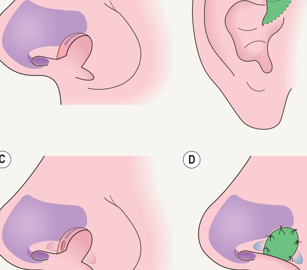
Fig. 148.8 Technique of interlocking composite graft placement.A A fullthickness nasal ala defect extending through both skin and cartilage requires repair. B Donor site at the crus of the helix. An area of skin ~5%–10% greater in area than the actual defect is marked out, with cartilaginous wings marked out on either side. After the graft is harvested, the skin is removed on both sides to expose the cartilaginous portion of the graft, such that two cartilaginous pegs with their overlying perichondrium (blue color) frame the lateral aspects of the graft. C A pocket is undermined on either side of the recipient site into which the cartilaginous pegs will be placed. D The cartilaginous pegs are inserted into the holes prepared within the alar tissue on either side of the defect. The graft is then sutured into place.