FREE CARTILAGE GRAFTS
Indications/Contraindications
Free cartilage grafts are used most commonly in dermatologic surgery to reconstruct the nasal ala, tip and sidewall, the ear, and the eyelid. These grafts may be used to restore the architecture of an anatomic site that has undergone significant cartilage loss. Free cartilage grafts are also useful in stabilizing the position and contour of free margins against the forces of contraction during wound healing.
Nasal Ala
Partial-thickness alar defects extending into deep soft tissue or approaching the alar rim often lead to rim collapse, producing a functional as well as a cosmetic deficit. Placement of cartilage grafts, consisting of cartilage with its overlying perichondrium, can be used to avert this potential problem. The grafted cartilage provides a rigid but flexible framework that braces the alar rim against collapse during inspiration and expiration. Cartilage grafts may be used in conjunction with flaps and FTSGs to maintain airway patency and to minimize the risk of alar retraction during healing (Fig. 148.9).
The usefulness of the free cartilage graft lies in its ability to prevent alar retraction. It is possible that long-term survival of the cartilage itself may not ultimately be important, as the mere presence of the rigid framework of the graft in the initial stages of wound healing seems to be sufficient to inhibit alar retraction.
Use of free cartilage grafts or composite grafts composed of skin and cartilage can help to prevent this complication. Courtesy Jean L. Bolognia, MD.
Nasal Sidewall and Tip
Deep nasal sidewall defects may, at times, involve loss of the upper lateral nasal cartilage. Nasal valve obstruction may result, which becomes noticeable with inspiration and resolves on expiration. Delayed nasal valve obstruction can also occur if unresisted scar contracture collapses the remaining cartilaginous structure of the nasal sidewall. Replacing lost cartilage at the time of reconstruction can avert these potential problems. Similarly, loss of cartilage at the distal nasal tip necessitates structural support replacement for optimal function and cosmesis.
Ear
Auricular defects involving cartilage loss are generally repaired for cosmetic, rather than functional, reasons. Cartilage grafts may be used as braces along the helical rim to minimize the risk of contracture, and may also be used in the conchal bowl to assist in hearing-aid placement.
Preoperative History and Donor Site Considerations
Potential donor sites for free cartilage grafts include the conchal bowl, the auricular helix, the antihelix, nasal septum, and ribs. The conchal bowl and antihelix are the donor sites most frequently used by dermatologic surgeons. Conchal cartilage is elastic, has a high degree of memory, and has varied contours that can be matched to the desired alar contour. Although an anterior approach to the conchal bowl may be used, the posterior approach results in better camouflage of the donor site scar and preservation of the shape of the ear. If the antihelix is used as a donor site, it is recommended that subtotal resection of the antihelical cartilage be performed, leaving a complete rim of intact helical cartilage to prevent distortion.
Description of Technique
Nasal ala
The techniques for alar batten cartilage grafting have been well described. The alar batten provides a rigid but flexible cartilaginous framework to brace against alar rim collapse. The length of the cartilage graft is determined by measuring the distance between the lateral and medial borders of the defect at the alar rim, and adding to that measurement an extra 4–5 mm. The conchal bowl donor site is incised anteriorly or posteriorly, whereas the antihelical donor site is incised anteriorly. The skin overlying the cartilage is then undermined with blunt-scissor dissection to expose the perichondrial surface. The desired length of cartilage is incised with a scalpel (Fig. 148.10A), and a second incision is made exactly parallel to the first, creating a cartilaginous strip that may be 3–6 mm in width, depending upon the desired width of the graft (Video 148.9). Alternatively, a larger disk or oblongshaped piece may be harvested to match the base of the defect. The cartilage is easily separated from the overlying skin with sharp-scissor dissection. The graft is placed in sterile saline while the donor site is reapproximated with non-absorbable sutures.
The cartilage graft is secured by undermining the soft tissue of the recipient bed medially and laterally with a hemostat or blunt-scissor dissection, and the ends of the graft are inserted into the undermined pockets such that the graft interlocks with its bed (Fig. 148.10B, Videos 148.10 and 148.11). The graft is anchored to its bed with one or two 5–0 absorbable sutures for additional security (Video 148.12). A disk- or oblong-shaped cartilage graft also requires suturing for secure placement.
After the graft has been anchored, a nasolabial transposition flap or FTSG is sutured into place to complete the closure (Fig. 148.10C,D, and see Videos 148.1, 148.2, 148.3, 148.4, 148.5, 148.6). A standard tie-over bolster is placed over the FTSG and secured with 5–0 non-absorbable sutures (see Videos 148.7, 148.8). Sutures are removed at 1 week.
Nasal sidewall and tip
Free cartilage grafts used for nasal sidewall and tip reconstruction are harvested in much the same way as free cartilage grafts for alar reconstruction. Nasal sidewall grafts tend to be wider and broader than alar grafts, since they must brace a broader area against the forces of inspiration. Alternatively, multiple cartilaginous strips can be placed perpendicular to the lateral nasal sidewall, and then secured with absorbable or non-absorbable sutures to brace the side of the nose against collapse. After the cartilaginous structure is in place, a flap or FTSG may be performed to cover the remaining cutaneous defect.
Nasal tip grafts for distal nasal reconstruction are organized to provide both proximal and distal structural support, thus optimizing the aesthetic and functional result. The proximal grafts consist of bilateral batten grafts secured to the lateral cartilages with 5–0 or 6–0 non-absorbable sutures. On top of these may be sewn a dorsal nasal cartilage graft. A columellar strut of cartilage often provides distal support, on top of which may be sewn an additional tip graft. Alar batten grafts secured to the columellar strut and lateral alar soft tissue provide alar rim support. A paramedian forehead flap or melolabial interpolation flap may be placed thereafter to provide coverage of the cutaneous defect.
Ear
If conchal cartilage is used to repair a large helical defect, the cartilage graft should match the defect as closely as possible in size and shape. Harvesting and placing a narrow strip of cartilage matching the helical defect alone will result in helical rim collapse under the forces of wound contraction. If necessary, a partial wedge closure of the defect may be performed to decrease the size of the defect requiring replacement of structural support. The cartilage graft is secured by sewing it to the intact cartilaginous framework with 5–0 absorbable or non-absorbable sutures. The graft is then generally covered with a pedicled retroauricular advancement flap.
Eyelid
Partial-thickness lower eyelid defects with loss of the tarsal plate, as well as full-thickness lower eyelid defects, may be repaired with cartilage grafts. Stabilization of the eyelid margin with free cartilage grafts provides structural support, minimizing the risk of ectropion and preventing corneal desiccation. The technique for performing free cartilage grafts to repair full-thickness and partial-thickness lower eyelid defects is beyond the scope of this chapter, but is well described by Otley and Sherris.
Postoperative Care
Regardless of the site of cartilage grafting, care must be taken postoperatively to minimize movement of the graft. Routine wound care is performed to the flap or skin graft overlying the free cartilage graft. Trauma to the area should be minimized.
Complications
Postoperative complications following cartilage grafting are rare. There is a risk of postoperative infection at conchal bowl donor sites, particularly with Gram-negative bacilli, which reside within the external auditory meatus. Appropriate bacterial and fungal cultures of any exudate should be obtained if infection is suspected. Empiric therapy with quinolone antibiotics should be initiated, and modified as sensitivities dictate. When prophylactic antibiotics and appropriate operative technique are used, the risk of suppurative chondritis is extremely low.
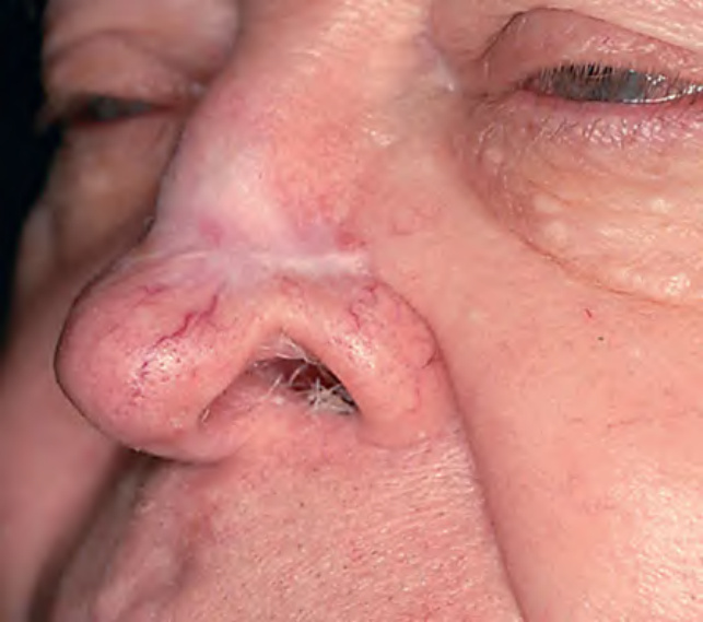
Fig. 148.9 Distortion of the nasal alar rim due to scar retraction from second intention healing following removal of a basal cell carcinoma.
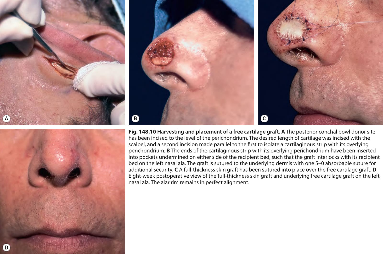
Fig. 148.10 Harvesting and placement of a free cartilage graft.A The posterior conchal bowl donor site has been incised to the level of the perichondrium. The desired length of cartilage was incised with the scalpel, and a second incision made parallel to the first to isolate a cartilaginous strip with its overlying perichondrium. B The ends of the cartilaginous strip with its overlying perichondrium have been inserted into pockets undermined on either side of the recipient bed, such that the graft interlocks with its recipient bed on the left nasal ala. The graft is sutured to the underlying dermis with one 5–0 absorbable suture for additional security. C A full-thickness skin graft has been sutured into place over the free cartilage graft. D Eight-week postoperative view of the full-thickness skin graft and underlying free cartilage graft on the left nasal ala. The alar rim remains in perfect alignment.