NAIL PLATE AVULSION
Nail avulsion is the most basic procedure in nail surgery and is a prelude to most other nail unit procedures. It allows visualization of the nail bed and matrix, thus providing good exposure for biopsy or excision (Fig. 149.7). Except for patients with retronychia, total nail avulsion should be discouraged because as the counterpressure exerted by the nail plate disappears, the distal wall and pulp raise upward in combination with
a shrinkage of the nail bed. The result is more bulky soft tissue at the end of the digit and an increased risk of distal embedding during nail regrowth. Partial nail avulsion is therefore preferred, with the remaining normal nail plate reducing the risk of distal embedding. Partial nail avulsion serves as an adjuvant therapeutic intervention in onychomycosis as it reduces the fungal mass, especially of a dermatophytoma (Fig. 149.8). In addition, nail avulsion is an early step in chemical matricectomy (see below), may be performed to drain an acute paronychia, and is a must when exploring pigmented lesions within the nail matrix. There are two approaches to nail avulsion – distal and proximal, although the latter is rarely used.
For distal nail plate avulsion, an elevator is first pushed under the proximal nail fold and slid back and forth from one side to the other in order to detach the fold from the plate (Fig. 149.9A). The elevator is then inserted under the free edge of the nail plate in a distal to proximal direction to free the plate from its bed (Fig. 149.9B). As the instrument reaches the matrix area where the attachment of the nail plate is looser, a decrease in resistance is felt. To loosen the entire plate, the instrument is inserted under the plate at several sites, advancing from the hyponychium to the matrix. Lateral motions across the nail bed should not be done to loosen the nail plate as this can injure the fragile longitudinal nail bed ridges. The plate is then grasped on one lateral edge by a hemostat or a nail puller, rotated (Fig. 149.9C), and pulled off the nail bed.
If the avulsion is performed to explore the entire matrix area, avulsion alone will not suffice. Two releasing incisions, ~1 cm in length, are made at each junction of the proximal and lateral nail folds, angling laterally and proximally. This allows the nail fold to be reflected so that the matrix can be inspected (Fig. 149.10). When performing a biopsy or excisional surgery, skin hooks or sutures help to maintain the reflection of the proximal nail fold. At the end of the procedure, the fold is placed back into its original position and secured with simple interrupted sutures or adhesive strips. The nail plate, if not needed for
Following two oblique incisions at the junction of the lateral and proximal nail folds, retraction of the proximal nail fold, and proximal lateral avulsion of the nail plate, the entire nail matrix is exposed. The glomus tumor within the matrix (arrow) is identified prior to removal.
pathologic examination or culture, should be repositioned to cover and protect the wound during the early stages of the healing process. It can be secured by sutures or adhesive. Although the plate will not reattach and is eventually shed, it provides protection and minimizes discomfort during the immediate postoperative period.
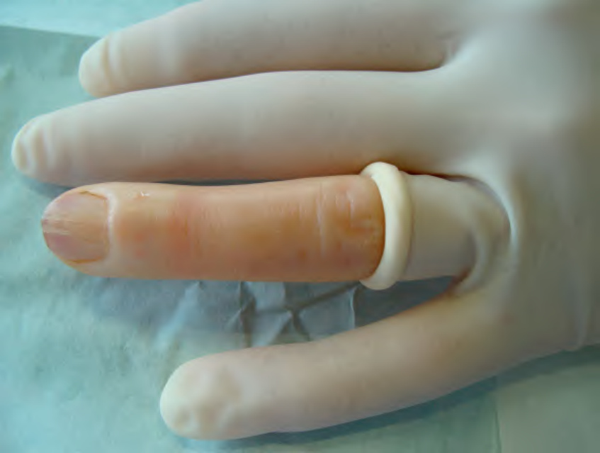
Fig. 149.6 Sterile glove used as a tourniquet and sterile field. One fingertip of the glove is pierced with scissors and rolled back to the base of the proximal phalanx.
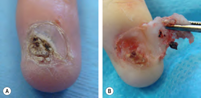
Fig. 149.7 Lateral avulsion of the plate to expose the nail bed.A Keratotic lesion suspicious for squamous cell carcinoma is extending from under the nail plate. B Lateral avulsion allows examination of the nail bed and biopsy of the tumor.
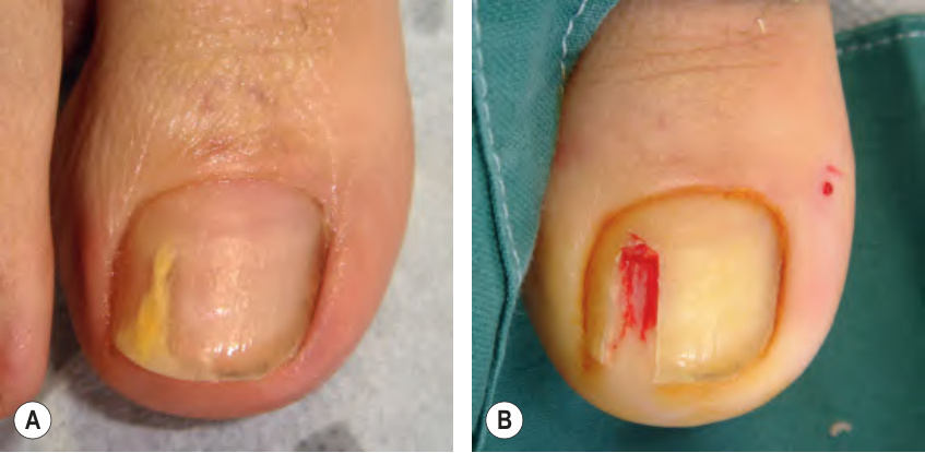
Fig. 149.8 Treatment of a dermatophytoma.A Yellow spike due to a dermatophytoma. B Partial avulsion of the nail plate and removal of the fungal mass.
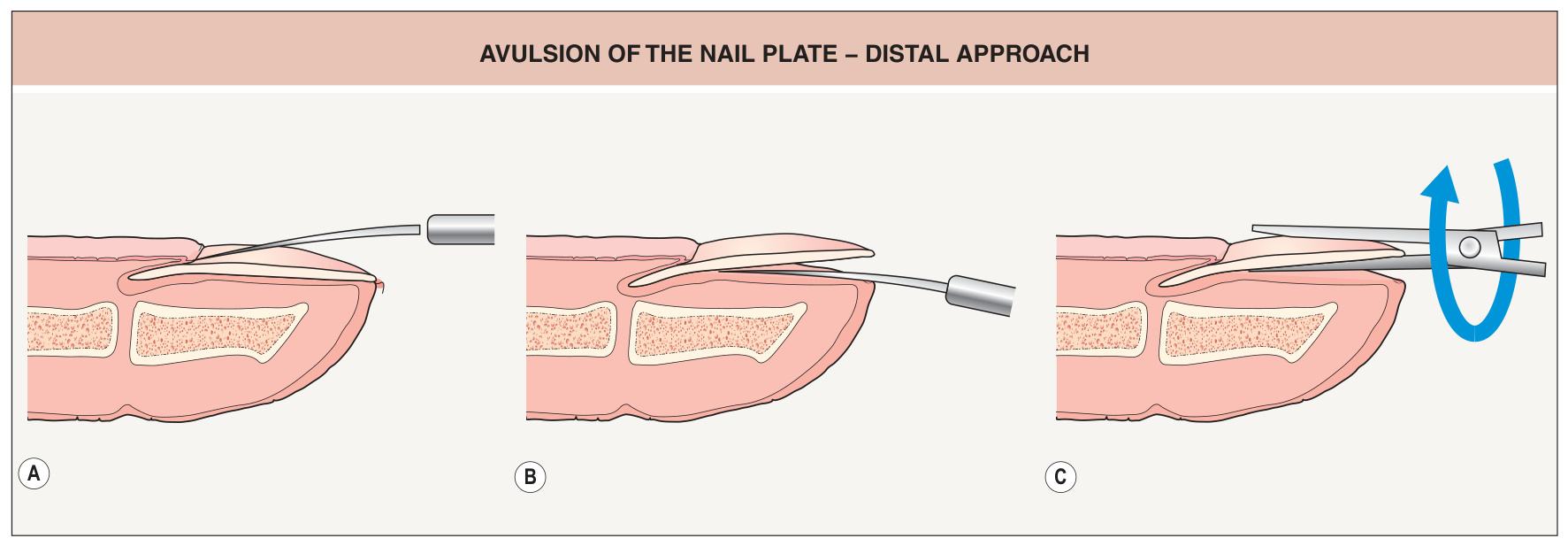
Fig. 149.9 Avulsion of the nail plate – distal approach.A An elevator is first pushed under the proximal nail fold, moving back and forth from one side to the other in order to detach the fold from the plate. B The elevator is then inserted under the free edge of the plate in a distal to proximal direction to free the plate from its bed. C The plate is then grasped by a hemostat or a nail puller and rotated off the nail bed.
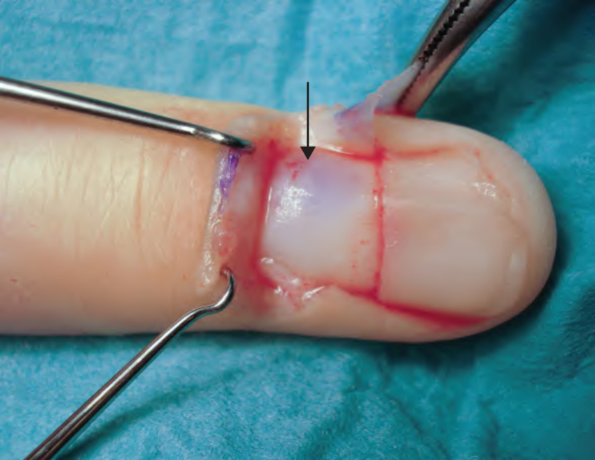
Fig. 149.10 Visualization of a glomus tumor within the proximal nail matrix.
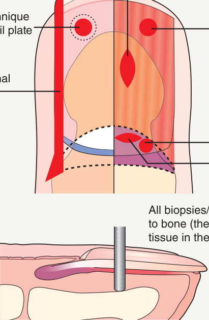
Fig. 149.11 Types and orientations of biopsies and excisions within the nail unit.