BIOPSY TECHNIQUES
To minimize scarring and offer the best functional and cosmetic outcome, nail surgical procedures should follow a few rules. Within the nail bed, the incision should be oriented longitudinally, whereas within the matrix it should ideally be oriented horizontally (Fig. 149.11). The surgical approach depends upon the location of the pathology within the nail unit.
Nail Bed Biopsy
Nail bed biopsy is a simple and safe technique for the diagnosis of a variety of nail bed disorders, from onycholysis to tumors (Table 149.2). The nail plate is usually partially avulsed prior to the nail bed biopsy (Fig. 149.12A). If the defect is >4 mm, re-approximation of the edges is recommended. However, the nail bed is fragile and suturing of the margins may be difficult. To facilitate closure of the wound, generous lateral undermining should be performed at the level of the periosteum. Nonetheless, it is amazing how very large defects of the nail bed (e.g. after Mohs surgery) may regenerate totally by second intention.
For the removal of tumors such as onychopapillomas, elliptical excisional biopsies of the nail bed along a longitudinal axis are recommended (Fig. 149.12B,C). Closure is performed with absorbable sutures (e.g. Vicryl®) (Fig. 149.12D).
Nail Matrix Biopsy
Nail matrix biopsy has one major indication: determination of the cause of longitudinal melanonychia (see Ch. 71). The biopsy must be taken from the nail matrix because that is where the pigment is generated. Fortunately, in ~95% of patients, the pigment’s origin is within the distal matrix. Since the latter synthesizes the ventral portion of the nail plate (see Fig. 149.2), the primary sequela is a nail plate thinned from below. If the pigment origin is located within the proximal matrix, then nail plate dystrophy will result, usually as a longitudinal fissure; this is because the proximal matrix generates the dorsal (upper) one-third of the nail plate.
In order to expose the entire matrix area, both retraction of the proximal nail fold and proximal nail avulsion are performed (Fig. 149.13). Once the matrix is exposed, the appropriate surgical technique is chosen based upon the width and shape of the pigmented band. The entire pigmented area is removed to ensure complete histologic examination.
●If the pigment origin is roundish and fits within a 3 mm punch biopsy trephine, it can be removed with that instrument. However, two caveats apply: (1) the trephine should be advanced down to bone, with the specimen severed at the level of the periosteum; and (2) the specimen should be harvested with delicate curved, fine-tipped scissors. Handling fragile matrix tissue with forceps will crush it and induce artifacts. The defect does not need to be sutured.
●If the pigment origin has a longitudinal orientation, then a narrow elliptical excision down to the bone is recommended. Delicate lateral undermining allows closure of the defect with 5-0 absorbable sutures.
●If the pigment origin involves a wide area of the matrix, a tangential excision is indicated. This technique virtually “shaves” the matrix, removing the matrix epidermis and a small portion of dermis. It allows the pathologist to examine the entire lesion, but the margins are usually difficult to assess. The resultant specimens have been shown to provide adequate tissue for accurate histopathologic diagnosis. In general, postoperative nail plate dystrophy is not observed even if nearly the entire distal matrix has been removed. In contrast, tangential excision of the proximal matrix results in nail thinning and brittleness in most patients, but rarely pterygium. It is noteworthy that this technique avoids mutilating surgery, especially since the cause of longitudinal melanonychia is often a benign process. Lastly, the nail plate is put back in place and secured to the lateral nail fold.
●When the pigmented streak is located within the lateral third of the nail plate, a lateral longitudinal biopsy (see next section) is appropriate.
Lateral Longitudinal Biopsy
This procedure permits examination of the entire nail apparatus including the proximal nail fold, matrix, nail bed, nail plate, and hyponychium. It is especially helpful in diseases involving the proximal portion of the nail apparatus (often presenting as alterations of the surface of the nail plate) and for inflammatory disorders in which all these structures are affected concomitantly. Obvious clinical involvement of the lateral aspect of at least one nail is required. The patient must be warned that this type of biopsy will narrow the nail permanently due to the partial amputation of the lateral horn of the matrix. In order to avoid postoperative lateral deviation, the specimen should be <3 mm in width.
The incision begins halfway between the cuticle and the crease of the DIP joint and extends distally through the proximal nail fold, then the nail plate and its bed until the hyponychium is reached. A second parallel incision is made in the lateral nail groove, joining the initial incision at the fingertip (Fig. 149.14A). Proximally, the incision curves laterally in order to remove the lateral horn of the matrix. The latter is especially important with the great toenail (see Fig. 149.2). The specimen is then carefully detached from the bone with delicate scissors. Detaching the matrix from the bone at the proximal tip of the biopsy specimen is a critical step, and care should be taken to avoid curving the scissors upward too soon and foreshortening the specimen. Residual lateral nail matrix tissue leads to nail spicule formation (Fig. 149.15). The defect is re-approximated with horizontal mattress sutures in order to recreate a lateral nail fold (Fig. 149.14B). This technique may be utilized for various tumors, including longitudinal melanonychia of the lateral third of the nail plate.
Proximal Nail Fold Biopsy
Lesions of the proximal nail fold can be biopsied via shave, punch, or incisional techniques as well as excised en bloc, similar to cutaneous lesions elsewhere. A Freer elevator may be inserted beneath the proximal nail fold to shield the matrix from inadvertent damage from the scalpel (Fig. 149.16).
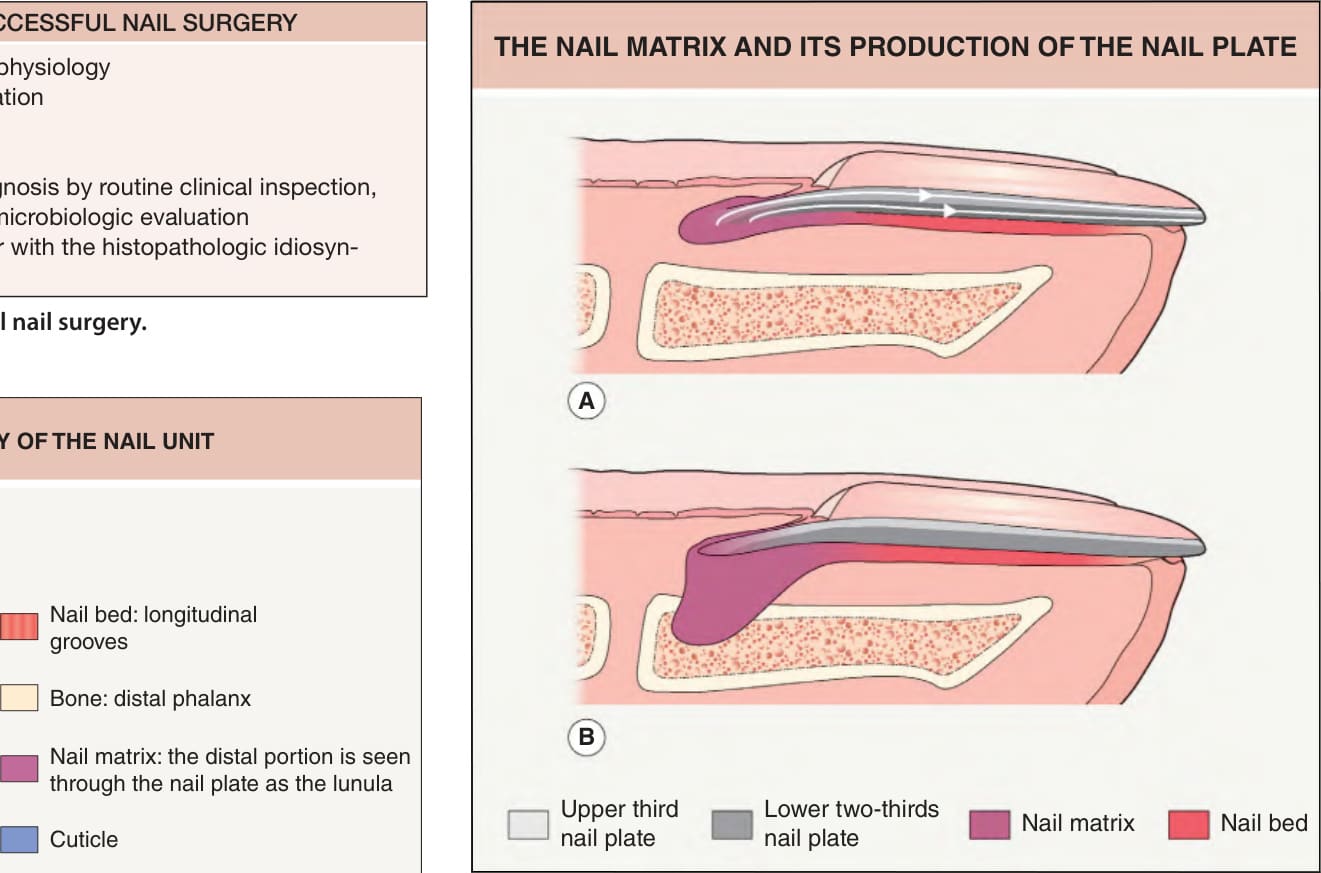
Fig. 149.2 The nail matrix and its production of the nail plate.A The proximal portion of the matrix produces the upper third of the nail plate (light gray) and its distal part the lower two-thirds (dark gray). B On the great toe, the lateral horns of the matrix can extend as far as the midline of the lateral aspect of the digit or even farther.
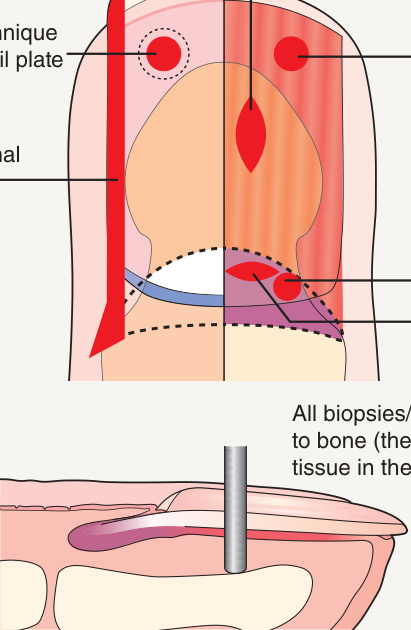
Fig. 149.11 Types and orientations of biopsies and excisions within the nail unit.
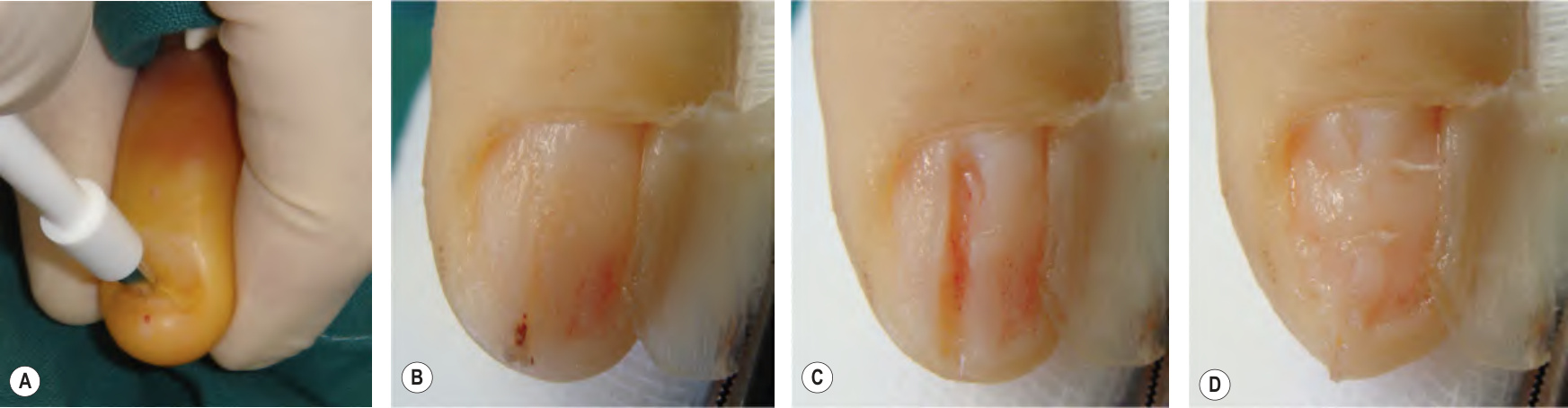
Fig. 149.12 Nail bed biopsy – punch biopsy versus longitudinal excisional biopsy.A After partial nail avulsion, the punch biopsy instrument is rotated with pressure until there is contact with bone. B Lateral avulsion of the nail plate exposes an onychopapilloma along the nail bed. C Longitudinal incision allows delicate dissection of the tumor from the bone. D Suturing of the nail bed; the nail plate will be returned to its original location and secured to the lateral fold.
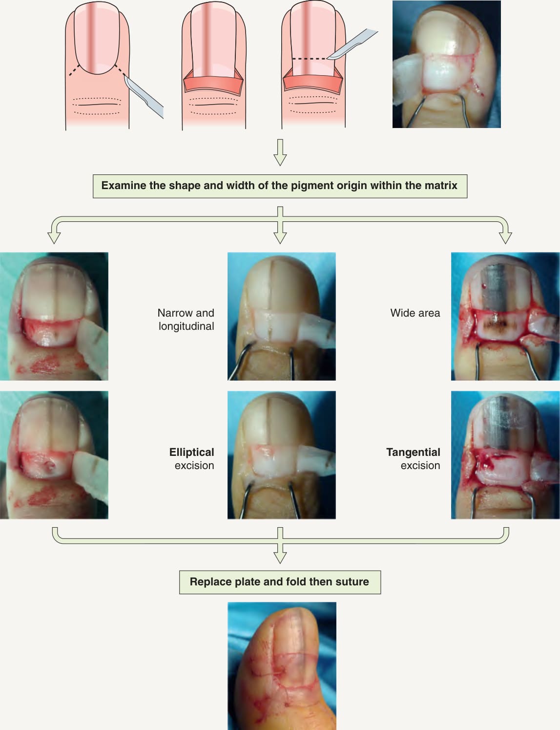
Fig. 149.13 Surgical approach to longitudinal melanonychia.
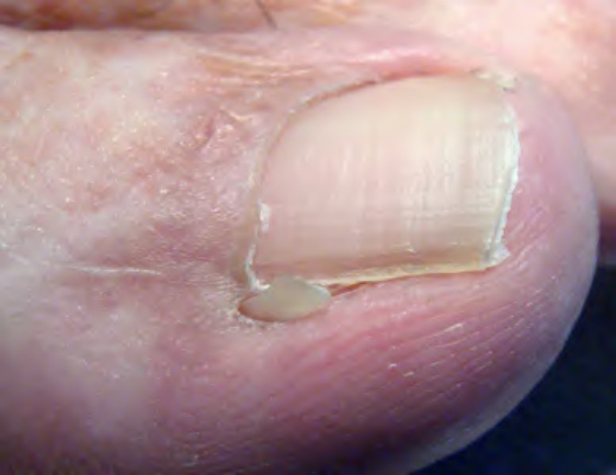
Fig. 149.15 Lateral nail spicule due to incomplete removal of the lateral horn of the matrix. Note the straight line of the incision and compare to Fig. 149.14.
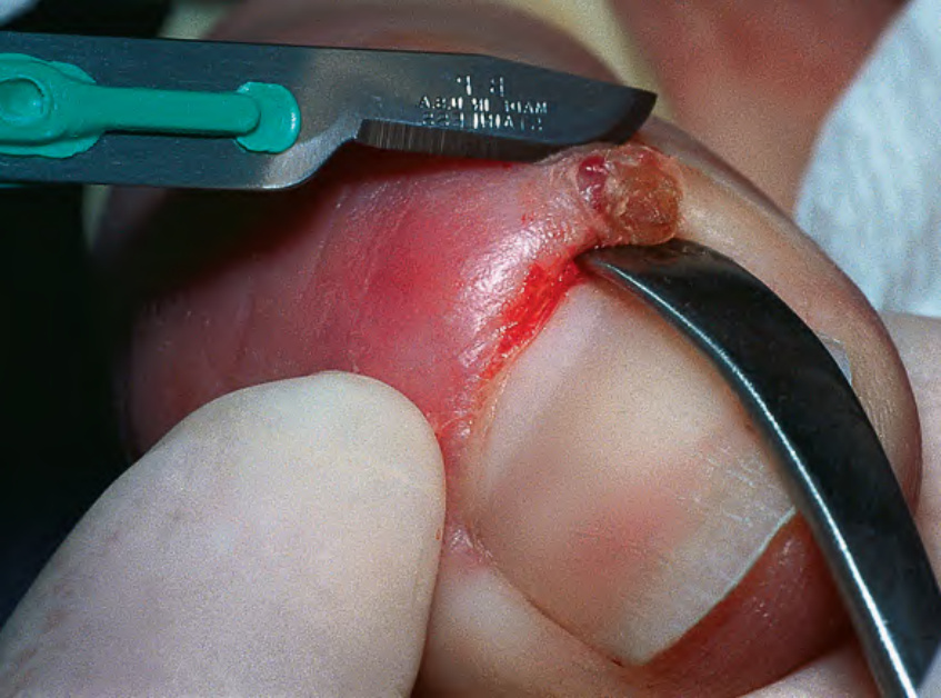
Fig. 149.16 Biopsy of the proximal nail fold via a crescent-shaped incision.
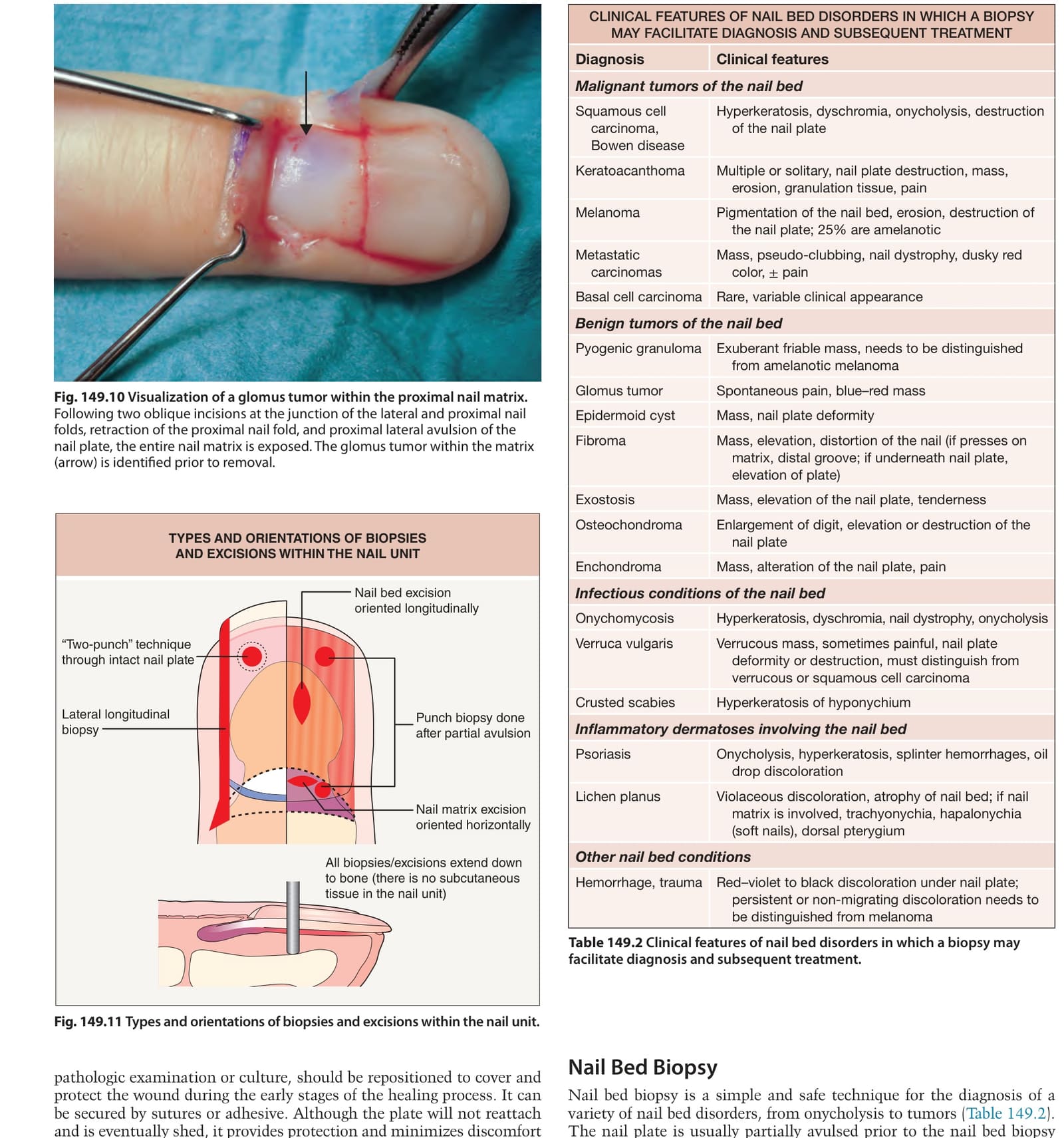
Table 149.2 Clinical features of nail bed disorders in which a biopsy may facilitate diagnosis and subsequent treatment.