DIFFERENTIAL DIAGNOSIS
The differential diagnosis of ACD includes many other forms of dermatitis, such as ICD, protein contact dermatitis, atopic dermatitis, stasis dermatitis, and seborrheic dermatitis, as well as the erythematous form of rosacea. Hand and foot ACD also needs to be distinguished from endogenous dermatitis, psoriasis, and tinea (see Table 13.4 & Fig. 15.7). Of note, these conditions may coexist, which can make clinical assessment complicated. In general, when evaluating regional dermatoses (e.g. eyelid, hand, and foot), other disorders common to the area need to be considered as well as allergens specific to that area (Table 14.2). If there is widespread disease, either because of widespread contact with an allergen or autosensitization, additional causes of erythroderma (see Ch. 10), e.g. Sézary syndrome, enter the differential diagnosis.
(ACD). Contact urticaria, both immunologic and non-immunologic, can also complicate the clinical picture.
Deciphering and differentiating these diseases can sometimes be a challenge. Distribution and history can be helpful (see Figs. 14.1, 14.4, 14.5), as can simple tests such as a KOH preparation. In addition, dermatoses are sometimes multifactorial, and ACD may be superimposed on atopic dermatitis or stasis dermatitis. However, in order to diagnose ACD, the clinician must first consider the possibility, then ask the right questions, and, finally, use the appropriate allergens to diagnose this condition. What follows will hopefully provide the clinician with an outline to the appropriate questions to ask and procedures to use when investigating ACD.
Patch Testing
Patch testing is a deceptively simple office procedure upon which the diagnosis of ACD often rests. Although the procedure is straightforward, deciding when and what to patch test requires training and experience. Unfortunately, patch testing is still underutilized. In 2010 in the US, only 22% of dermatology program directors and 27% of chief residents reported having a specific patch testing rotation.
The T.R.U.E. TEST®, which is approved by the US Food and Drug Administration (FDA), consists of panels with pre-impregnated
allergens, allowing for increased ease of use and perhaps resulting in increased patch testing. However, it currently screens for 35 allergens, in addition to a negative control, and although this is helpful, extended testing beyond these allergens has been shown to improve diagnostic accuracy. With extended testing, 37%–76% more positive reactions were detected, and 47% of the patients had positive reactions only to non-screening allergens (these latter allergens come in multi-use syringes or tubes [see Appendix]). Lastly, when patch testing was performed with 28 T.R.U.E. TEST® allergens, only 27.6% of patients had completely detected allergens.
Prior to the application of the patch tests, the clinician should ask questions about exposures both at home and at work, and attempt to understand the mechanics of the work environment. The effect of vacations and time away from work or home should also be ascertained. In addition, all personal care products should be inventoried and hobbies explored. The information gained can help to direct allergen selection more appropriately.
Technique
Although the T.R.U.E. TEST® now has 35 allergens (plus control) and the European Baseline Series 30 allergens plus 13 recommended additions (Table 14.3), referral centers with patch test clinics routinely patch test to an “expanded” series of 60+ allergens. Such panels include the American Contact Dermatitis Society Series (updated in 2020), the North American Contact Dermatitis Group 70 (which includes allergens “of interest” primarily for research purposes), and
(2021), the European Baseline Series (2019), and the North American Contact Dermatitis Group (NACDG) 70 (2017–18). The concentrations and vehicles are used in the ACDS Series unless otherwise noted. aq, aqueous; Conc, concentration; HPC, hydroxypropyl cellulose; pet, petrolatum; ppm, parts per million; PV, polyvidone; SOF, soluble organic fraction.
the Australian Baseline Series (updated in 2021). This is in addition to more specific allergen panels, e.g. the hairdressing tray, dental tray or florist tray, which contain allergens unique to a particular occupation. The companies from which these allergens and other supplies can be obtained are listed in the Appendix. Although these expanded series of allergens are not FDA-approved, they are often required to establish the precise etiology of the patient’s dermatitis.
Substances brought by patients to the dermatologist should not be tested in a blinded fashion. The physician should be aware of the chemical ingredients of the product, or severe irritation such as a burn or ulceration could occur. Therefore, no unknown product or chemical should be applied as a patch test. Material safety data sheets (MSDS) can sometimes help in further evaluating the chemical. However, not all ingredients are listed on these forms: those chemicals that represent a small percentage and fall below a certain threshold do not need to be listed, even though they may be the causative allergens. Identification of the latter requires communication with the manufacturer, so that full disclosure of the chemical ingredients can be obtained.
When patients bring all their personal care products to the office for patch testing, special attention is required. The general rule regarding testing of these products is that products intended to be left on the skin (so-called “leave-on” products), such as moisturizers and make-up, may be tested “as is”. Products that are intended to be diluted by water or rinsed off (so-called “rinse-off” products), such as soaps and shampoos, need to be diluted prior to patch testing (often to 1%–10% in water). There are helpful guides for determining appropriate patch test concentrations for numerous chemicals. When these nonstandard allergens are tested, controls (including vehicles) must be tested to evaluate for the possibility of ICD.
After allergen selection has been finalized, appropriate technique is necessary to ensure adequate testing. The most common site is the upper back. The patient should not have a sunburn in this area and should not have applied topical corticosteroids to the sites of patch testing for 1 week. Systemic and longer-lasting injectable corticosteroids should also be avoided for at least 1–2 weeks. (If necessary for disease control, the daily oral AM dose of corticosteroids should not exceed the equivalent of 20 mg of prednisone during testing.) Any one of these factors may decrease the individual’s ability to elicit a reaction when challenged by an allergen, resulting in a false-negative test. Other immunosuppressants (e.g. methotrexate, mycophenolate mofetil) and targeted immune modulators (“biologics”) may also impact patch testing results.
A nurse or technician in the office can be trained to apply the patches, and this leads to improved efficiency. Either the pre-packaged allergens are placed on the back as in the case of the T.R.U.E. TEST®, or the allergens are dispensed into chambers. There are a number of different patch test systems, including Finn Chambers® (SmartPractice®) which are adhered to Scanpor® tape (Norgesplaster, Norway; available in the US from SmartPractice®); allergEAZE® chambers (SmartPractice®; Fig. 14.12); and IQ, IQ Ultra™, or IQ Ultimate™ chambers (Chemotechnique Diagnostics; distributed by Dormer Laboratories, Inc.). These patches
are applied to the back, reinforced with more Scanpor® tape if required, and the patient is sent home with instructions to keep the back dry and the patches secured until the second visit at 48 hours. Patients should also be told to avoid excessive sweating and to avoid heavy lifting, as the patches may come loose. Antihistamines can be prescribed, as they will not affect the outcome of the testing. A map of where the allergens were placed should be constructed for future reference.
When the patient returns at 48 hours, the patches need to be examined to ensure that the testing technique was adequate. Initial inspection can determine that the patches are still in place. Confirmation comes from observing whether the chambers have adhered adequately so as to leave an impression in the skin (see Fig. 14.14G). As the patches are removed, their sites of application should be marked in order to identify the location of the particular allergens (Fig. 14.13). Two types of marking pen are recommended for this procedure: either a permanent surgical marker or a fluorescent highlighter. Highlighters are less messy and do not rub off as easily as the permanent marker. Because of the latter property, permanent markers can soil clothing and make interpretations at the second reading more difficult. Any positive reactions are scored according to the International Grading System (Table 14.4; Fig. 14.14). The patient is again asked to keep the back dry until the second reading, which can be performed from 72 hours to 1 week after the initial application of patches.
When the patient returns for the second reading, the map is used to identify any positive reactions. If a fluorescent marker has been utilized, a Wood’s lamp may be needed to identify the markings. Positive reactions are again graded according to the standard system (see Table 14.4). This later reading is necessary as patch test responses to some allergens such as gold, neomycin, and corticosteroids may be delayed. The actual products the patient uses in his or her work and/ or home environment are examined and the ingredients compared with the positive reactions. In so doing, products can be divided into groups: those that are free of the suspected allergen(s) and are safe to use and those that contain these chemical(s) and should be avoided. There will usually be some products with no ingredients listed, and these will have to be further investigated by the patient or physician by contacting companies and inquiring about ingredients.
Interpreting the test
Through the process of reviewing exposures and products, the clinical relevance of positive test results can be determined. Allergens may have past relevance. For example, a patient who presents with a known allergy to nickel and reactions to costume jewelry is found on patch testing to have a positive reaction to nickel. The nickel may have past relevance to the patient’s problem with costume jewelry but no current relevance to the chronic hand dermatitis. Relevance may also be determined to be current. For example, a positive reaction to tuliposidase A (see Ch. 17) in a florist who has hand dermatitis and contact with Alstroemeria when arranging flowers would have present relevance. In some individuals, relevance may be unknown, as in a patient with an eyelid dermatitis and a positive reaction to thimerosal but no history of current or past exposure; sensitization may have occurred from a vaccine.
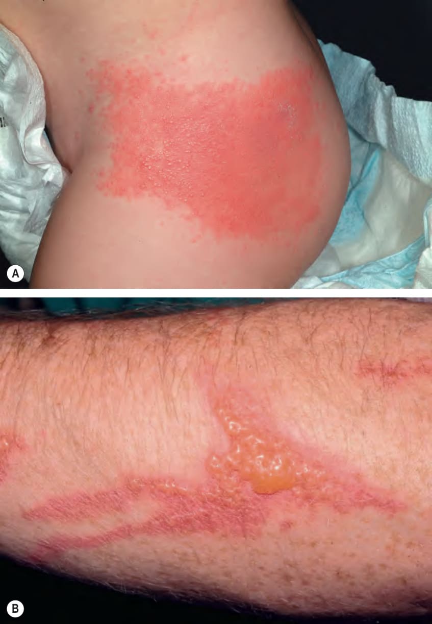
Fig. 14.1 Allergic contact dermatitis (ACD).A This erythematous plaque with vesiculation developed in a 14-month-old boy following the application of neomycin ointment. B Erythematous streaks with linear vesicles caused by ACD to poison ivy. A, Courtesy Anthony J. Mancini, MD; B, Courtesy Joyce Rico, MD.
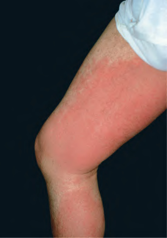
Fig. 14.10 Allergic contact dermatitis due to cashew nut shell oil. This represented an occupational exposure.
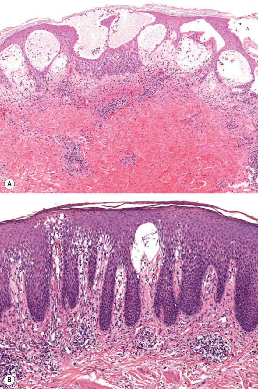
Fig. 14.11 Allergic contact dermatitis (ACD) – histologic features.A Acute ACD with marked spongiosis leading to intraepidermal vesiculation. There is a perivascular infiltrate of lymphocytes and eosinophils as well as exocytosis of these cells into the epidermis. B Chronic ACD with irregular psoriasiform epidermal hyperplasia and significantly less spongiosis. There is also a primarily perivascular infiltrate of lymphocytes admixed with eosinophils. Courtesy Lorenzo Cerroni, MD.
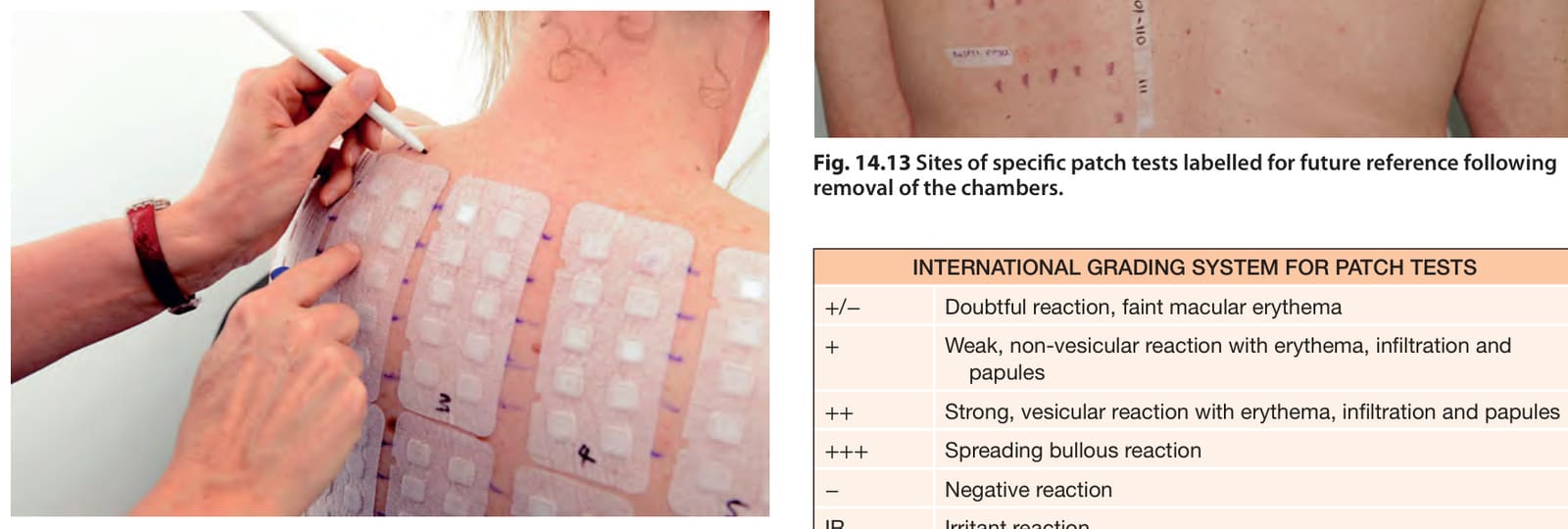
Fig. 14.12 Placement of allergens to the patient’s back utilizing allergEAZE® chambers.
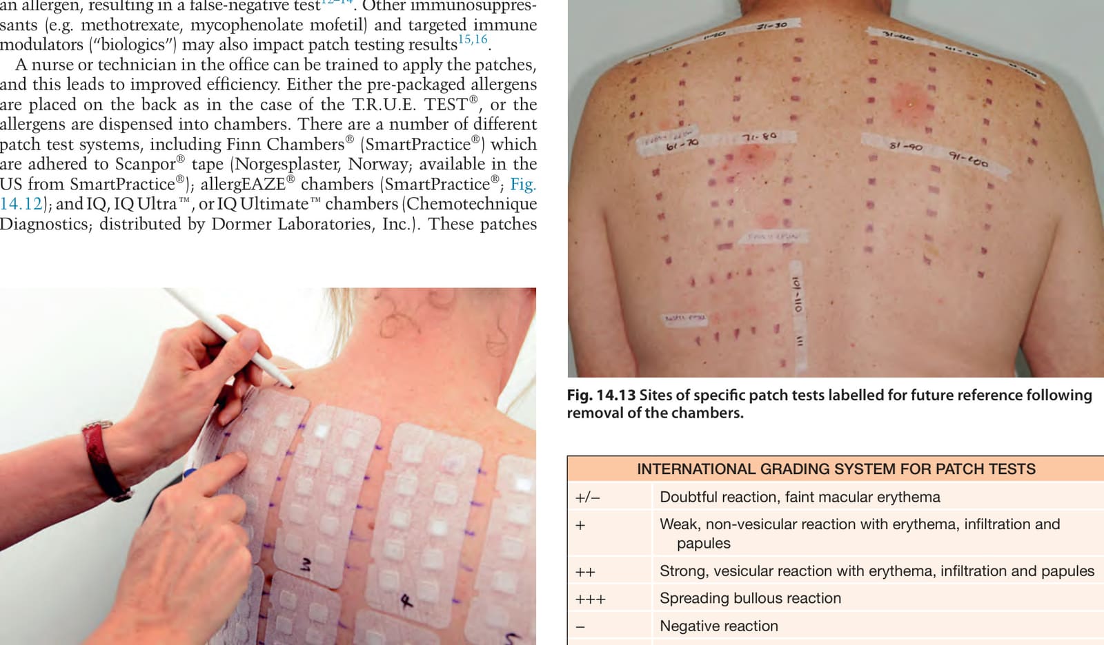
Fig. 14.13 Sites of specific patch tests labelled for future reference following removal of the chambers.
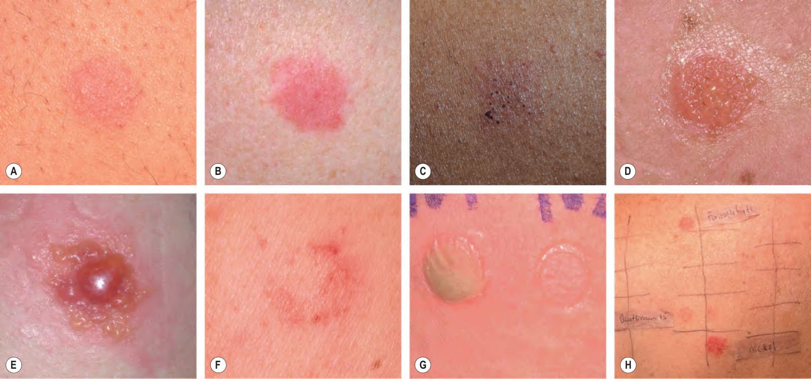
Fig. 14.14 Patch test reactions.A+/− to + reaction. B, C+ reaction. D++ reaction. E+++ reaction. F Erythematous papules at the edge of the Finn chamber application site (rim or edge effect). G Pustular irritant reaction at the site of the application of a metalworking fluid. The adjacent skin impression of a chamber reflects good adherence. H Three different patch test reactions: +/− to quaternium-15, + to formaldehyde, and ++ to nickel.
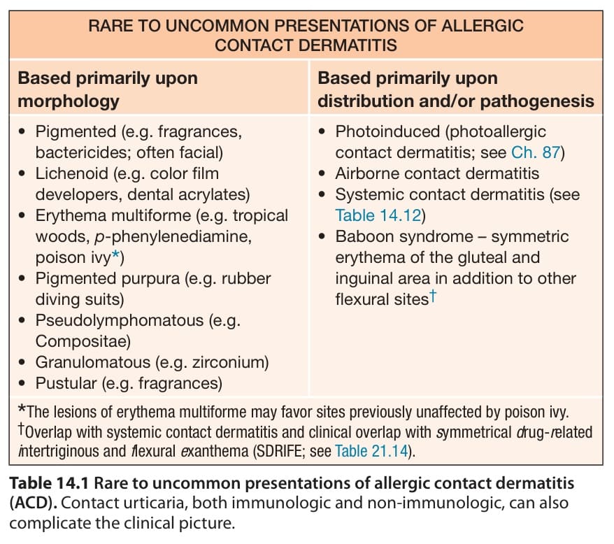
Table 14.1 Rare to uncommon presentations of allergic contact dermatitis
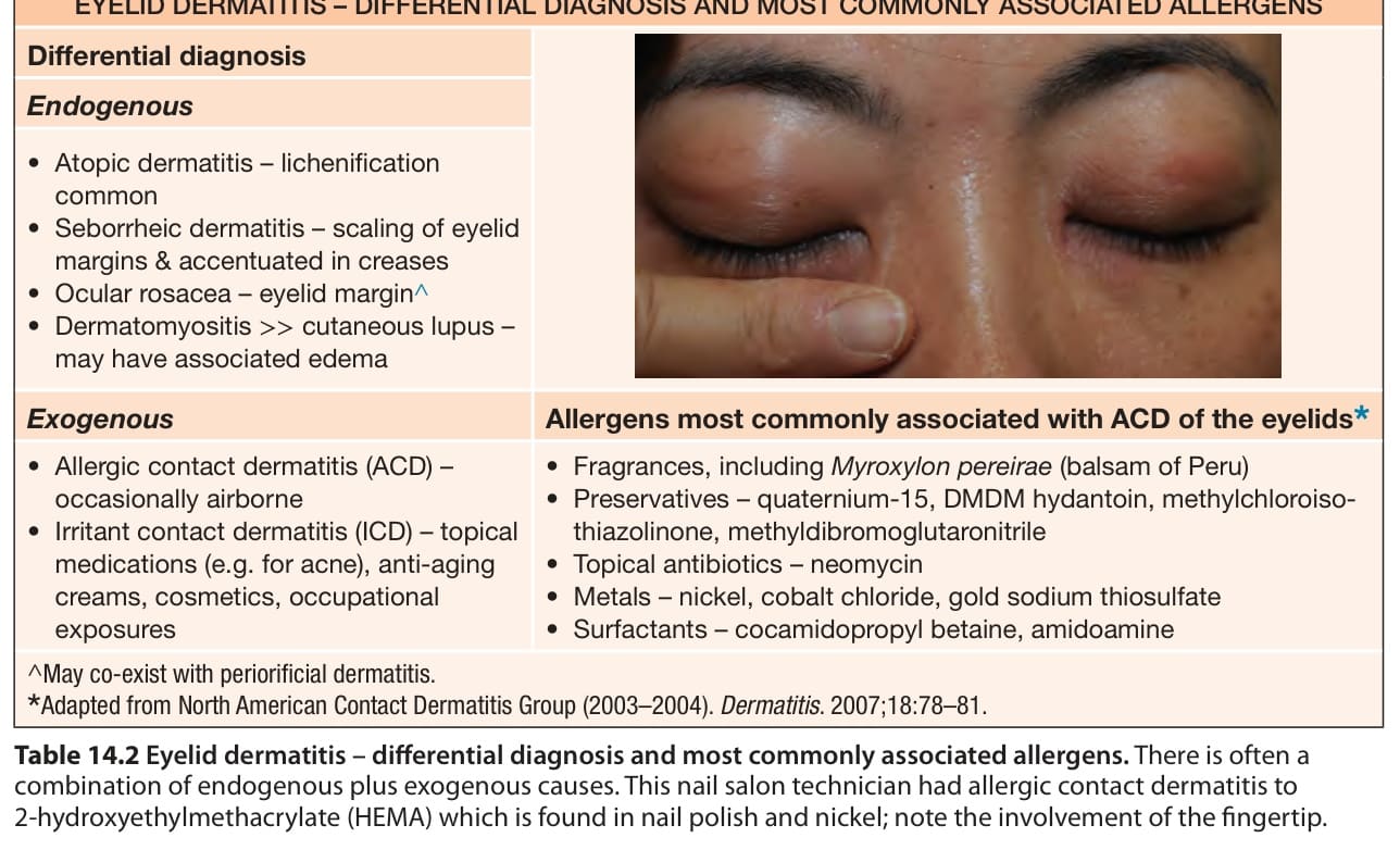
Table 14.2 Eyelid dermatitis – differential diagnosis and most commonly associated allergens. There is often a combination of endogenous plus exogenous causes. This nail salon technician had allergic contact dermatitis to 2-hydroxyethylmethacrylate (HEMA) which is found in nail polish and nickel; note the involvement of the fingertip.
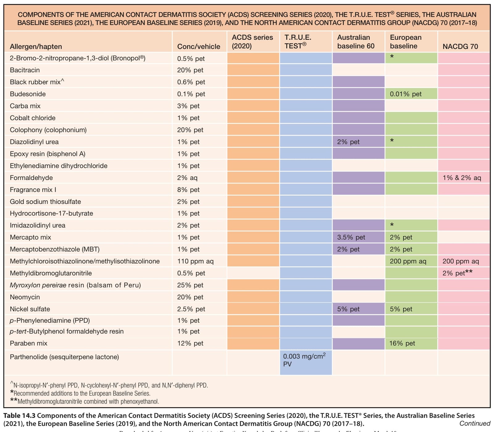
Table 14.3 Components of the American Contact Dermatitis Society (ACDS) Screening Series (2020), the T.R.U.E. TEST® Series, the Australian Baseline Series (2021), the European Baseline Series (2019), and the North American Contact Dermatitis Group (NACDG) 70 (2017–18). Continued
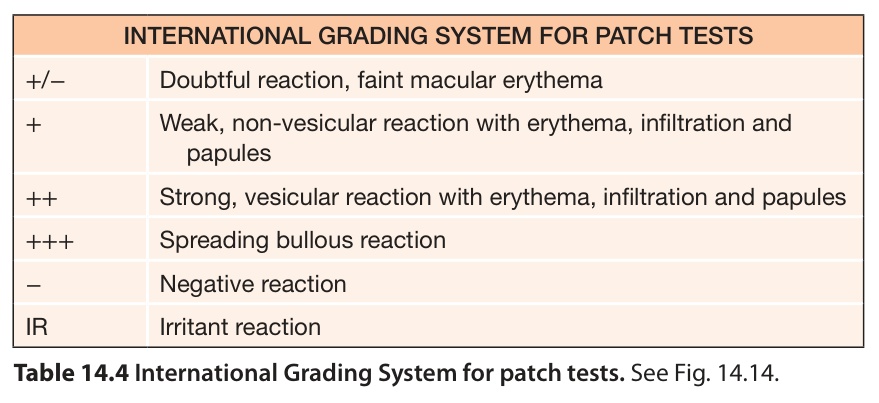
Table 14.4 International Grading System for patch tests. See Fig. 14.14.