INTRODUCTION
Mohs micrographic surgery (MMS) is a specialized method for skin cancer removal that combines surgery with pathology resulting in an excellent cure rate. The goals are to remove only the cancerous tissue while viewing the complete margin, thereby preserving the maximum amount of normal tissue (i.e. tissue-sparing). From a functional, aesthetic and reconstructive perspective, tissue-sparing is especially important for skin cancers in the periorbital, nasal, auricular, perioral, digital, and anogenital areas. MMS is also the preferred treatment method for skin cancers that are clinically poorly defined, recurrent, and/or have a higher risk of recurrence because of histologic features or anatomic location (Table 150.1).
There are four major components: surgical excision, histopathologic examination, precise mapping, and wound management. The surgical component consists of removing a disc of tissue with a beveled surgical margin (Fig. 150.1A). The disc of tissue, or “layer”, ensures that the entire 360° of tumor margin is circumferentially removed and examined. The histopathologic component involves cryopreserving this disc and cutting it in a horizontal fashion, then mounting sections onto glass slides, followed by staining and microscopic examination for residual tumor. This processing technique differs from standard excisions in which specimens are formalinfixed, paraffin-embedded (FFPE) and cut in a “bread-loaf” fashion (Fig. 150.1B). Precise mapping entails marking any residual tumor on the map of the excision with the remaining positive area of tumor reexcised as indicated. This three-step process is repeated until there is no residual tumor identified microscopically. During this procedure, the Mohs surgeon has complete control of the excised tissue, including laboratory processing and interpretation of the tissue sections, as well as precise mapping. In the fourth step, wound management, the tumor-free defect is surgically closed or allowed to heal by second intention.
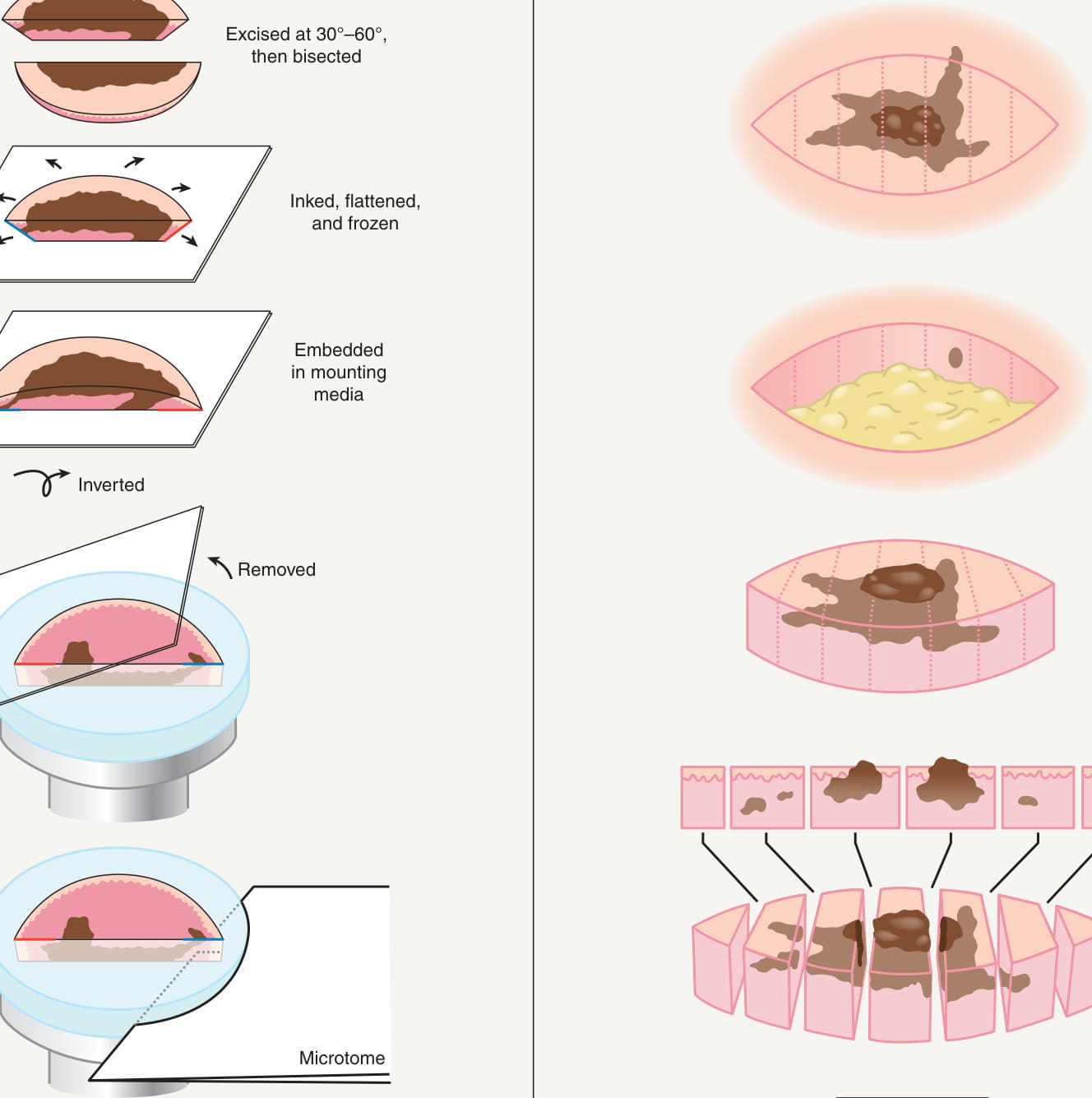
Fig. 150.1 Surgical excision of a tumor and pathologic examination of tissue margins.A Mohs micrographic surgery in which 100% of the total margin is examined. Following excision with a narrow margin, the specimen is flattened so that when the tissue is horizontally sectioned, the skin edge is in the same plane as the deep margin. Note the inversion of the tissue prior to sectioning. B Standard elliptical excision with vertical tissue sectioning (“bread loafing”). Histologically, the margins may appear clear even though there is still residual tumor.
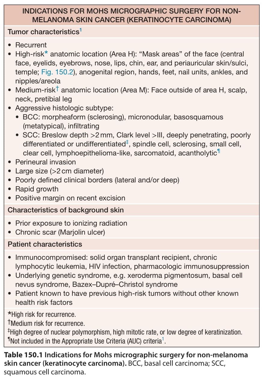
Table 150.1 Indications for Mohs micrographic surgery for non-melanoma skin cancer (keratinocyte carcinoma). BCC, basal cell carcinoma; SCC, squamous cell carcinoma.