INDICATIONS
Most skin cancers are locally invasive and, depending on their location, can invade vital structures such as the eye, nose, lips and ears, leading to significant tissue destruction. With time and neglect, metastasis may develop. The majority of non-melanoma skin cancers are low-risk and do not fulfill the criteria outlined in Table 150.1. For such tumors, cost-efficient superficial ablative techniques (e.g. electrodesiccation and curettage) or simple excision can be performed. Currently, ~25% of skin cancers are treated with MMS in the US. MMS is well suited for skin cancers at high risk for recurrence and for those tumors located in regions requiring tissue conservation and complete margin control. This includes large lesions in cosmetically sensitive areas.
Factors that identify a “high risk” skin cancer include clinical and histologic characteristics of the tumor, anatomic site, and history of prior treatment(s) (see Table 150.1). Because of the complete histologic examination of surgical margins, MMS allows tracking of unpredictable superficial or deep projections of tumor (see Fig. 150.1A). MMS is very useful for tracking tumors that involve embryonic fusion planes, the periorbital region, nerves, and bone. It is these deep and irregular tumor extensions that are primarily responsible for the higher recurrence rates observed with other techniques. Importantly, the indications for MMS are not absolute, and any treatment decision needs to be made in collaboration with the patient’s preferences (e.g. local vs. general anesthesia
for extensive procedures), performance status, and understanding of alternative treatment options and associated cure rates.
The use of MMS for the more commonly managed cutaneous malignancies will be reviewed first, followed by its use for unusual neoplasms (Table 150.2).
Basal Cell Carcinoma
For primary BCCs, cure rates of 87% to 95% can be obtained with superficial ablative therapies and excision, especially on the trunk and extremities. In a meta-analysis by Rowe et al., the 5-year cure rate for primary BCC treated with MMS was 99%, as compared to 90% to 93% using standard treatments (Table 150.3). For recurrent BCC treated with MMS, the 5-year cure rate was 90%–94% as compared to 80% for standard treatments (surgical excision, radiotherapy, cryotherapy, electrodesiccation and curettage).
MMS provides superior cure rates for high-risk BCCs that are more likely to recur, including those that are large in size (>2 cm) or have aggressive histologic subtypes (e.g. basosquamous carcinoma) or perineural invasion.
Squamous Cell Carcinoma
MMS offers the highest cure rates for patients with high-risk primary or recurrent SCCs. The local recurrence rates, based on a retrospective review of MMS versus non-MMS modalities for primary SCC, are outlined in Table 150.3.
MMS is indicated for management of SCCs with one or more risk factors for recurrence or metastasis (see Table 150.1). These indications include large SCCs (>2 cm), recurrent tumors, incompletely excised SCCs, and tumors with ill-defined clinical margins, rapid growth, an aggressive histologic subtype, and/or perineural invasion (Fig. 150.3), as well as SCCs arising in chronic scar tissue (Marjolin ulcer). Immunosuppressed patients, including those with solid organ transplants and chronic lymphocytic leukemia, as well as patients receiving medications such as voriconazole and unopposed vemurafenib, can develop rapidly growing SCCs that benefit from MMS.
In addition, SCCs arising in high-risk anatomic sites may be managed with MMS. For example, tumors of the lip have a high propensity for recurrence and metastasis with cure rates as high as 92% achieved with MMS. MMS is also well suited for SCCs occurring in anogenital areas and on the digits, where tissue conservation is important. When
compared with conventional methods, MMS for the treatment of penile SCC offers comparable survival rates. Prior to MMS, SCCs involving the nail unit were often treated with amputation or radiation. MMS provides an excellent choice for treatment of periungual and subungual SCC without osseous involvement, with cure rates of up to 96%.
Erythroplasia of Queyrat (EQ) is a term used for SCC in situ (SCCIS) arising in anogenital mucosae, especially in men. An erythematous thin plaque is typically seen on the inner surface of the foreskin, glans penis, and/or coronal sulcus of an uncircumcised man (see Chs. 73 & 108). In women, differentiated vulvar intraepithelial neoplasia (dVIN) and high-grade squamous intraepithelial lesion (HSIL) are the preferred terms, with oncogenic human papillomaviruses (HPVs) often playing an etiologic role in the latter. HPV infection also increases the risk of anogenital SCC in immunosuppressed individuals (e.g. HIV-infected).
MMS is an effective treatment for EQ, with a 5-year cure rate of 90%. When excising EQ of the glans penis, Mohs layers should be very thin in order to minimize entry into and resection of the corpus spongiosum and cavernosum. If either is entered, achieving hemostasis can be challenging. EQ can often extend into the urethra and differentiation of SCCIS from transitional epithelium is often difficult on frozen sections. With deep extension along the urethra, collaboration with a urologist and completion of the surgery under general anesthesia are recommended.
Keratoacanthomas (KAs) classically grow quickly and then can spontaneously involute. However, management of KAs remains controversial. Microscopically, some KAs are difficult to distinguish from SCC and there are reports of non-regressing and metastatic KAs, implying that the tumor was actually an SCC. Currently, most KAs are considered to be well-differentiated SCCs. MMS may be particularly beneficial for large lesions, as well as those occurring in anatomically sensitive areas.
Verrucous carcinoma is considered a variant of SCC, and it can be associated with HPV infection. Tumors most commonly occur in the oral cavity, on the plantar foot, or on the penis (see Ch. 108). While generally considered a low-grade carcinoma, there are reports of metastasis. Histopathologic interpretation of frozen sections can be challenging because the tumor cells are very well differentiated with minimal nuclear atypia, making distinction from pseudoepitheliomatous hyperplasia, verruca vulgaris, or condyloma acuminatum challenging. In patients with a clear-cut clinical diagnosis verified by histologic examination, the irregular architectural pattern of verrucous carcinoma may aid in evaluating the margins of excision by MMS.
Cutaneous Melanoma Melanoma in situ (lentigo maligna) of the head and neck region
Treatment of melanoma in situ (MIS), in particular lentigo maligna (LM), of the head and neck region is a subject of debate. Controversies include width of surgical margins, MMS versus standard surgical excision, and use of pre- or postoperative topical imiquimod. Even within MMS, there are different techniques, from classic MMS with frozen sections to “slow Mohs” utilizing “rush” permanent sections. Reasons for the debate include: (1) the difficulty in distinguishing atypical melanocytes of photodamaged skin from melanoma cells (Fig. 150.4), especially in frozen sections stained with H&E or antibodies that recognize melanosomes; and (2) the natural history and prognosis of LM. The first can lead to residual tumor or unnecessary excision of photodamaged skin while the second can complicate determining an impact on survival.
Current NCCN guidelines recommend surgical treatment with margins of 0.5–1.0 cm, allowing for narrower margins depending upon anatomical and functional considerations. However, LM treated with surgical excision alone may require wider margins as lesions tend to be ill-defined with subclinical spread and a local recurrence rate of 6%–20% has been reported with standard excision. Although still employed in a minority of patients, the use of MMS to treat cutaneous melanoma has increased over the past two decades, from 2.6% (2001) to 7.9% (2016).
There are no randomized controlled trials comparing wide local excision (WLE) versus MMS to treat MIS of the head and neck region. Database studies, including a retrospective review of a prospective database that compared H&E-based MMS with WLE, reported comparable rates of overall and melanoma-specific survival. In the retrospective review, there was also no difference in recurrence rates. Studies from single institutions have also observed low recurrence rates, e.g. <1% with a mean follow-up of 2.8–3.7 years.
As noted previously, distinguishing atypical melanocytes of photodamaged skin from melanoma cells in frozen sections can prove challenging. In addition to similar cellular features, processing artifacts (e.g. freeze-induced vacuolization within keratinocytes that resembles vacuolization in melanocytes) may lead to misinterpretation. Detection of a transition zone between photodamaged skin and MIS in FFPE vertical sections can assist in determining clear margins, but this is not possible with en face processing of LM margins. To address this potential problem, a prospective cohort study (n = 167) was conducted that examined the accuracy of MIS margin assessment with MMS and H&E-stained frozen sections; the authors reported an accuracy rate of 95%. The latter was determined by excising an additional 1 mm margin followed by its examination by a dermatopathologist. Other approaches include: (1) vertically processing the central debulk plus a narrow margin; (2) obtaining shallow biopsies from the patient’s normal-appearing adjacent skin or both perilesional and distant skin; and (3) examination of FFPE tissue margins to confirm that the final MMS margin is negative.
Potential improvements include immunohistochemical (IHC) staining with antibodies that recognize melanocyte-specific transcription factors (e.g. MITF) rather than melanosomal proteins (e.g. Melan-A/
MART-1) (Table 150.4). This prevents staining of melanosome-containing keratinocytes and overestimating the density of melanocytes within photodamaged skin as well as falsely diagnosing pigmented actinic keratoses as LM. Of note, in a survey of Mohs surgeons, only 36% felt comfortable interpreting MART-1-stained frozen sections and a need for additional education was noted as was recognition that formal training is not offered in all fellowship training programs.
AE1/AE3, pankeratin marker; Ber-EP4, stains cellular glycoproteins in
BCCs, sweat glands, and some hair follicles; BCC, basal cell carcinoma; CEA, carcinoembryonic antigen; CK7, cytokeratin 7; DFSP, dermatofibrosarcoma protuberans; EMPD, extramammary Paget disease; MAC, microcystic adnexal carcinoma; MART-1, melanoma antigen recognized by T cells; MITF, microphthalmia transcription factor (nuclear stain); PAS, periodic acid Schiff; SCC, squamous cell carcinoma.
Photomicrograph: Courtesy Luis Requena, MD.
Invasive melanoma
Based on large randomized controlled trials (RCTs), WLE with recommended margins is the gold standard for treatment of invasive melanoma (see Ch. 113). In patients undergoing sentinel lymph node biopsy, the WLE is routinely performed in tandem. Cutaneous melanomas in general are well-defined, with the exception of some cases of LM and acrolentiginous melanoma. For locally recurrent melanomas, ill-defined or amelanotic melanomas, or melanomas in anatomic sites such as the genitalia, digits, eyelids, nose and ears, a multidisciplinary approach that includes MMS can be considered. There is also some evidence suggesting the utility of MMS for thin melanomas.
Staged excisions (“slow Mohs”) for melanoma
One variant of MMS is staged excisions (“slow Mohs”) in which the tissue specimen is sent for “rush” FFPE sections that are read by a dermatopathologist the next day. The wound is kept open and covered with a pressure dressing. Some surgeons debulk the visible tumor and determine its depth via vertical processing while assessing the peripheral margin in an en face fashion, whereas others cut the tissue radially, allowing the pathologist to appreciate the transition from tumor to actinically damaged skin. If there is residual melanoma, another layer is removed until a negative (tumor-free) margin is achieved. The obvious major disadvantage is the inconvenience of a multi-day procedure. Reported recurrence rates range from <1% to 7.3%, with follow-up ranging from 3 to 57 months.
Additional staged-excision techniques include the square technique, geometric staged excision, and the spaghetti technique. In the square technique, two closely spaced, parallel blades are used to create a square-shaped defect (thin and frame-like) with the tumor and pre-sumed adequate margins left intact. If the excised peripheral tissue has clear margins, then the central square is removed surgically. When the square technique was used to treat 834 melanomas, the mean margin required for histologic clearance was 9.33 mm for MIS and 13.7 mm for invasive melanoma. With a median follow-up of 9.3 years, the projected recurrence rate was 2.2% at 10 years.
In the geometric staged excision, the entire specimen is excised in a geometric pattern; the margins are inked and strips are cut off along the margins. The cut strips are oriented, processed and cut en face while the central portion is cut vertically to determine tumor depth. As a result, the entire peripheral margin is examined, which is where the tumor may extend a significant distance from the clinically apparent lesion. In addition, the vertical sections of the central portion confirms or adjusts the tumor thickness. Some dermatopathologists prefer this method because it may be easier to process the tissue for examination of the entire peripheral epidermal and dermal margin. In the spaghetti technique, thin strips at the peripheral margin of the visible tumor are excised in stages until the whole tumor is mapped out, then the central lesion is removed. This allows for closure between each stage. A local recurrence rate of 4.76% at a median follow-up of 25 months has been reported.
A potential disadvantage of the square and geometric pattern techniques is that a few extra millimeters of skin are sacrificed in order to provide the straight lateral margins. For small tumors, this difference is marginal but if several layers are required and/or the tumor is irregularly shaped, reconstruction can become more complex.
Currently, evidence-based support for the use of MMS to treat cutaneous melanoma is limited by single institution experiences, inclusion of heterogenous tumor types, variable (and sometimes limited) observation periods, and outcome measures based upon local recurrence rates. Given the natural history of LM, the latter can be problematic. Whether a multi-center, randomized, controlled, prospective trial comparing WLE with MIS will ever be conducted remains uncertain.
Uncommon or Rare Cutaneous Neoplasms
Due to the relative rarity of these uncommon tumors, the efficacy of MMS is based primarily on case series from a single institution or case reports.
Atypical fibroxanthoma/pleomorphic dermal sarcoma
Atypical fibroxanthoma (AFX) is a dermal-based mesenchymal neoplasm that develops within photodamaged skin and has minimal, if any, metastatic potential (see Ch. 116). Tumors with similar pathologic features but which demonstrate subcutaneous invasion, necrosis, and/ or lymphovascular or perineural invasion are associated with a worse outcome and are referred to as pleomorphic dermal sarcoma (PDS). Typically, AFX presents as a nodule in the head and neck region of an elderly person with significant actinic damage. Its histologic features are said to be relatively easy to identify in frozen sections. For AFX, MMS has been reported to result in lower recurrence rates and greater tissue preservation whereas in PDS, the data is limited.
Dermatofibrosarcoma protuberans
Dermatofibrosarcoma protuberans (DFSP) is a slowly growing, locally aggressive soft tissue sarcoma that most often arises on the trunk of young to middle-aged adults (see Ch. 116). It typically extends in an irregular fashion beyond the clinically visible tumor margins. Based upon pooled raw data collected over a 20-year period, recurrence rates following WLE have ranged from 6% to 60% versus 1% to 3% following MMS. A possible explanation for this difference is the inclusion of patients with primary DFSP, recurrent DFSP, and DFSP with fibrosarcomatous changes, which can lead to biased results as patients with more aggressive disease tended to have WLE (compared to MMS). Of note, a recent retrospective, multicenter study comparing MMS vs. WLE for DFSP on the trunk and extremities found no difference in outcomes while emphasizing the need for complete resection with precise margin control. In the head and neck region, MMS can be advantageous given the potential for conserving tissue while offering margin control.
DFSP is often a large-sized tumor and as a result, MMS can be a timeconsuming procedure. If tumor identification proves difficult in frozen sections, staged excision (“slow Mohs”; see above), utilizing anti-CD34 IHC staining, can be performed.
Merkel cell carcinoma
Merkel cell carcinoma (MCC) is a very aggressive malignancy, often arising in older adults with photodamage or who are immunosuppressed (see Ch. 115). Clinically, the tumor usually presents as a pink–red to violaceous papulonodule that favors the head and neck region. Microscopically, the tumor cells are aggregated into large sheets, allowing them to be easily identified in frozen sections. NCCN guidelines recommend WLE plus sentinel lymph node biopsy (SLNB) followed by radiotherapy to the primary site and regional lymph nodes (unless the lesion is low risk [≤1 cm on the trunk or an extremity with no lymphovascular invasion and negative surgical margins in the setting of a negative SLNB]; (see Fig. 115.19). However, a database study found that patients who underwent MMS were less likely to have a sentinel lymph node biopsy performed. That said, when 22 MCCs were removed via MMS, positive margins were noted in 57% of the cases despite the surgical margins taken meeting or exceeding NCCN guidelines of 1–2 cm margins; however, 92% of the positive cases involved the deep margin.
Microcystic adnexal carcinoma
Microcystic adnexal carcinoma (MAC) is a locally aggressive tumor that typically involves the face of older adults (Fig. 150.5). Histologically, it has an infiltrative growth pattern with frequent perineural invasion (Fig. 150.6). With an overall recurrence rate reported in the literature approaching 40%, MMS offers an advantage. Based upon several studies, a five-year cure rate of 90% has been observed following MMS (see Table 150.2). In one of these studies, defects following MMS were four times larger than clinical estimates.
Sebaceous carcinoma
Sebaceous carcinoma can develop in any site that contains sebaceous glands, but it has a predilection for periocular skin. MMS is often employed for tumors in the latter location. Based upon a review of the literature, WLE of sebaceous carcinomas was associated with a local recurrence rate of 4%–37% and a nodal metastatic rate of 3%–28%. In comparison, MMS had a local recurrence rate of 11% (2 of 18 patients) with nodal metastases developing in 1 patient. A more recent
retrospective review of 45 sebaceous carcinomas treated with MMS found no local recurrences, metastases, or disease-specific deaths, with a mean follow-up of 3.6 years. One challenge associated with these tumors is discontiguous growth, which can thwart any method of histologic margin evaluation. The removal of an extra safety margin of tissue for permanent sections is a reasonable strategy.
Other adnexal carcinomas
This group consists of the uncommon tumors that have sweat gland (eccrine, apocrine), sebaceous gland, or follicular differentiation and includes mucinous carcinoma, eccrine porocarcinoma, adenoid cystic carcinoma, papillary adenocarcinoma, and trichilemmal carcinoma (see Ch. 111). These tumors are usually locally destructive with extensive unpredictable local tissue invasion and therefore have a high rate of local recurrence following excision. Typically, their microscopic features are readily recognizable in frozen sections so they are amenable to MMS. Rapid IHC staining can aid in diagnosis (see Table 150.4).
Miscellaneous tumors
MMS has been utilized for other malignant neoplasms, including angiosarcoma, lymphoepithelioma-like carcinoma, leiomyosarcoma and extramammary Paget disease, as well as cutaneous breast metastases. Occasionally MSS is employed for benign tumors that have a predilection for recurrence such as infantile digital fibroma, desmoplastic trichoepithelioma, and granular cell tumor. Rarely, it is used to eradicate drug-resistant cutaneous fungal infections.
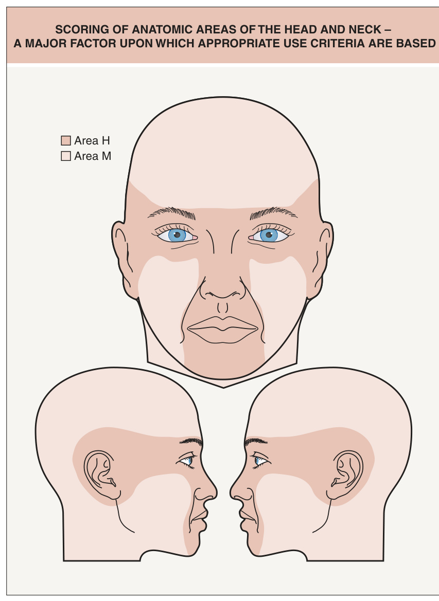
Fig. 150.2 Scoring of anatomic areas of the head and neck – a major factor upon which appropriate use criteria are based. Area H represents the anatomic sites with the highest risk for recurrence and the greatest need for tissue sparing, followed by Area M. The majority of the trunk and extremities is labeled as Area L (lowest need for tissue sparing and risk of recurrence), with the hand, foot, ankle, nail unit, nipple/areola, and anogenital region representing additional Area H sites and the shins an Area M site. See www.aad.org/education/appropriateuse-criteria/mohs-surgery-auc as well as the Mohs AUC app.
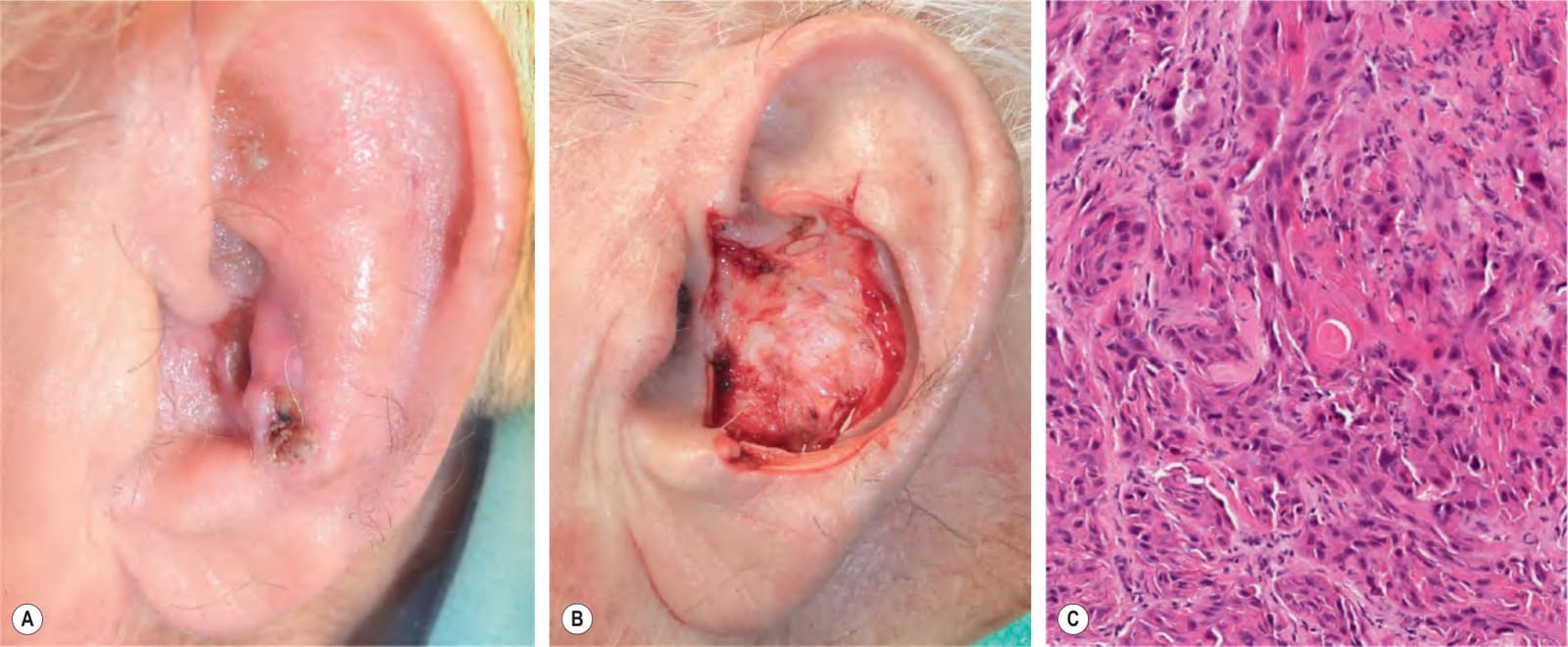
Fig. 150.3 Recurrent, biopsy-proven, moderately differentiated squamous cell carcinoma (SCC) of the left conchal bowl.A Prior to Mohs micrographic surgery. B Tumor extirpation resulted in a through-and-through defect of the ear. C Histologic features of this aggressive SCC include nests and cords of polygonal cells with enlarged, pleomorphic nuclei and only small foci of keratinization.
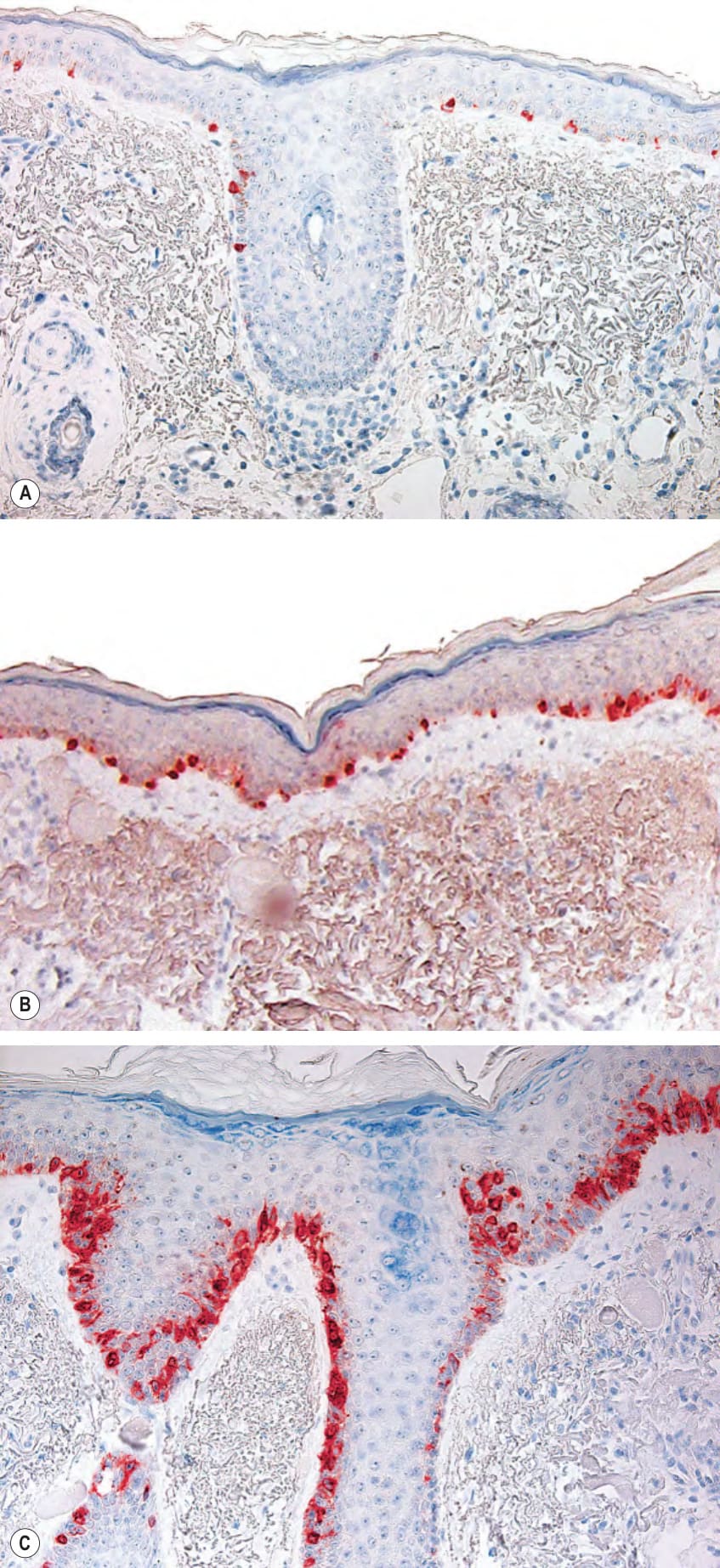
Fig. 150.4 Immunostaining with Melan-A/MART-1.A Normal density of melanocytes. B Melanocytic hyperplasia with scattered atypical melanocytes in sun-damaged skin. C Lentigo maligna with atypical melanocytic proliferation, some pagetoid spread, and adnexal extension. Courtesy Clark C. Otley, MD, and Randall K. Roenigk, MD.
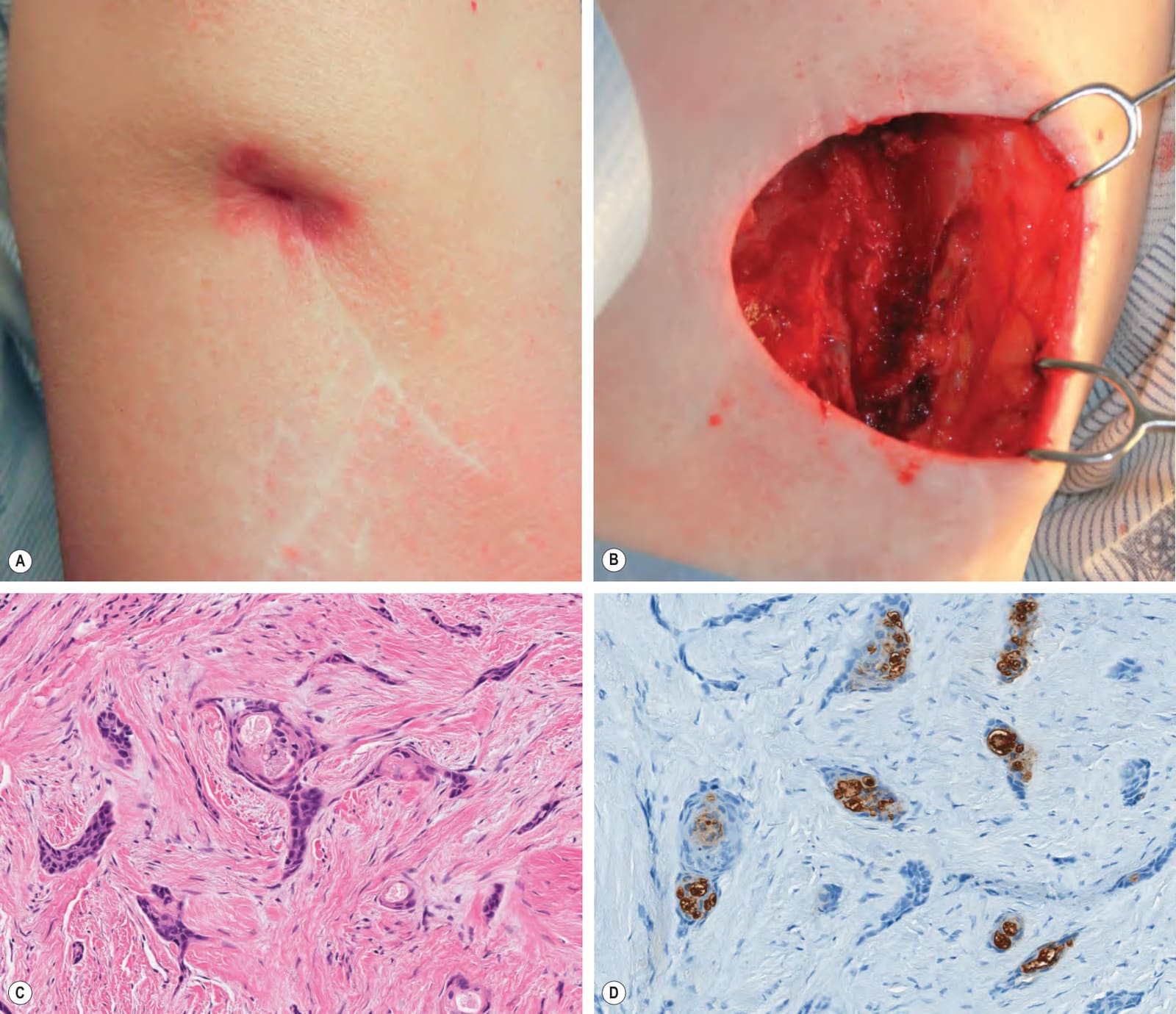
Fig. 150.5 Recurrent basal cell carcinoma (BCC) plus microcystic adnexal carcinoma (MAC).A Recurrent, biopsy-proven BCC of the right upper lateral arm that had been previously treated four times (electrodesiccation and curettage; excision; Mohs micrographic surgery [MMS] twice) over a twenty-year period. An indurated nodule was present at the scar site. B Defect after two MMS stages; there was deep infiltration of the subcutaneous tissue, muscle, and periosteum of the humerus. C Histologically, small infiltrative basaloid tumor strands embedded within a desmoplastic stroma were seen; there was also ductal differentiation. Because the possibility of a coexisting MAC was raised, the tissue was thawed and submitted for formalin-fixed paraffin-embedded (FFPE) sections. D Immunohistochemical staining of FFPE sections demonstrated CEA-positivity of ductal cells, supporting the diagnosis of MAC.
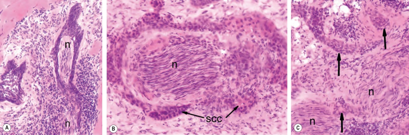
Fig. 150.6 Perineural involvement by various cutaneous tumors (frozen sections).A Basal cell carcinoma with a halo of basal cells enveloping a cutaneous nerve (n). B Squamous cell carcinoma (SCC) surrounding nerve fiber (n). C Microcystic adnexal carcinoma with perineural invasion by dominant pilar structures (arrows) in the deep subcutaneous portion of the tumor; n, nerve. Reproduced with permission from Snow S. Atlas of Mohs Surgery Frozen Sections. Madison, WI: Scope Publishing, 2010.
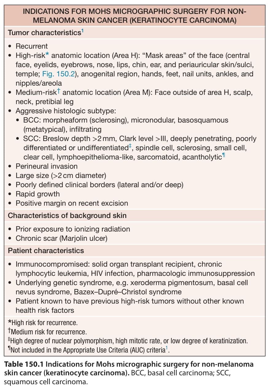
Table 150.1 Indications for Mohs micrographic surgery for non-melanoma skin cancer (keratinocyte carcinoma). BCC, basal cell carcinoma; SCC, squamous cell carcinoma.
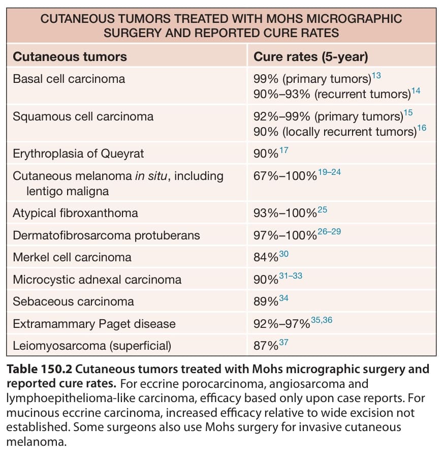
Table 150.2 Cutaneous tumors treated with Mohs micrographic surgery and reported cure rates. For eccrine porocarcinoma, angiosarcoma and lymphoepithelioma-like carcinoma, efficacy based only upon case reports. For mucinous eccrine carcinoma, increased efficacy relative to wide excision not established. Some surgeons also use Mohs surgery for invasive cutaneous melanoma.
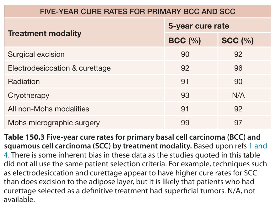
Table 150.3 Five-year cure rates for primary basal cell carcinoma (BCC) and squamous cell carcinoma (SCC) by treatment modality. Based upon refs 1 and
4. There is some inherent bias in these data as the studies quoted in this table did not all use the same patient selection criteria. For example, techniques such as electrodesiccation and curettage appear to have higher cure rates for SCC than does excision to the adipose layer, but it is likely that patients who had curettage selected as a definitive treatment had superficial tumors. N/A, not available.
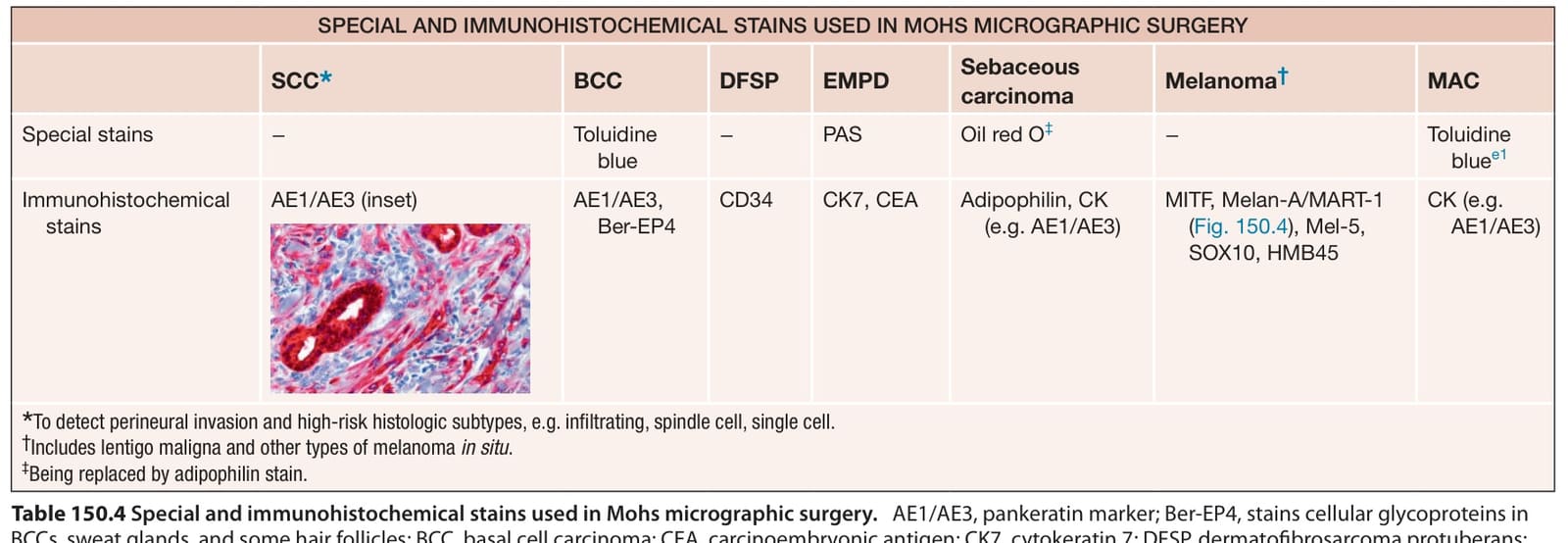
Table 150.4 Special and immunohistochemical stains used in Mohs micrographic surgery.