CHALLENGES AND PITFALLS
The success of the procedure is dependent upon the precision and meticulousness of each step. Recurrences following MMS have been linked to inherent tumor characteristics such as morphology, size or location as well as errors in the procedure. Errors encountered during MMS can be divided into technical (e.g. excess epidermal tissue in the central portion of the layer pushed down to cut margin and interpreted as tumor, incorrect bevel angle or relaxing incisions) and pathologic interpretation (Fig. 150.8). A limitation of MMS arises when the invasiveness of the tumor prevents continued tracking and consultation with the appropriate surgical specialist is indicated.
There are several clinical scenarios in which it is appropriate to send tissue for FFPE including: (1) a second opinion is required during removal of a cutaneous melanoma; (2) further assessment is needed of an aggressive, deep, or histologically unusual tumor; (3) confirmation of the presence of a different tumor than was diagnosed preoperatively (Figs. 150.5 and 150.9); (4) a significant amount of tissue is available for complete staging of the primary tumor; (5) unusual histologic findings in the frozen sections require a second opinion; (6) frozen section interpretation is insufficient to assess the tissue margin reliably; (7) a tumor diagnosed solely by frozen section prior to surgery, i.e. no pre-operative biopsy was sent for routine histology; or (8) special stains
are required that cannot be performed reliably on frozen sections. In addition, when there is histologic evidence of a second neoplasm or the histologic features of the tumor being removed via MMS and the pre-operative diagnosis differ, the original FFPE sections should be reviewed.
Although infrequent, cutaneous neoplasms can invade bone. This may be the consequence of previous inadequate treatment, anatomic site (e.g. embryonic fusion planes, areas with thin overlying skin such as the scalp, sternum, and distal phalanx), tumor biology, and patient comorbidities (e.g. chronic lymphocytic leukemia, solid transplantrelated immunosuppression). In addition, tumors with perineural invasion can gain access into bone via bony foramina. When tumors invade bone, the following strategies are used to control its growth and potentially effect a cure: (1) establish the diagnosis and mode of invasion of the tumor histologically; (2) obtain radiologic imaging to determine the macroscopic extent of bone involvement and status of the regional lymph nodes (see above); and (3) refer to a related subspecialist for bone
Continued
resection as indicated. In difficult tumors with perineural invasion, additional Mohs layers may be taken to assure a tumor-free plane.
Small foci of sinus, nasal, or scalp bone may be excised locally with a bone rongeur or a chisel and mallet. Even when bone is exposed during tumor surgery, second intent healing is possible. If more than 1 cm of bone is exposed, burring, abrasion, or chiseling of spots 1 cm apart to reach bleeding spots can facilitate granulation. The procedure should be done with the patient recumbent to minimize the extremely rare risk of air embolus into the cavernous sinus (to date 2 cases reported). If sufficient granulation tissue can be stimulated to cover the exposed facial or scalp bone relatively quickly, cellulitis and osteomyelitis rarely occur. Other options include coverage with an acellular dermal matrix or a split-thickness skin graft. These three methods of wound management are frequently preferred over more complex flaps that may delay detection of recurrent tumor. Close clinical surveillance is imperative.
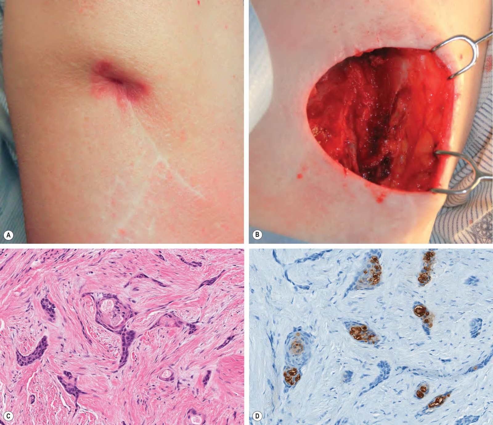
Fig. 150.5 Recurrent basal cell carcinoma (BCC) plus microcystic adnexal carcinoma (MAC).A Recurrent, biopsy-proven BCC of the right upper lateral arm that had been previously treated four times (electrodesiccation and curettage; excision; Mohs micrographic surgery [MMS] twice) over a twenty-year period. An indurated nodule was present at the scar site. B Defect after two MMS stages; there was deep infiltration of the subcutaneous tissue, muscle, and periosteum of the humerus. C Histologically, small infiltrative basaloid tumor strands embedded within a desmoplastic stroma were seen; there was also ductal differentiation. Because the possibility of a coexisting MAC was raised, the tissue was thawed and submitted for formalin-fixed paraffin-embedded (FFPE) sections. D Immunohistochemical staining of FFPE sections demonstrated CEA-positivity of ductal cells, supporting the diagnosis of MAC.
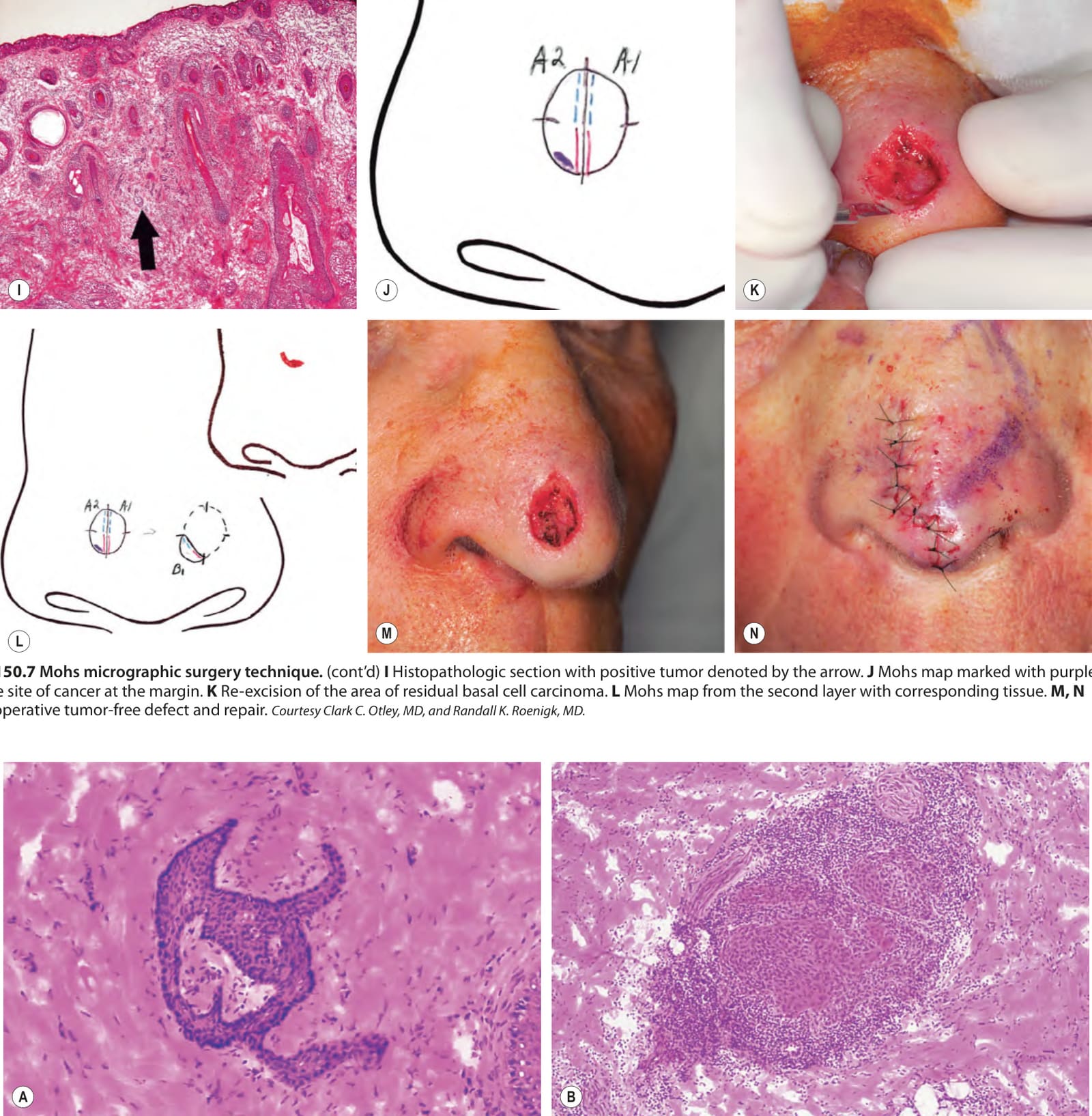
Fig. 150.7 Mohs micrographic surgery technique.A Preoperative appearance of a basal cell carcinoma on the nose. B Debulking of the tumor via curettage. C Beveled excision (with scalpel at a 45° angle) of all clinically evident tumor plus a small margin of normal-appearing skin. D Placement of skin notches at 3, 6, 9 and 12 o’clock positions in the specimen and adjacent surrounding tissue. E Orientation of the excised specimen on an anatomic card. F Specimen sectioned into two pieces and flattened. G Marking of the cut margins of the specimen with red and blue inks. H Matching Mohs map and tissue specimen ready for histologic preparation. A red mark on the gauze denotes the location of the first specimen with the second specimen always placed below. Different systems are used but precision is common to all.
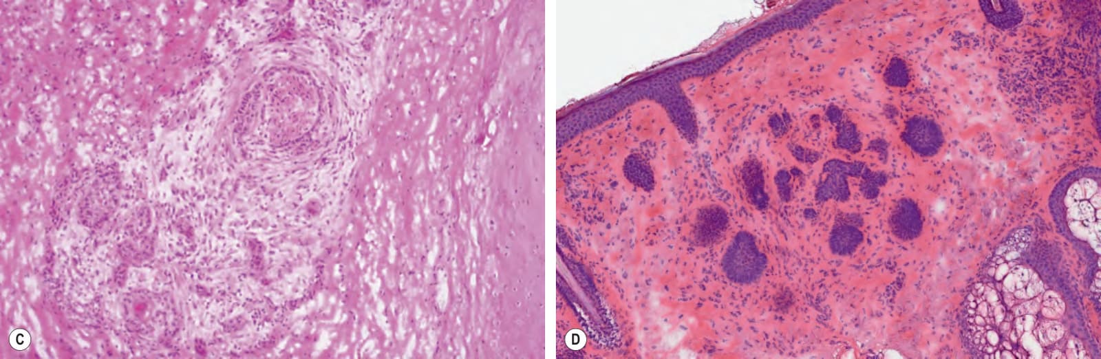
Fig. 150.8 Histopathologic challenges in Mohs surgery.A Folliculocentric basaloid proliferation may mimic basal cell carcinoma; however, the lack of clefting adjacent to the cells, apoptosis, or mitotic figures as well as the presence of a fibrous sheath permits distinction. B Dense inflammation can obscure tumor cells, as seen here with a squamous cell carcinoma in a patient with chronic lymphocytic leukemia. C Careful evaluation is required to identify subtle foci of perineural squamous cell carcinoma. D Follicular remnants can mimic basaloid islands. A–C, Courtesy Clark C. Otley, MD, and Randall K. Roenigk, MD.
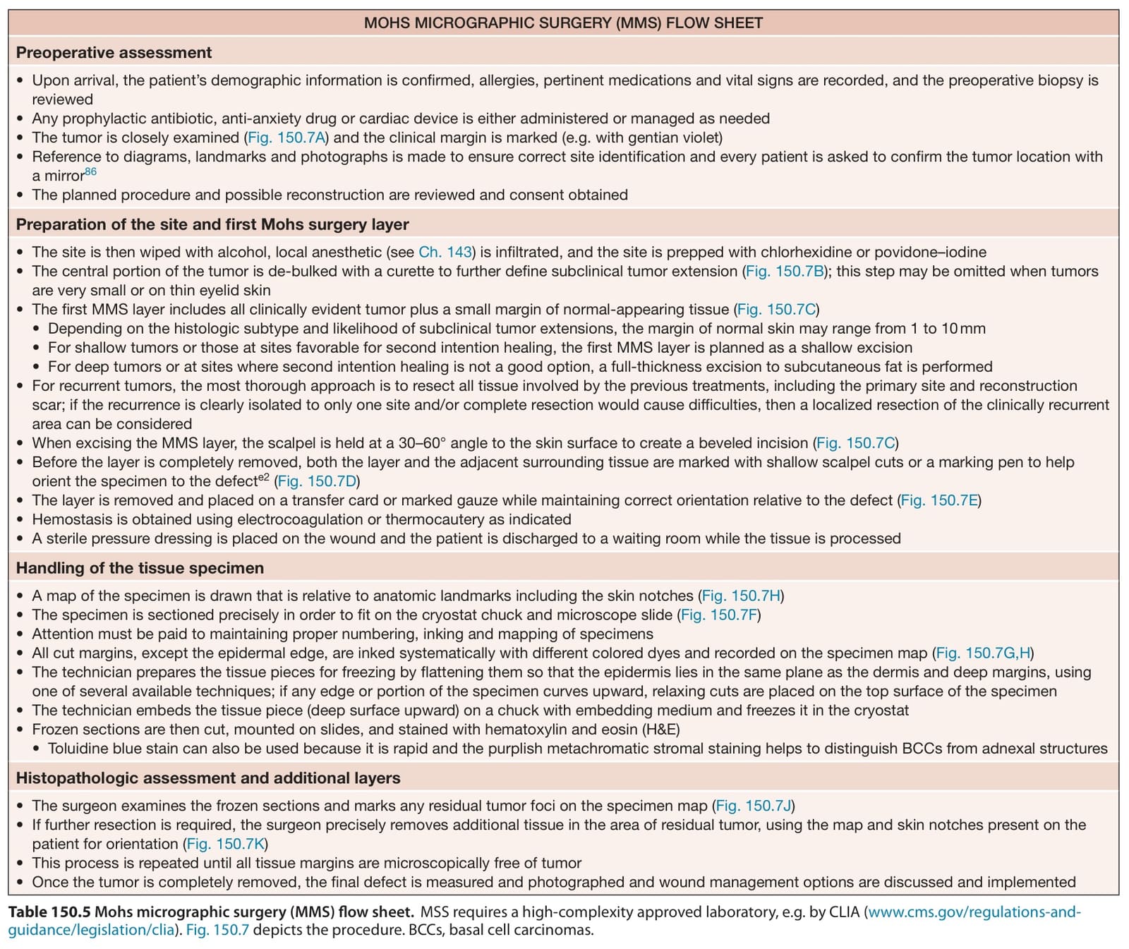
Table 150.5 Mohs micrographic surgery (MMS) flow sheet.MSS requires a high-complexity approved laboratory, e.g. by CLIA (www.cms.gov/regulations-andguidance/legislation/clia). Fig. 150.7 depicts the procedure. BCCs, basal cell carcinomas.