DISCUSSION
Preoperative Assessment
A thorough preoperative assessment is a valuable tool for identifying conditions that could lead to surgical complications. A simple preoperative questionnaire can be completed by the patient at the time of the initial evaluation. “Yes” or “no” answers to specific questions can indicate a need for further evaluation or for a change in the operative plan (Fig. 151.2).
Potential for bleeding
Although some bleeding is expected during any surgical procedure, excessive bleeding is a common complication. The discovery of a bleeding disorder prior to surgery allows for correction of the condition if it is reversible, or changing plans for surgery if it is not. The bleeding disorder may be due to abnormalities in the coagulation cascade, platelets, or both. Platelet disorders result from abnormalities in production, survival, or function.
If the history does not suggest a bleeding disorder and the patient is otherwise well, laboratory testing is usually unnecessary. Appropriate screening tests should be checked if there is suspicion for a hereditary or acquired bleeding disorder. A platelet count only identifies a quantitative abnormality. The bleeding-time test measures platelet function, but accurate determination of results is operator-dependent. An in vitro bleeding-time assay, the platelet function analyzer 100 (PFA-100), provides an easy method for rapidly assessing platelet function. Unlike the bleeding time, the PFA-100 can be performed on a collected blood specimen. Additional screening tests include a PT (prothrombin time) and an aPTT (activated partial thromboplastin time) to determine defects in the extrinsic and intrinsic coagulation pathways, respectively. The international normalization ratio (INR) is discussed in the next section.
The preoperative assessment should include a review of medications and dietary supplements (see Table 133.3), in particular antiplatelet and anticoagulant medications. When these drugs are prescribed for cardiac or neurologic indications, discontinuation can potentially lead to serious events (e.g. coronary artery stent thrombosis). In general, surgeons should not discontinue these medications but should instead focus on intraoperative precautions, such as meticulous hemostasis and minimal undermining.
Aspirin and aspirin-containing products are well-documented sources of bleeding problems. Platelet aggregation is irreversibly altered when aspirin acetylates cyclooxygenase. Patients taking aspirin for general health benefits, rather than specific medical indications, may discontinue the aspirin from ≥10 days before until 5–7 days after surgery. Nonsteroidal anti-inflammatory drugs affect the same enzyme, although the block is neither as severe nor irreversible. If possible, the patient should stop these medications 1–4 days prior to surgery, depending on the half-life of the drug.
Dipyridamole suppresses platelet aggregation by inhibiting phosphodiesterase and increasing cyclic adenosine monophosphate (cAMP) levels within platelets. It is used in combination with aspirin for secondary prevention of thromboembolic events. The antiplatelet effect of dipyridamole is less than that of aspirin, and it does not appear to increase bleeding complications.
Thienopyridines (e.g. clopidogrel, ticlopidine, prasugrel) irreversibly inhibit platelet aggregation via inhibition of P2Y, a platelet ADP receptor. Cilostazol is a selective type 3 phosphodiesterase inhibitor with weaker P2Y inhibition; it is typically used for claudication. Normal platelet function usually returns 5–7 days after discontinuation. Despite the slightly increased risk of complications in patients receiving P2Y inhibitors, these medications can typically be continued without significant issues.
Several studies have been published in the vascular, oral, orthopedic, and dermatologic surgery literature supporting the viewpoint that surgery can be performed without stopping warfarin. An INR should be checked prior to surgery. Most authors advocate an upper limit of 3 or 3.5. The safety of dermatologic surgery with INRs >3.5 has not been well studied, though one study found a poor correlation between bleeding complications and INR levels, with complications also occurring in the setting of in-range INRs. If warfarin is discontinued in consultation with the prescribing physician, it should be held for 4–5 days preoperatively. If a patient on warfarin develops significant postoperative bleeding, the anticoagulation can be reversed with one or more of the following: oral or intravenous vitamin K, fresh frozen plasma, and four-factor prothrombin complex concentrate.
Factor Xa inhibitors apixaban and rivaroxaban are often used as alternatives to warfarin while the direct thrombin inhibitor dabigatran is occasionally used as an alternative; this group of drugs is referred to as DOACs (direct oral anticoagulants; see Table 22.8 and Fig. 22.6). Literature remains sparse regarding these agents in dermatologic surgery. One large randomized trial of invasive, non-dermatologic procedures found similar rates of bleeding complications with warfarin
versus dabigatran, but both medications were held preoperatively. A more recent single-center, retrospective analysis found that patients receiving a DOAC at the time of Mohs micrographic surgery had a significantly greater risk for developing postoperative hemorrhagic complications compared to patients treated with traditional oral anticoagulants. The authors recommend similar intraoperative precautions with these medications (see above). Idarucizumab is approved for reversing dabigatran, and andexanet alfa is a specific antidote for apixaban and rivaroxaban, but this is less of an issue because of their shorter half-lives of ≤12 hours.
Heparin acts as an antithrombin factor. Intravenous, unfractionated heparin has a short half-life of ~1 hour and can be rapidly reversed by intravenous protamine sulfate. Low-molecular-weight heparin (LMWH) has a half-life of 3–5 hours and is administered subcutaneously. Either form can be used to bridge patients in whom discontinuing warfarin preoperatively has been deemed necessary. LMWH is discontinued 24 hours prior to surgery, whereas unfractionated heparin is stopped five hours preoperatively. Either form of heparin is then restarted, together with warfarin, 24 hours post-procedure, with the heparin discontinued after a therapeutic INR has been attained.
Complementary and alternative therapies can inhibit platelet function or enhance anticoagulation. Common agents include vitamin E, garlic, and eicosapentaenoic acid (fish oil). Table 133.3 has a more comprehensive list of dietary supplements that have effects on platelet function and coagulation. Patients should discontinue these supplements prior to surgery. More recently, enhanced bleeding has been observed in patients receiving ibrutinib, a Bruton tyrosine kinase (BTK) inhibitor used to treat lymphomas and chronic lymphocytic leukemia. This is thought to be related to the role of BTK and other kinases in platelet signaling pathways. This is less of an issue with second- and third-generation BTK inhibitors, e.g. acalabrutininib, zanubrutinib, pirtobrutinib.
Ethanol is a potent vasodilator that inhibits platelet aggregation and platelet granule release, as well as accentuating the increase in bleeding time caused by aspirin. Excessive alcohol consumption may also decrease the patient’s attention to optimal wound protection. Patients should be advised to avoid alcohol consumption during the immediate perioperative period.
Wound healing
The principles of wound healing are covered in detail in Chapter 141. To forestall complications due to poor wound healing, close attention should be paid to pre-existing medical conditions and medications that affect wound healing. Postponing surgery may be a wise decision for some patients. If the procedure cannot be postponed, the patient should be forewarned that the postoperative healing phase could be prolonged. Any chronic debilitating illness can predispose patients to secondary infections.
Systemic corticosteroids can delay healing and predispose patients to secondary infections. Patients on other immunosuppressive medications such as cyclosporine may also be prone to infections and prolonged healing. Inhibitors of vascular endothelial growth factor (VEGF), including monoclonal antibodies (e.g. bevacizumab) and oral tyrosine kinase inhibitors (e.g. sorafenib), have been associated with delayed healing and wound dehiscence (see Fig. 102.16). These agents should be held for at least 4–6 weeks prior to surgical procedures.
Cigarette smoking interferes with wound healing in a dose-dependent manner, and flaps or grafts are particularly susceptible. Ideally, patients should avoid smoking for at least 3 weeks before surgery, but one study found that over 85% of smokers continued smoking preoperatively despite instructions to stop. At a minimum, patients should be strongly encouraged to decrease consumption to less than half a pack per day or switch to nicotine replacement patches for at least 2 weeks preoperatively and 1 week postoperatively. Compared to cigarettes, the patches do not result in the high peak blood nicotine levels that lead to peripheral vasoconstriction and reduced wound tissue oxygen tension.
There is literature to support delaying cosmetic procedures for up to one year after cessation of isotretinoin therapy due to the risk of poor wound healing and the formation of excessive granulation tissue. However, most reported problems have been individual case reports involving resurfacing procedures after courses of isotretinoin for acne therapy. More recent data from larger cohort studies suggest that procedures, including laser hair removal, dermabrasion, and laser resurfacing for acne scars, may be safe to perform during or shortly after a course of isotretinoin therapy.
Reactivation of herpes simplex virus (HSV) infection can dramatically interfere with the postoperative course. Patients with a history of herpes labialis or other facial herpetic infections should receive prophylactic antiviral therapy prior to dermabrasion, laser resurfacing, chemical peels, or surgery involving known trigger area(s). Antiviral therapy, such as oral valacyclovir 500 mg twice daily, should start on the day of the procedure and continue for 10 days postoperatively or until the skin has re-epithelialized. Alternative regimens include oral famciclovir 250 mg three times daily or oral acyclovir 200 mg five times daily.
Prophylactic antibiotics
Antibiotic prophylaxis is administered to prevent surgical site infection, bacterial endocarditis, or infection in implanted prosthetic devices. The risk and consequences of infection must be weighed against the risk and cost of the antibiotic.
Wounds are placed into four general categories (Table 151.1). The majority of dermatologic surgical procedures are clean or clean-contaminated and do not need prophylaxis. However, prophylactic antibiotics may be helpful in certain clean-contaminated wounds. For example, antibiotics can be considered for procedures involving the oronasal mucosa, axilla, or anogenital region, although there are no controlled studies. Procedures that approach the ear canal may be at higher risk for infection with Gram-negative organisms. Long procedures are also more likely to develop wound infections: the infection rate almost doubles with each hour of operation. Prophylactic antibiotics are recommended for procedures at increased risk for surgical site infection, including surgery on the lower extremities or in the groin, wedge excisions of the lip or ear, flaps on the nose, all skin grafts, and in patients with extensive inflammatory skin disease. When possible, prophylactic antibiotics should be prescribed in anticipation of these types of repairs. Unless the patient is in a special group (see below), some surgeons limit prophylactic antibiotics to just excisions on the lower extremities and ears, especially if the cartilage is exposed. Antibiotics are therapeutic, not prophylactic, for contaminated and infected wounds.
Two special patient groups should be considered for antibiotic prophylaxis: (1) those at risk for infective endocarditis; and (2) those at risk for bloodborne infection of total joint replacements (Table 151.2). In patients with total joint replacements or specific cardiac diseases, no prophylaxis is indicated if the surgical procedure is in a non-infected cutaneous site. In general, prophylactic antibiotics are no longer recommended by the American Academy of Orthopedic Surgeons and American Dental Association for patients with prosthetic joint replacements undergoing dental procedures; possible exceptions, including mucosal incision in a patient with a history of joint surgery complications, are outlined in Table 151.2. However, to date, recommendations specific to dermatologic procedures are lacking. Table 151.3 summarizes suggestions for antibiotic prophylaxis regimens. If any question arises, consider consultation with the patient’s cardiologist, internist, or orthopedic surgeon.
Implantable electronic medical devices
The electrosurgical instruments often used in cutaneous procedures have the potential to interfere with the function of pacemakers and implantable cardioverter-defibrillators (ICDs), though the risk is quite low with modern devices (see Ch. 140). Alternatives to electrosurgery include battery-operated and AC-powered thermal cautery units that do not produce high-frequency electromagnetic interference (EMI) or electrical currents traversing the body. Bipolar instruments (e.g. forceps) concentrate the current across the tips, minimizing the chance of interference. If conventional electrosurgery is used, the active tip should not be applied within 5 cm of the implanted device whenever possible. Electrocoagulation should also be kept at as low a setting as possible and delivered in bursts lasting <5 seconds to minimize the chance of prolonged inhibition.
If conventional electrosurgery is used in a patient with an ICD, a contingency plan should be in place to deal with an arrhythmia should it occur, and an AED should be immediately available. If the device is deactivated or its function altered for the procedure, it is the surgeon’s responsibility to ensure that patients return to their cardiologists within a reasonable time period for readjustment and evaluation.
There are few reports of patients undergoing dermatologic surgery who have an implanted deep brain stimulator (DBS). These devices are used to treat Parkinson disease and can be turned off with an external magnet if the resultant tremor does not interfere with surgery. Bipolar forceps or electrocautery are preferred to limit EMI. If a monopolar device is necessary, the dispersive plate should be positioned so that the pulse generator and lead wires are not located between the plate and the surgical site.
Cochlear implants (CIs) provide a sense of sound by direct stimulation of the auditory nerve. They consist of an external part that sits behind the ear and another part implanted below the skin. While potential for EMI exists, there are limited studies examining electrosurgery in patients with CIs. The British Cochlear Implant Group and CI manufacturer guidelines recommend against the use of monoterminal electrosurgery in the head and neck region; biterminal electrosurgery can be used safely if not within 1 to 2 cm of the CI. A conservative approach may be prudent, with use of electrosurgical instruments below the clavicles and only electrocautery in the vicinity of the implant.
Implantable nerve stimulators (e.g. vagal nerve, sacral nerve, phrenic nerve, spinal cord stimulators) are now available for a variety of indications. There are also implantable neurostimulators (e.g., NeuroPace® RNS® System) for epilepsy. Some of these devices can be temporarily turned off during electrosurgery without adverse effects. Electrocautery is recommended when possible to minimize risk of EMI. Other techniques to minimize interference include use of bipolar forceps and positioning of dispersive pads away from the implanted device. When in doubt, the authors advise consultation with the prescribing physician for further recommendations.
Allergies
True allergic reactions to local anesthetics may be type I (immediate) or type IV (delayed-type hypersensitivity) (see Ch. 143). Patients with a type IV sensitivity to one major group of local anesthetics (ester or amide) are usually not allergic to members of the other group. Type I reactions are more commonly associated with the ester anesthetics and their para-aminobenzoic acid (PABA) metabolite. Although the lack of cross-reactivity between ester and amide anesthetics is true for type IV sensitivity, it has not been adequately studied for anaphylaxis. If there is a possible history of a type I amide allergy, referral to an allergist for a progressive challenge protocol can be considered. It is important to consider the possibility that the allergic reaction is due to the preservative (e.g. methylparaben, metabisulfite). Use of preservative-free anesthetics can resolve this problem.
Epinephrine (adrenaline) is a vasoconstrictive agent commonly added to local anesthetics to reduce bleeding and prolong the duration of anesthesia. Patients occasionally report a history of an “allergy” to local anesthetic, describing their symptoms as palpitations, headache, tachycardia, tachypnea, or tremor (see Ch. 143). Such reports likely represent reactions to epinephrine given in high doses or inadvertently injected intravascularly. Since the half-life of epinephrine in serum is short, specific therapy for these reactions is usually not necessary. In order to avoid them, the lowest dosage and concentration of epinephrine (<1:100 000) should be used, and injection of significant amounts intra-vascularly should be avoided using proper infiltration technique.
Another common systemic side effect often mistaken as an allergic reaction is the vasovagal reaction. The patient recalls becoming pale, sweaty, and lightheaded, perhaps even fainting. Hypotension can occur, but in contrast to a true toxic or allergic reaction, the pulse is slow and regular (see Table 143.2). Urinary incontinence and abnormal limb movements may occur during a vasovagal reaction and are not diagnostic of seizure activity. Treatment consists of keeping the patient supine or in the Trendelenburg position and giving reassurance. Once the patient has recovered, they can gradually sit up and ambulate.
Chlorhexidine gluconate is a frequently utilized surgical skin preparation (see Table 146.6). It is a broad-spectrum antibacterial of the halogenated phenol class. Despite its very weak sensitizing potential, it has been reported to cause allergic contact dermatitis. Similar reactions have occurred with the iodophor povidone–iodine (Betadine®). Both compounds can be skin irritants, especially when occluded. Care must be taken to remove all traces of cleanser prior to dressing application.
Topical antibiotics can cause allergic contact dermatitis. Although neomycin is the most common sensitizer, bacitracin can also cause sensitization. Co-reactivity against both neomycin and bacitracin is common. Cross-sensitivity between neomycin, gentamicin, kanamycin, streptomycin, and tobramycin can occur. Mupirocin or erythromycin ointment are alternatives if the patient is allergic to both neomycin and bacitracin. As there is no convincing evidence that topical antibiotic ointments prevent infection, plain petrolatum ointment may be the best agent for wound dressing.
Informed consent
The lines of communication should be open between the surgeon and the patient before the procedure begins. The physician should explain the condition, how it affects overall health, risks and benefits of the procedure, and treatment alternatives in layman’s terms. Common complications (e.g. bleeding, infection, allergic reactions, scarring) and strategies for dealing with them should also be discussed. In turn, the patient must accept responsibility for preoperative preparation and postoperative wound care. When complications arise, as they inevitably will, the surgeon should be honest with the patient about treatment options and outcomes.
Intraoperative Considerations
Contamination
The risk of infection in cutaneous surgery is very low. Two often-cited studies estimated the risk at 2.3%–2.4%, but more recent data suggest the rate is likely <1%. Wound infections usually do not become evident until 4–8 days postoperatively, but in most cases the infection truly begins at the time of surgery. The surgical team should prepare the surgical field and wear the correct protective equipment to reduce the incidence of infection.
The skin cannot be sterilized, but the majority of the resident flora and pathogenic bacteria can be removed with mechanical cleansing and antiseptic agents. The extent of skin preparation varies with the invasiveness and complexity of the proposed surgical procedure. A 2-second wipe with 70% isopropyl alcohol or a 10-second cleansing with an iodophor swab is adequate for superficial biopsies. Chlorhexidine is the most frequently utilized preoperative preparation for skin surgery. It produces rapid destruction of a wide range of Gram-positive and Gram-negative bacteria, and it binds with the protein of the stratum corneum, providing residual action. However, chlorhexidine is irritating to the conjunctiva and toxic to the cornea, tympanic membrane, and middle ear; caution must be exercised in these areas. Iodophors are another excellent antiseptic; one advantage is that the prepared area is readily apparent because of the brownish-orange color. They are effective against Gram-positive and Gram-negative bacteria and some fungal spores, but iodophors have a slower and shorter duration of action than chlorhexidine. As noted earlier, they can cause irritant or allergic contact dermatitis. There are also rare reports of anaphylaxis to chlorhexidine used as a surgical scrub.
Use of a surgical mask may help protect the patient from surgical personnel with active upper respiratory tract infections. The mask protects the surgical team from contamination by saliva droplets or blood from the patient. It is also essential to protect the eyes from accidental splashes of body fluid. Appropriate personal protective equipment is advocated in all surgical situations as part of universal precautions. In cases where sterility is not necessary, non-sterile examination gloves can be used. Recent data suggest that Mohs micrographic surgery can be performed as a clean procedure with low infection rates.
For all surgical procedures, proper handling of sharps is essential. Needles should never be recapped, and blades should not be changed by hand. Despite best intentions, needlestick injuries may still occur, and surgeons must have a plan for testing and treatment when appropriate (Table 151.4). The current recommendations for treating needlestick injuries are available online from the National Institute for Occupational Safety and Health (OSHA) or from the Clinicians’ Post-Exposure Prophylaxis Hotline (PEPline) at 1-888-448-4911.
Bleeding and hemostasis
Hemostasis can be approached based on the amount of bleeding encountered (Fig. 151.3). Bleeding that occurs during shave biopsies can be controlled through the use of mechanical pressure, topical hemostatic agents such as aluminum chloride, or electrodesiccation. Bleeding during more extensive procedures may require suturing, use of other hemostatic agents (Table 151.5), electrocoagulation, or a combination of techniques.
Intraoperative bleeding can obscure the surgical site and can be bothersome to both the surgeon and the patient. If bleeding continues after closure, it may result in a hematoma that can lead to infection, increased tension on wound edges, dehiscence, flap necrosis, and graft loss (see Fig. 151.1). A careful history assists in identifying patients prone to bleeding. When not contraindicated (e.g. procedures on toes or fingers in patients with peripheral vascular disease), use of epinephrine with the anesthetic is helpful (see Ch. 143). Suction provides good visualization in an otherwise bloody field. An assistant can place traction across the wound surface or pressure at the periphery of the site. Frequent blotting with sponges or cotton-tipped applicators also helps to keep the field clear.
Bleeding should be controlled promptly; individual small vessels should be isolated and precisely cauterized (to avoid excessive char) or ligated. It may be prudent to ligate visible vessels lying adjacent to the wound bed as they might be nicked or partially cut during the procedure. Large vessels noted in the surgical wound should be tied off or cauterized at both visualized ends if the surgeon is sure there is adequate collateral circulation to the area. Tying off visible vessel ends using a figure-of-eight technique with an absorbable suture provides reliable hemostasis (see Fig. 146.2). Of note, bleeding, which may not be apparent at the time of the closure, may develop once the effect of epinephrine diminishes. The surgeon must ensure that the surgical field is as dry as possible prior to closure. Particular attention should be paid to apices, where visualization can be more difficult, or cut muscle that can steadily ooze.
If, at the beginning of the procedure, bleeding is more than usual, undermining should be minimized, and a linear closure should be
considered over a flap or graft. Blood easily accumulates unnoticed in non-visualized “dead space”. Drain placement should be considered for larger flaps or complex multilayered linear closures to avoid hematoma formation. Drains can be open or closed systems. An open system, such as a Penrose drain, lacks an enclosed network or collecting system. It is a passive drain that allows fluids to flow by gravity into gauze. For dermatologic surgery, a fenestrated small Penrose drain can often suffice (Fig. 151.4). It should be removed after 24–48 hours, once the chance for hematoma formation has diminished. Closed systems, such as the Mini-Flap wound suction drain and the Jackson–Pratt drain, are sealed,
Courtesy Thomas Stasko, MD.
with a draining tube connected to a container. Active suction draws the fluid from the tissue cavity into the container. In open wounds that bleed despite adequate attempts at hemostasis, hemostatic agents such as gelatin foams (Gelfoam®), oxidized cellulose (Oxycel®, Surgicel®), microfibrillar collagen (Avitene™, Collastat®), or topical thrombin can be applied to the wound. These agents provide a matrix to speed coagulation (see Table 151.5). Caution should be exercised when using oxidized cellulose because excessive amounts can result in foreign body reactions.
Tissue injury
Some tissue injury is unavoidable during invasive procedures. Gentle handling of the wound edges minimizes trauma and allows the viable tissue to heal more rapidly. Skin hooks or single-toothed forceps should be used to grasp tissue without crushing it. Indiscriminate and excessive electrocoagulation should be avoided. Large areas of charred and necrotic tissue within the wound increase the risk of poor healing and infection.
Tension
Excessive tension at the closure site can lead to tissue necrosis and dehiscence (see Fig. 151.1). Postoperatively, tension can cause increased discomfort for the patient once the local anesthesia has dissipated. Surgeons tend to tighten surface sutures when wounds gape open, but such action can result in “train track” scars. Closures should be planned to minimize wound tension. Buried absorbable sutures should bear the tension, minimizing the need for tight surface sutures. Adequate under-mining and appropriate orientation of the closure will help achieve maximal tissue movement. Instead of a linear closure, a flap or graft can minimize tension in appropriate circumstances. Relaxing incisions
can also be performed 2–3 cm from the wound edge, parallel to the incision site, to share tension over a larger area. If the defect cannot be completely closed due to excessive tension, partial closure can be performed, and the remainder of the wound allowed to heal secondarily. If the site is chosen well (usually in concave areas), this method yields excellent cosmetic results. Intraoperative or preoperative tissue expansion may also allow for the safe closure of large defects. If a wound appears too tight at the time of closure, it probably is.
Necrosis
Necrosis is usually the result of a hematoma, an infection, or wound tension compromising blood flow to the skin edge (see Fig. 151.1). Although pale tissue at a flap or wound edge may be a warning of compromised perfusion, actual necrosis will not become evident until the postoperative period (Fig. 151.5). The factors leading to necrosis, however, begin preoperatively. The hazards of smoking have been discussed previously, but they cannot be overemphasized. Smoking decreases perfusion and increases the risk of necrosis. Patients should be counseled repeatedly to stop, or at least decrease, their exposure to tobacco and nicotine-containing products (e.g. e-cigarettes).
The blood supply to the skin arises primarily from the subdermal plexus. Extensive superficial undermining may damage this plexus and the segmental arteries supplying it. Hematoma formation and even normal postoperative edema can compromise blood flow via pressure on this plexus. Sutures tied too tightly may also decrease perfusion. The use of a loop stitch, leaving space between the first and second loops of the surgeon’s knot, allows for tissue expansion from postsurgical edema, decreasing the risk of tissue necrosis.
Surgical dogma long held that epinephrine was contraindicated in digits and other areas of limited collateral circulation. However, several reviews and one large series of 3110 consecutive patients have demonstrated that injecting anesthesia with epinephrine into digital skin is safe. It would be reasonable, though not requisite, to use a more dilute epinephrine concentration in these areas (e.g. 1:500 000 instead of 1:100 000). Caution should still be exercised in patients with Raynaud phenomenon or other peripheral vascular disease.
Flap tips can become necrotic because of an inadequate blood supply. Exceeding the 3 : 1 length-to-base ratio decreases circulation to the distal tip. When correcting redundancies caused by rotation at the flap base, the surgeon must ensure that the pedicle is not narrowed excessively. Correction of the redundancy can be delayed to protect the vascular supply.
Grafts may become necrotic if an adequate blood supply from the wound bed is not established. In general, full-thickness skin grafts require more nutrients than split-thickness grafts (see Ch. 148). Areas with a poor or absent vascular supply, such as exposed bone, exposed cartilage, or sites of previous therapeutic radiation, often cannot support full-thickness grafts and may not fully support even splitthickness grafts. In particular, sites with greater than 1 cm2 of exposed bone or cartilage have a greater incidence of graft failure. Delaying graft placement for about 10 days may improve graft take.
The graft must also be well-apposed to the wound bed. Strict hemostasis is required to prevent blood from accumulating in the space between the graft and the wound. However, excessive electrocoagulation should be avoided because of the resultant necrotic debris. Absorbable hemostatic agents may also interfere with engrafting. The placement of basting sutures and tie-down bolsters increases contact between the graft and the wound bed (see Ch. 148).
Nerve deficits
The preoperative evaluation should include a quick survey of neurologic function in the areas potentially affected by the proposed procedure (e.g. lifting eyebrows, tightly closing eyelids, smiling). During surgery, sensory and motor nerves may be injured or cut (see Ch. 142). It is essential to document – and, more importantly, make the patient aware of – pre-existing deficits before infiltrating the anesthetic (see Ch. 143). Local anesthesia or nerve blocks can cause temporary deficits lasting 6–12 hours. However, full evaluation of nerve injuries may not be possible until several weeks after surgery.
Skin surgery will result in the transection of cutaneous nerves. Patients frequently note temporary sensory deficits at the site of primary closures or flaps and almost all will experience hypoesthesia in skin grafts. Most areas have diverse sensory innervation, and unless a major nerve is injured, there are very few significant permanent deficits. Sensory nerves often do regenerate, but this may take months to occur. The patient should be aware preoperatively that there may be paresthesias in the area as it heals. Occasionally, normal sensation never returns. Areas most prone to noticeable sensory defects are the digits, the forehead, and the scalp.
Injury to motor nerves can be more alarming, since this can result in paralysis of the denervated muscles. The closer the injury to the nerve root, the more severe the consequence. Thankfully, most of the larger branches of the facial nerve are deep to the muscle fascia and well protected. As a result, injuries to the main trunk of the facial nerve (or its zygomatic and buccal branches) rarely occur in dermatologic surgery.
The area of highest consequence on the cheek lies lateral to a line drawn from the lateral canthus of the eye to the angle of the mouth and medial to where the nerve divides into five major branches near the surface of the parotid gland (Fig. 151.6). Medial to this area, innervation is usually diverse enough to render the results of transection of a single nerve mild. Lateral to this area, the main trunk lies deep within the parotid gland and is relatively well protected. Unless the procedure is a deep excision of an infiltrative skin cancer, the majority of cutaneous surgeons are unlikely to cause significant motor nerve injury except in the following important danger zones:
●The temporal branch of the facial nerve courses very superficially as it crosses the zygomatic arch and traverses the temple. Here it is covered only by the superficial temporalis fascia, with very little
subcutaneous fat. The nerve lies deep to the superficial temporal artery. Paralysis of the frontalis muscle, manifesting as an inability to elevate the forehead, results from transection of the nerve in this location (Fig. 151.7A). This loss is often of only cosmetic concern; however, if there is pre-existing brow ptosis, the paralysis may result in a severe ptosis that interferes with vision and requires surgical correction with a browpexy.
●The zygomatic branch of the facial nerve. Damage to this nerve, although less common than damage to the temporal branch, impairs the function of the orbicularis oculi muscles. This can lead to an inability to completely appose the upper and lower eyelids and subsequent corneal desiccation.
●The mandibular branch of the facial nerve is subject to injury as it crosses the mandible, near the facial artery and vein. It is covered at this point only by skin and thin, variably present, platysma muscle. This nerve innervates the lip depressors, and injury can cause asymmetry visible when the patient smiles or grimaces. In addition, mouth function may be compromised, resulting in drooling.
●The spinal accessory nerve can be damaged as it exits from behind the sternocleidomastoid muscle at Erb’s point in the posterior triangle of the neck. Damage paralyzes the trapezius muscle leading to winging of the scapula and difficulty abducting the arm. Pain frequently accompanies injury to the spinal accessory nerve. The surgeon must be aware of these danger zones (see Fig. 151.6). Injury is sometimes unavoidable, especially if a neoplasm involves the nerve. Early consultation, even preoperatively, with the appropriate specialties (neurology, neurosurgery, radiation oncology) may allow for optimal management of the problems. Regeneration of motor nerves is possible but unpredictable. When major motor branches are cut, surgical reapproximation or nerve grafts may be necessary. Muscle stimulation may prevent atrophy while awaiting return of function. At times it may be practical to simply address the functional problems created, such as placing gold weights to allow eye closure (Fig. 151.7B).
Unsatisfactory scarring
Although scars do not acquire their final appearance until many months following surgery, operative techniques can greatly affect the appearance. Though scar spreading may be unavoidable in certain anatomic areas (e.g. upper trunk, shoulders), use of longer-lasting absorbable or non-absorbable sutures in the deeper layers of the closure can reduce the risk.
A trapdoor or pincushion-like appearance may develop in transposition flaps (Fig. 151.8). Wide undermining at the time of repair, squaring off the corners, and basting sutures may reduce the incidence of this complication. Removing excess subcutaneous tissue underneath the flap and trimming the flap to minimize redundancy may also help.
Postoperative Considerations
Before the patient leaves the office, detailed wound care instructions should be reviewed with the patient and caregiver(s). The surgeon must provide easily understood, written postoperative wound care instructions that include answers to the most commonly asked questions – for example, “How do I know if it is infected?”, “How much bleeding is too much bleeding?”, “When can I take a shower/bath?”, “When can I play golf/tennis?”. The patient should also have a direct method of contacting the physician or a suitable representative 24 hours a day if questions or concerns arise. A little foresight and education can greatly reduce the number of midnight calls and allow rapid identification and correction of complications. It is imperative that the contact physician be available and responsive. It is helpful for the physician or designee to call every patient postoperatively to check in and answer questions.
Bleeding/hematoma/ecchymosis
Some cases of postoperative bleeding will occur despite exhaustive preoperative evaluation, preparation, and careful intraoperative hemostasis. Most bleeding occurs within the first 24 hours, with the majority occurring within 6 hours. During this period, clots are fragile, and movement can easily dislodge them. As epinephrine from the local anesthetic dissipates, small vessel bleeding may increase. A pressure dressing applied immediately after the procedure helps prevent this problem. The patient should be instructed to leave the dressing in place for at least 24 hours. If the surgical procedure was more involved than a superficial shave or punch biopsy, the dressing should consist of multiple layers: petrolatum, then a non-adherent contact layer (e.g. Telfa™, CURAD® Non-Stick Pads), followed by a bulky absorbent layer (e.g. gauze, cotton, sponges, eye pads). An outer layer of tape or stretchable wrap (e.g. Ace Wrap®, Coban®) then covers all of these. The bandage should provide pressure but not cause ischemia.
A small amount of bleeding is normal; however, bleeding which soaks the dressing needs attention. The patient should be instructed to remove the old dressing, as saturated dressings no longer provide meaningful pressure, and apply firm pressure without release for 15–20 minutes as timed by the clock. If pressure stops the bleeding, then the dressing should be reinforced with additional gauze and tape. If bleeding persists at a reduced level, pressure should be reapplied for an additional 15–20 minutes. If bleeding persists, then the patient needs to be evaluated by the surgeon.
The surgeon can once again attempt direct pressure. If this fails, the wound should be exposed and the area re-anesthetized and explored.
If a single bleeding vessel is identified, it can be electrocoagulated or ligated. Frequently, a single source is not identified. Bleeding can occur in multiple areas and each must be addressed. A drain may be helpful if a very dry surgical field cannot be achieved. If possible, surgical procedures should be scheduled early in the day to prevent late-night bleeding events.
If blood collects in the dead space of a wound, a hematoma forms. Hematomas can lead to dehiscence or necrosis due to pressure, and they may act as a nidus for infection (see Fig. 151.1). Hematomas consist of gelatin-like clots that are usually too firm to evacuate without removing sutures. The presenting symptom may be an acute, throbbing pain (large hematomas) or a pressure sensation (smaller hematomas). Early, expanding hematomas should be evacuated after re-anesthetizing the site. After evacuation (Fig. 151.9), the area should be irrigated, and any residual bleeding controlled. The wound may be re-closed, with particular attention to closing any dead space. Placing a drain is often appropriate. Many surgeons advocate empiric antibiotics after hematoma evacuation, because of an increased risk of infection.
If the hematoma goes unnoticed and untreated for several days, the clot becomes organized (Fig. 151.10A). It is then thick, fibrous, and adherent to the surrounding tissue. If large or actively expanding, it should still be evacuated (Fig. 151.10B). In most circumstances, the resulting wound should be allowed to heal by second intention with possible delayed closure or scar revision (Fig. 151.10C).
Small, organized hematomas can be allowed to resorb without evacuation. If, around 1–2 weeks later, there is significant liquefaction leading to a fluctuant mass, the fluid can be aspirated with a large bore (16- to 18-gauge) needle. Warm compresses applied for 30- to 60-minute intervals several times daily speed resolution.
Bromelain (bromelin), found in the pineapple plant, has long been used as a medicinal. One placebo-controlled study found that oral bromelain decreased the time required for resorption of surgeryassociated hematoma. The recommended dose was 500 mg immediately postoperatively and then three to four times a day on an empty stomach until the hematoma resolved. Patients not allergic to pineapple reported no side effects.
Ecchymoses occur when there is leakage of a small amount of blood into the interstitial space. Significant bruising is common following surgical procedures involving areas with loose distensible tissue, such as the periorbital area, the neck, or the upper chest in the elderly. It can be alarming to the patient who is not counseled to expect this possibility. The involved area changes color from deep purple, black and blue, to green, and then to yellow as the hemoglobin is degraded to bilirubin. The ecchymosis can extend widely, usually migrating to dependent areas. The concern is mainly cosmetic as ecchymoses are temporary with most resolving without sequelae.
Infection
Although most cases of infection begin at the time of surgery, postoperative contamination is possible without good wound care. Blood-soaked dressings are breeding grounds for bacteria. Patients and caregivers
should be instructed to wash their hands carefully prior to changing any dressings or cleaning the wound site. Excessive exposure or manipulation of the wound site during the first 24–48 hours after the procedure should be avoided.
Infection usually does not become evident until 4–8 days after surgery. The patient may note increasing erythema (Fig. 151.11) and worsening pain and tenderness. Ascending red streaks (lymphangitis), swelling, and a purulent discharge may develop. Systemic symptoms of fever and chills indicate spread of the infection.
The area should be treated with rest, elevation, and heat. If the infection appears significant, sutures should be removed, and the defect left open. Abscesses should be drained, and the discharge sent for culture, primarily bacterial and occasionally also fungal. Deep cavities should be packed lightly with iodoform gauze with the packing changed daily until drainage stops. Antibiotics should be initiated based on the most likely causative organism(s) and then adjusted according to culture results.
Staphylococcus aureus is the most frequent culprit in the skin. A first-generation cephalosporin, a β-lactamase-resistant penicillin, or a penicillin/β-lactamase inhibitor combination would be an appropriate choice (see Ch. 127). If Pseudomonas aeruginosa is suspected, as in postoperative chondritis of the external ear, a fluoroquinolone would be appropriate. Rapid spread, systemic symptoms, or extensive lymphangitis may require parenteral antibiotics, more aggressive wound care, and possible hospitalization.
Given the rise in the incidence of methicillin-resistant S. aureus (MRSA), the surgeon should be aware of the rates of MRSA in the local community. After culture of the site, if there is a high index of suspicion for MRSA infection, empiric coverage with oral doxycycline, trimethoprim–sulfamethoxazole, or clindamycin should be considered.
Penicillins and cephalosporins are considered safe during pregnancy by the American Academy of Pediatrics (formerly FDA category B). When indicated, safe second-line alternatives include erythromycin and clindamycin. Tetracyclines (previous category D) are contraindicated beyond 15 weeks of gestation due to fetal risk of discoloration of teeth and inhibition of bone growth.
Contact dermatitis, candidiasis, or dermatophyte infections may mimic bacterial infection. Contact dermatitis due to topical antibiotics frequently presents with pruritus rather than pain and with erythema in all the areas of cream or ointment application (Fig. 151.12). Contact dermatitis from bandage adhesives will usually be strikingly limited to the area of adhesive contact. Inflammatory suture reactions can occur without infection (Fig. 151.13).
Necrosis
The earliest sign of necrosis may be pallor or cyanosis at the periphery of the flap or graft. Early intervention could include judicious suture removal or replacement to reduce tension, elevation to reduce edema and thereby improve blood flow, and gentle heat to the area to improve circulation. Treatment with hyperbaric oxygen may be considered in the face of early necrosis of a large or vital reconstruction.
Once necrosis is established, minimal cleaning and debridement should be undertaken until the full extent of necrosis is clearly demarcated. Vigorous debridement may extend the process further. An eschar eventually forms and then separates from the wound base as the latter heals by second intention (Fig. 151.14). If infection is present, it should be treated appropriately. Delayed scar revision is an option if the site does not heal with an acceptable appearance.
Dehiscence
Wound edges separate because of excessive tension, infection, or necrosis (see Fig. 151.1; Fig. 151.15). Dehiscence most often occurs at the time of suture removal. Scar tensile strength never exceeds 80% of the strength of uncut skin, and maximum tensile strength is not achieved until months after the procedure. At 2 weeks after surgery, tensile strength is less than 10% of normal (Fig. 151.16). Patients should be told specifically which activities they may or may not undertake (i.e. no lifting more than “x” pounds, no running, no jogging, no sit-ups, etc.).
If prolonged support is needed, sutures may be removed in stages. Adhesive strips provide some short-lived support for 1–2 days after suture removal. If the wound opens at suture removal because of excessive tension or too much activity (Fig. 151.17), the surgeon can consider resuturing if there is no infection, hematoma, or necrosis. If dehiscence is due to another underlying complication, such as infection or hematoma, that condition should be treated first.
Wound appearance
Postoperative wound appearance may be a source of anxiety for both patient and surgeon. Problems range from simple (e.g. a spitting subcutaneous suture) to more complex (e.g. a trapdoor deformity or spread scar). A key to dealing with most of these problems is continued and open communication. The patient who does not feel free to express concerns in the office may all too readily do so elsewhere.
Buried sutures should be absorbed uneventfully by the body within the time period specified by the manufacturer. Unfortunately, nature does not always adhere to package inserts. As digestion of the suture takes place, pustules reflecting sterile abscesses or papules due to granulomatous inflammation can appear along the suture track (Figs. 151.18 & 151.19). In addition, sutures may extrude or be “spit” from the skin
intact (Fig. 151.20); while some authors believe this is more likely to occur if the sutures are placed too superficially within the dermis, even deeply placed sutures can be extruded in some patients. These events may occur anywhere from 1 to 4 months postoperatively, most commonly at about 6 weeks. The pustules can be opened with a sterile needle and the remaining suture gently removed. Sometimes, the papules are deeper. If the appearance is consistent with a suture reaction, the patient can be reassured then instructed to gently massage the site. If the suture is protruding, it can be removed. The ultimate appearance of the scar is usually unaffected by this process.
In sites with severe photodamage (e.g. the shins in women), atypical squamous proliferations can appear at the margins of completely
excised skin cancers (Fig. 151.21). These lesions are likely triggered by inflammation and growth factors involved in wound repair. A biopsy can be performed, especially if the original tumor was a squamous cell carcinoma or keratoacanthoma. Treatment options include intra-lesional injections of triamcinolone, 5-fluorouracil, and methotrexate (see Table 108.9) or oral acitretin for more extensive involvement.
Suture tracks (railroad tracking) are caused by tight sutures left in place too long (Fig. 151.22). The best way to avoid this appearance is to minimize wound tension with proper placement of buried sutures in order to allow for early suture removal.
Some anatomic sites are more prone to the development of keloids and hypertrophic scars (Fig. 151.23), and some individuals may have
Courtesy Thomas Stasko, MD.
a genetic predisposition to keloid development. Intervention at the early stages of a hypertrophic scar can yield a more acceptable result. High-potency topical or intralesional corticosteroids may decrease the thickness of the scar and the accompanying pain and pruritus. However, there is a risk of telangiectasias, atrophy, or widening of the scar. Anecdotally, smaller hypertrophic scars may respond to simple massage, though the ideal duration and frequency have not been defined. Silicone gel sheeting applied to the scars and worn for
12–24 hours per day for at least 2 months has been shown to improve the appearance of hypertrophic scars and keloids. Simple occlusion and hydration may be just as effective without silicone. One study using topical onion extract found that it was ineffective in improving erythema or pruritus of postsurgical scars, while petrolatum-based ointment alone did improve appearance.
Scar appearance can be improved by dermabrasion or ablative laser resurfacing. When performed during the 4- to 8-week interval following the initial injury, it superimposes the mechanisms of second intention wound healing on the remodeling phase of the primary scar. This appears to re-establish a normal-appearing epidermis across initial scar boundaries. Lasers (e.g. carbon dioxide (CO) laser, 585 nm pulsed dye laser) may be used to improve the appearance of scars as well as in the treatment of hypertrophic scars and keloids (see Ch. 137).
Keloids develop more frequently on the earlobes, neck, and trunk. In predisposed individuals, even superficial shave biopsies can result in keloids. Patients at risk should be counseled about this prior to the procedure. There are several treatment options for keloids and they have varying degrees of success (see Ch. 98).
Even well-approximated wound edges can produce spread scars. Scar spreading is more common on the back, chest, and shoulders, especially in younger patients. Patients who need excisions in these areas should be counseled preoperatively about the possibility that an initially thin scar may widen over time. Most widening occurs within 6 months after the surgery.
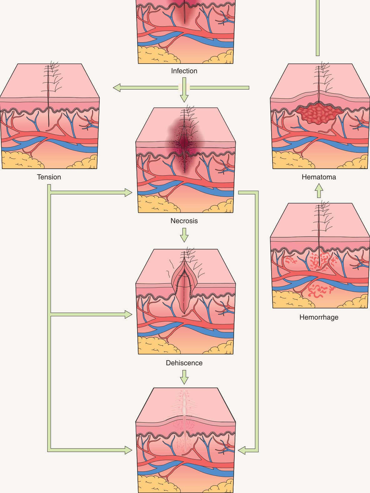
Fig. 151.1 Interrelated surgical complications.
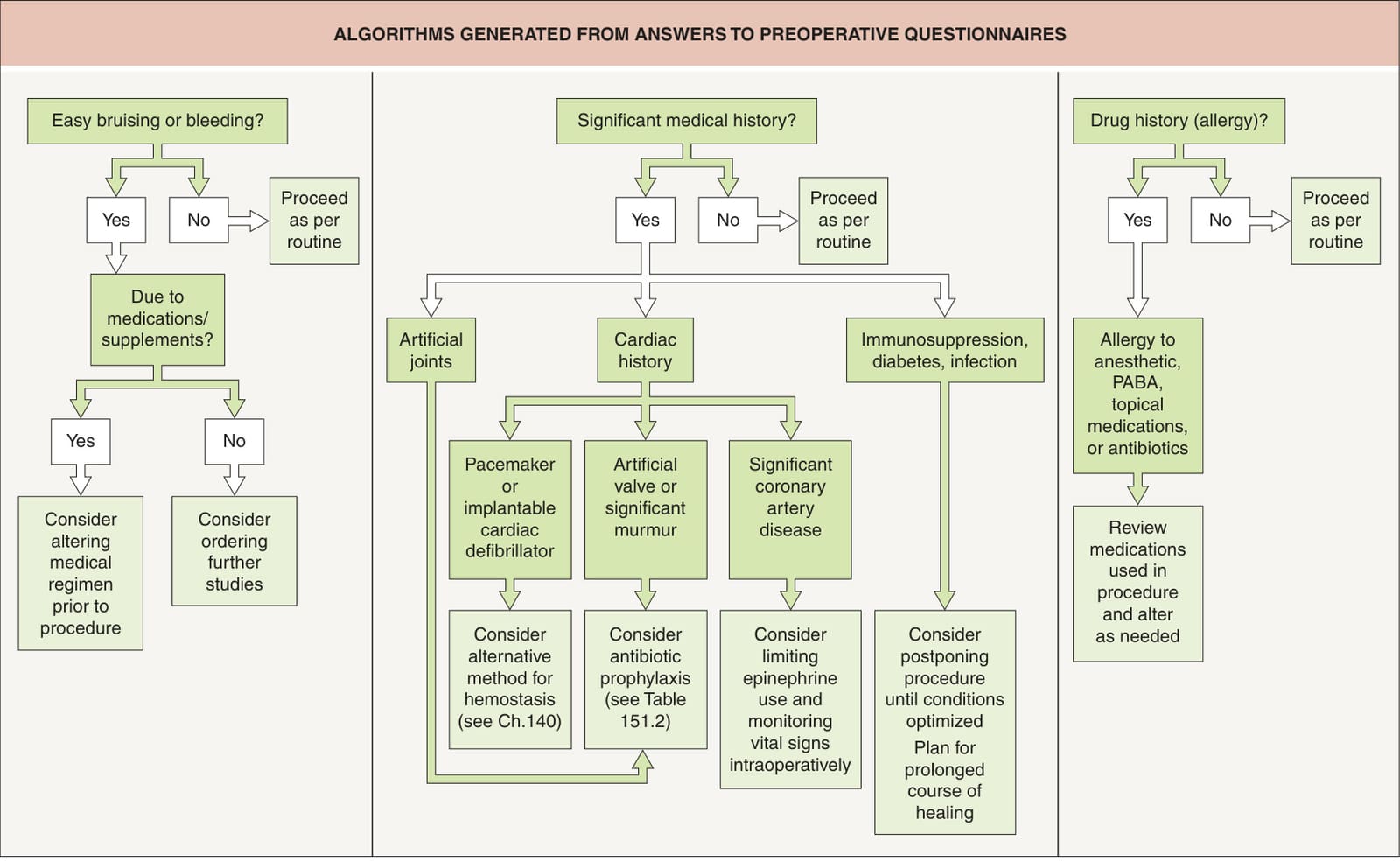
Fig. 151.2 Algorithms generated from answers to preoperative questionnaires. PABA, para-aminobenzoic acid.
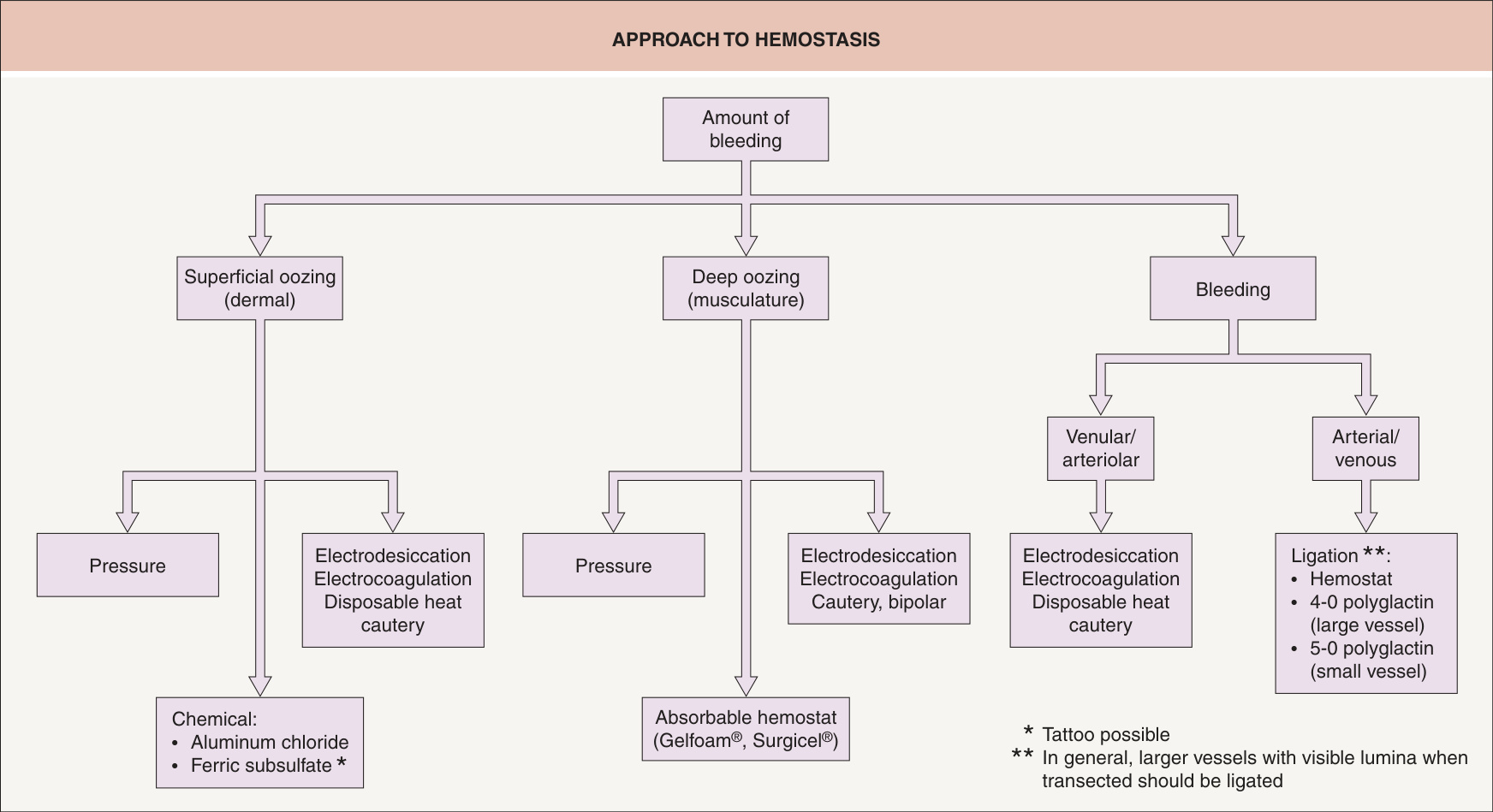
Fig. 151.3 Approach to hemostasis.Courtesy Glenn Goldman, MD.
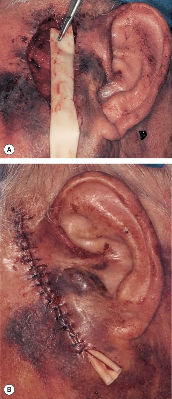
Fig. 151.4 Placement of Penrose drain because of excessive bleeding.
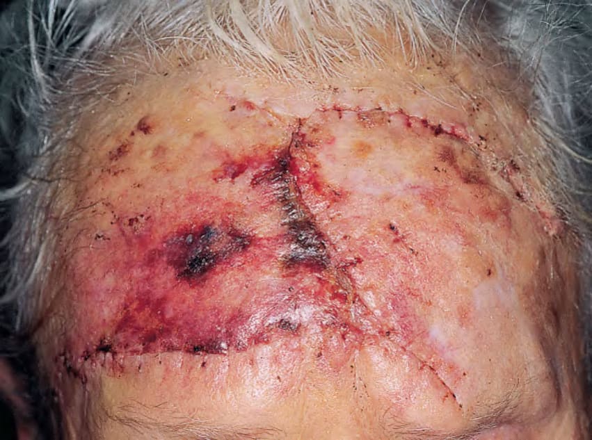
Fig. 151.5 Postoperative appearance of wound closed under tension with resulting necrosis.Courtesy Thomas Stasko, MD.
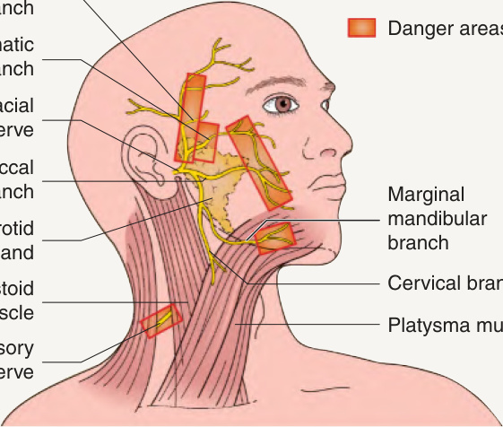
Fig. 151.6 Motor nerve danger areas.
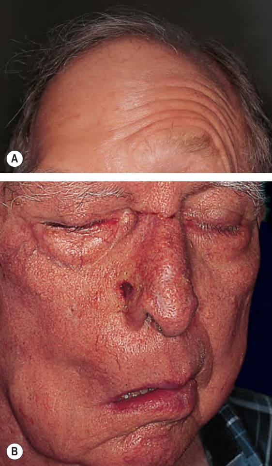
Fig. 151.7 Damage to the facial nerve.A Loss of the ability to elevate the right forehead due to transection of the right temporal branch. B Loss of function of the main branch of the right facial nerve. The ability to close the right eye has been restored by the placement of a gold weight in the eyelid. Courtesy Thomas Stasko, MD.
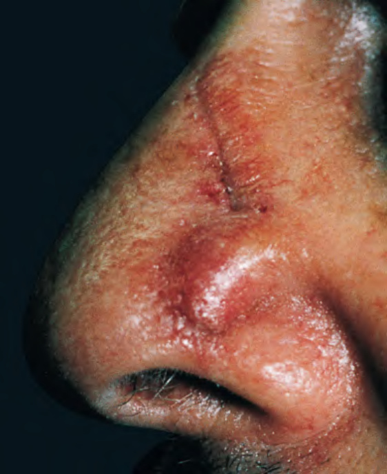
Fig. 151.8 “Trapdoor” or “pincushion” appearance of a flap reconstruction.Courtesy Thomas Stasko, MD.
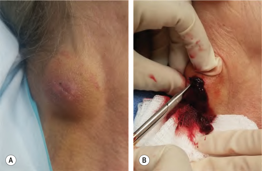
Fig. 151.9 Hematoma.A Postoperative hematoma formation. B Evacuation of gelatin-like clot.
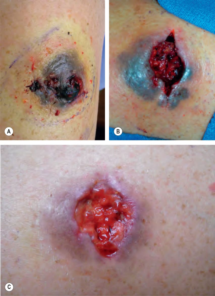
Fig. 151.10 Evacuation of an organized hematoma. Prior to (A) and immediately after (B) evacuation of the clot. C After 2 weeks of healing by second intention.
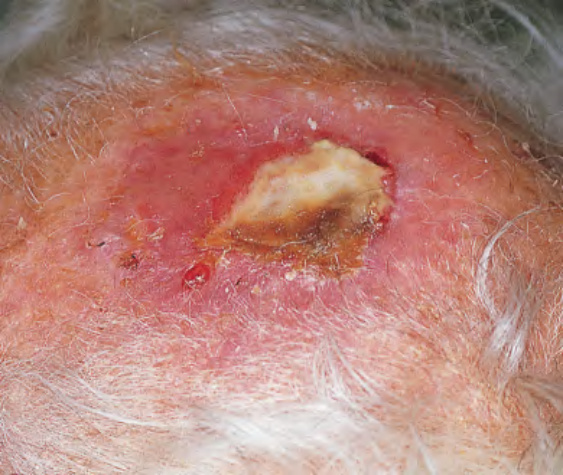
Fig. 151.11 Wound infection and necrosis at full-thickness skin graft site.Courtesy Thomas Stasko, MD.
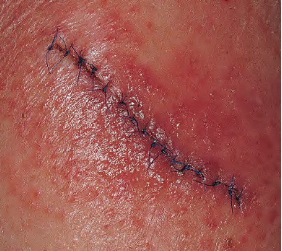
Fig. 151.12 Contact dermatitis to antibiotic ointment.Courtesy Thomas Stasko, MD.
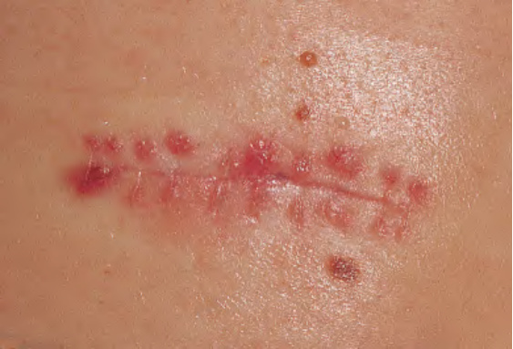
Fig. 151.13 Inflammatory suture reaction.Courtesy Thomas Stasko, MD.
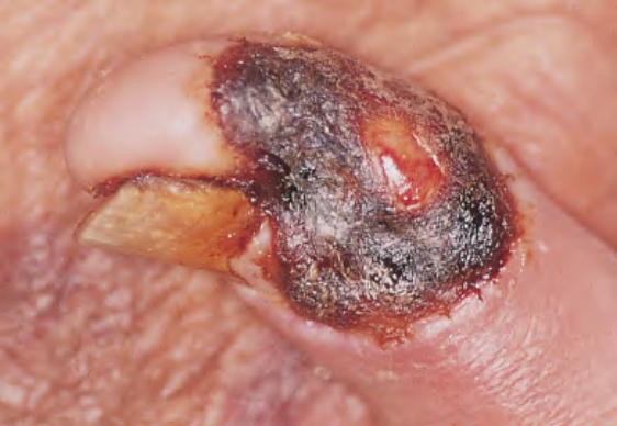
Fig. 151.14 Necrosis of a full-thickness skin graft. Two weeks after placement. Courtesy Thomas Stasko, MD.
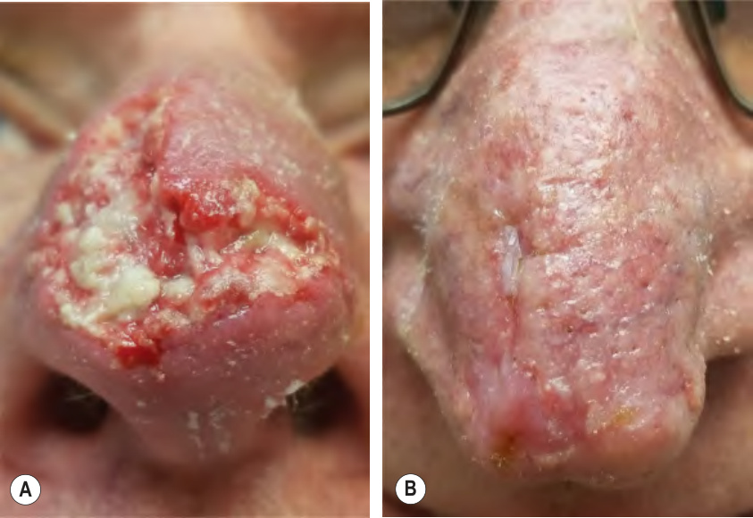
Fig. 151.15 Dehiscence and necrosis of a bilateral advancement flap of the nasal tip.A Initial dehiscence. B Follow-up after second intention healing with application of petrolatum; no additional interventions were performed.
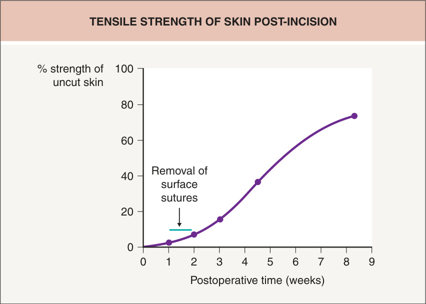
Fig. 151.16 Tensile strength of skin post-incision.
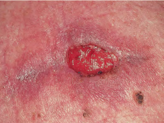
Fig. 151.17 Wound dehiscence after suture removal.
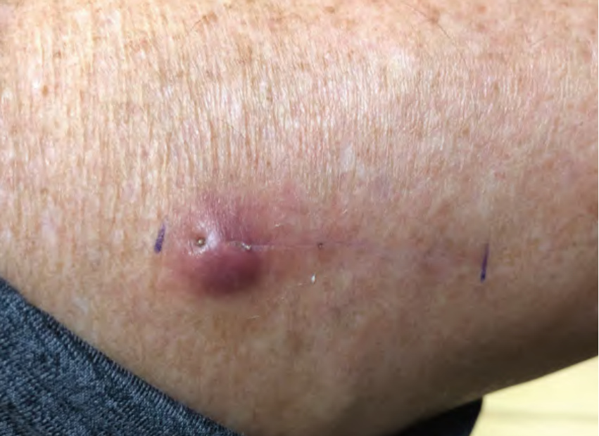
Fig. 151.18 Granulomatous reaction to suture placement. Dull pink nodule with obvious spitting suture at the end of the incision line. Courtesy Jean L. Bolognia, MD.
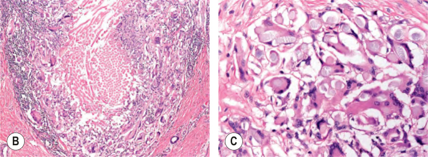
Fig. 151.19 Histologic features of a granulomatous reaction to braided suture material.A, B The suture material present within the dermis is cut both en face and longitudinally. On higher power, histiocytes and multinucleated giant cells surround the suture (B) and the giant cells have engulfed the suture material (C). Courtesy Lorenzo Cerroni, MD.
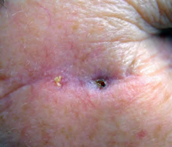
Fig. 151.20 Spitting absorbable suture within the incision line.
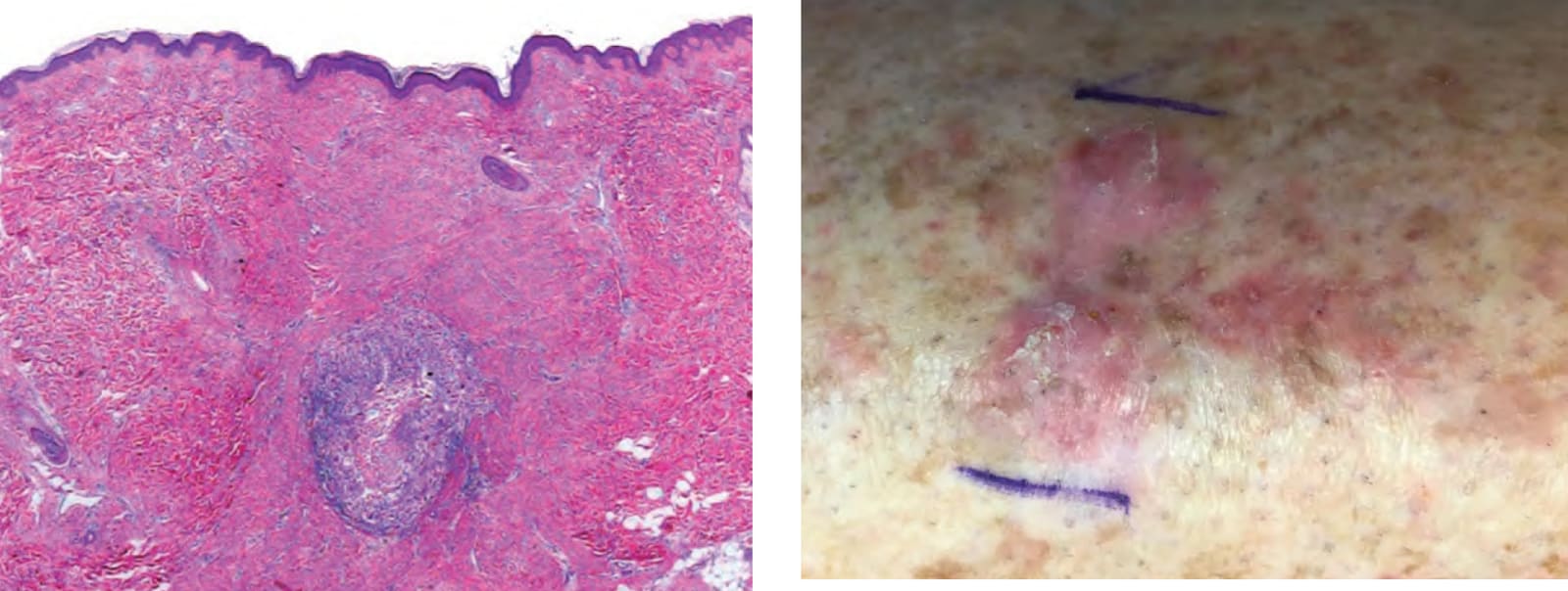
Fig. 151.21 Atypical squamous proliferations on the shin of a woman with severe photodamage. The nodules arose following complete surgical excision of a squamous cell carcinoma. Biopsy of one lesion confirmed the clinical diagnosis and the nodules resolved following two intralesional injections of triamcinolone and topical halbetasol. Courtesy Jean L. Bolognia, MD.
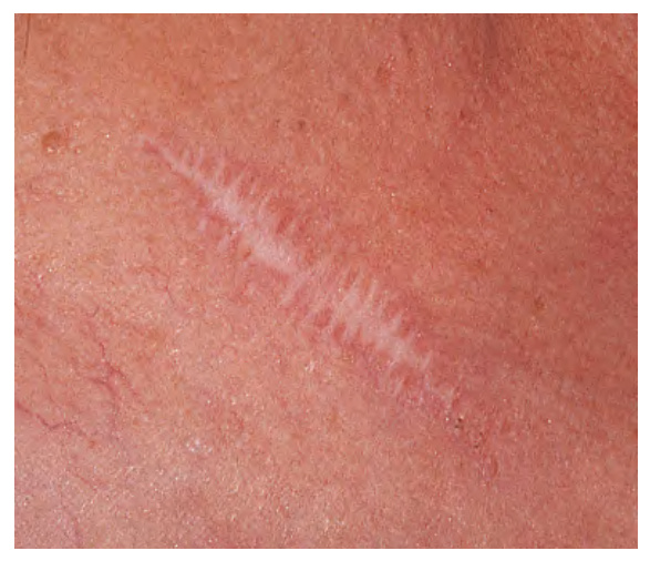
Fig. 151.22 Suture “tracks” in a spread scar.
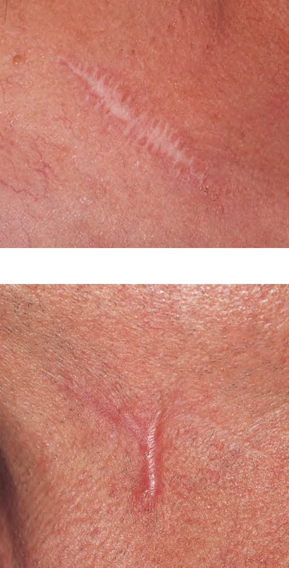
Fig. 151.23 Hypertrophic scar. One month after excisional surgery. Courtesy Thomas Stasko, MD.
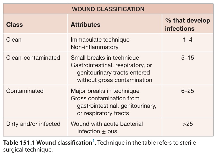
Table 151.1 Wound classification. Technique in the table refers to sterile surgical technique.
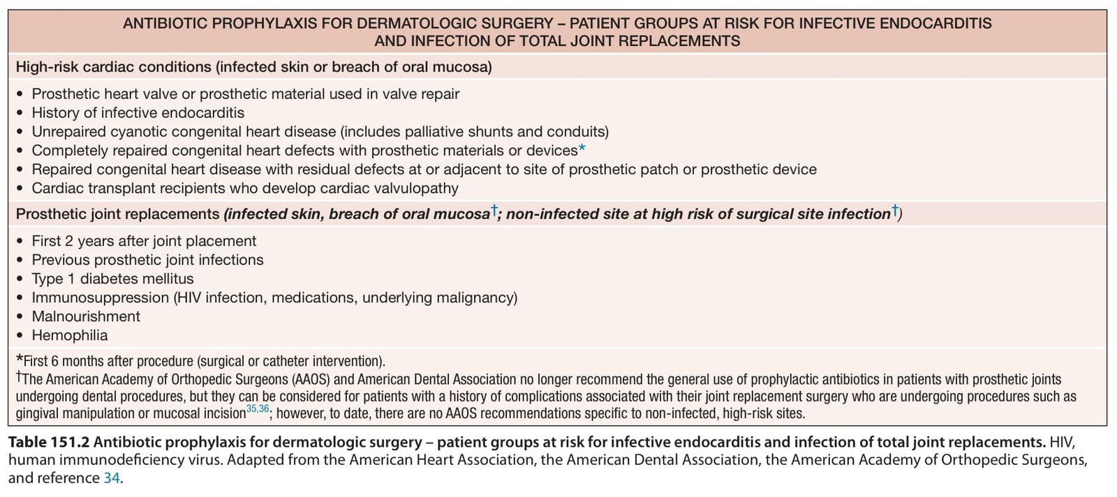
Table 151.2 Antibiotic prophylaxis for dermatologic surgery – patient groups at risk for infective endocarditis and infection of total joint replacements. HIV, human immunodeficiency virus. Adapted from the American Heart Association, the American Dental Association, the American Academy of Orthopedic Surgeons, and reference 34.
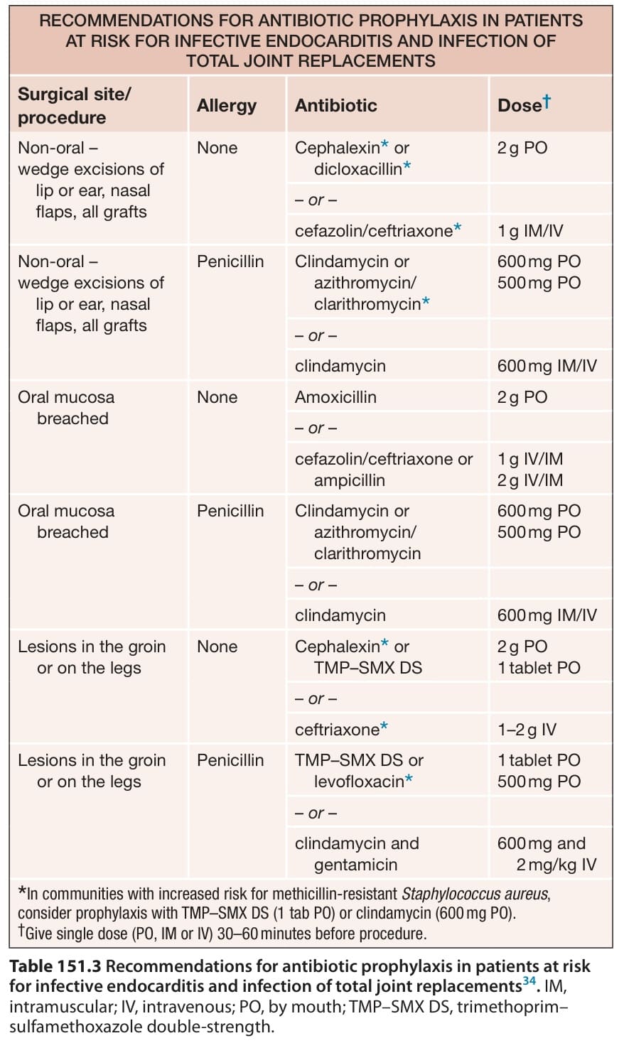
Table 151.3 Recommendations for antibiotic prophylaxis in patients at risk for infective endocarditis and infection of total joint replacements. IM, intramuscular; IV, intravenous; PO, by mouth; TMP–SMX DS, trimethoprim– sulfamethoxazole double-strength.
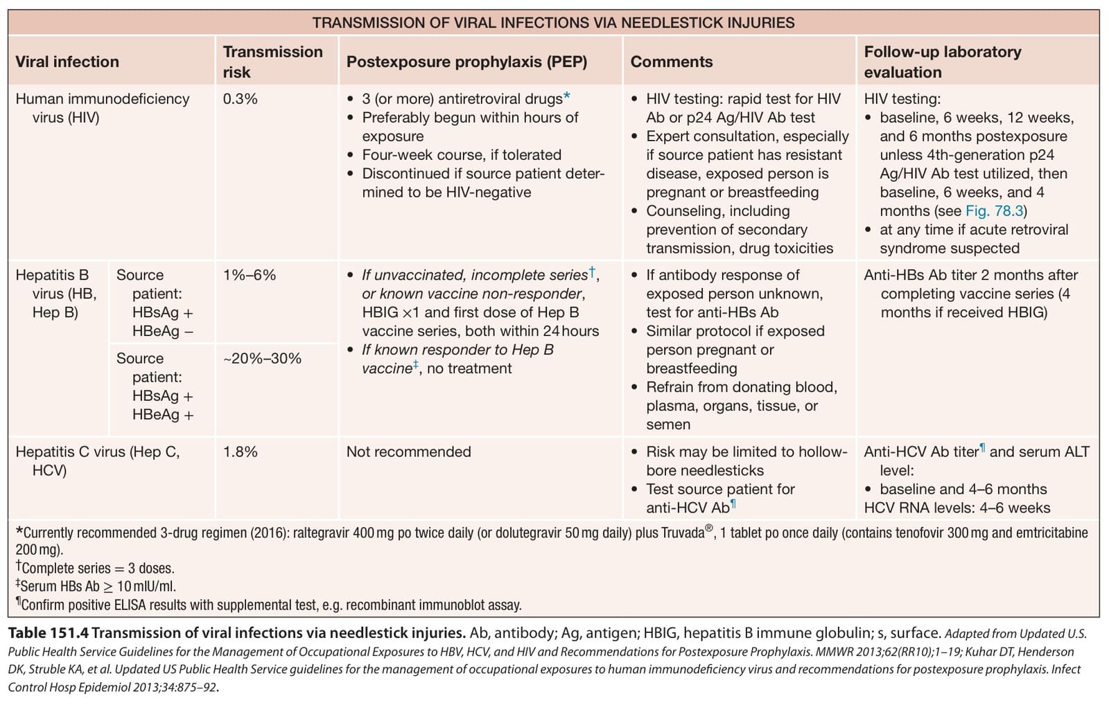
Table 151.4 Transmission of viral infections via needlestick injuries. Ab, antibody; Ag, antigen; HBIG, hepatitis B immune globulin; s, surface. Adapted from Updated U.S. Public Health Service Guidelines for the Management of Occupational Exposures to HBV, HCV, and HIV and Recommendations for Postexposure Prophylaxis. MMWR 2013;62(RR10);1–19; Kuhar DT, Henderson DK, Struble KA, et al. Updated US Public Health Service guidelines for the management of occupational exposures to human immunodeficiency virus and recommendations for postexposure prophylaxis. Infect Control Hosp Epidemiol 2013;34:875–92.
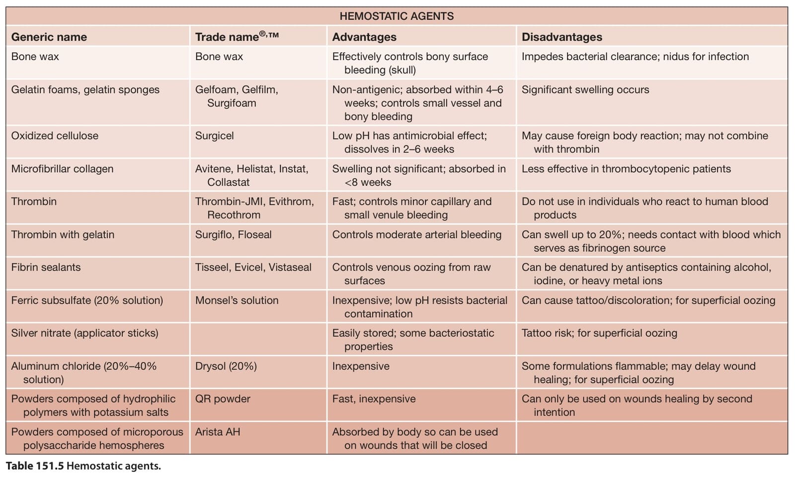
Table 151.5 Hemostatic agents.