COMPLICATIONS
There are three major considerations when anticipating and managing complications of sclerotherapy (Table 155.7). The first is that fastidious technique will minimize the incidence of side effects. Second is an appropriate understanding of maneuvers that may be instituted if something is thought to have gone wrong. Finally, an appreciation of the management options available for adverse sequelae can minimize the intensity of complications when they occur.
Postsclerotherapy Pigmentation
The incidence of postsclerotherapy hyperpigmentation (Fig. 155.12) varies from 5% to 30%. The purple–brown discoloration has been shown to be due to extravascular deposits of hemosiderin. Hypercoagulability syndromes such as antithrombin III, protein C or protein S deficiencies, use of nonsteroidal anti-inflammatory drugs (NSAIDs), high total body iron stores, use of minocycline, or vessel fragility, commonly noted in the elderly, may predispose to this adverse sequela. Methods to minimize the incidence of postsclerotherapy pigmentation are outlined in Table 155.8. Although various treatment modalities have been tried for this complication, only the Q-switched ruby laser has shown any degree of reproducible success. The hyperpigmentation usually resolves over 6–12 months. If it persists, the dermatologic surgeon should search for a vessel with persistent reflux into the area or an underlying predisposition.
Telangiectatic Matting
Blushes or nets composed of very fine telangiectasias may occur around sites of previously injected veins or sites of previous surgical interruption, e.g. ligation or ambulatory phlebectomy (Fig. 155.13). They may occur from days to months after treatment and are seen in 5%–15% of patients. Sites of predilection include the thighs, medial malleolus, and medial and lateral calves. Risk factors include obesity, use of estrogen-containing
medications, and pregnancy. Excessive postsclerotherapy inflammation may also predispose individuals to this complication, due to inadequate compression or exceeding the MSC of the sclerosing solution.
Generally, telangiectatic matting resolves within 3–12 months without specific treatment. Persistence should lead the dermatologic surgeon to attempt to locate a source of reflux in the area (usually a feeding reticular vein), although matting may be a sign of underlying saphenous vein insufficiency. Low injection pressure utilizing the MSC is the most effective way to minimize this complication. Additional measures include the use of no more than 1 ml of sclerosant per injection site, limiting the injection blanch to 1–2 cm, encouraging normalization of weight, and discontinuation of oral contraceptives for 1 month prior and 2 months following sclerotherapy. Short-wavelength lasers (595–600 nm) and intense pulsed light sources as well as reinjection of vessels utilizing a glycerin solution are the most effective ways of managing this problem (see Table 155.4).
Ulceration
Cutaneous necrosis may occur with any sclerosing agent (although the authors have never seen this with glycerin). It may be the result of extravasation of a sclerosing agent into the perivascular tissues, which
NSAIDs, nonsteroidal anti-inflammatory drugs.
is especially common with the use of hypertonic saline, non-foamed sodium tetradecyl sulfate, or polidocanol that is >1% in concentration; injection into a dermal arteriole or an arteriovenous anastomosis; reactive vasospasm of such a vessel; or excessive cutaneous pressure created by compression techniques. If there is extravasation of hypertonic saline into the perivascular tissue, infiltration of the area with normal saline may dilute the sclerosant. A hemorrhagic bulla may form at the site within 12–24 hours and later progress to an ulcer. If a porcelain-white area appears within the skin immediately after injection, this indicates arteriole injection or spasm. To reduce the likelihood of ulceration, the physician may rub a small amount of 2% nitroglycerin ointment into the area until a reactive hyperemia is seen. Since most ulcerations are less than 4 mm in diameter, second intention healing usually results in an acceptable scar. Larger ulcerations may be excised (as early as possible) and closed for faster healing and a smaller scar. Application of topical silicone gel (e.g. Stratacel® [Stratpharma]) may reduce the pain associated with the ulcer and speed the rate of healing.
Systemic Allergic Reaction
True allergic reactions following sclerotherapy treatments are extremely rare. Delayed hypersensitivity reactions, which may be urticarial or morbilliform in nature, can occur from 8 to 16 hours after treatment. Type I immediate reactions, varying from urticaria to bronchospasm to anaphylaxis eventuating in cardiovascular collapse, have been rarely reported. Intrinsically, only hypertonic saline has no potential to act as an allergen. However, the presence of additives such as benzoate or lidocaine may lead to the development of hypersensitivity reactions.
Sodium tetradecyl sulfate has a surprisingly low allergic potential (<0.01%). The most commonly reported hypersensitivity reactions are morbilliform eruptions developing 30–90 minutes after injection. Reactions to polidocanol are similarly rare (<0.01%). Both urticarial and morbilliform reactions have been reported, as well as an extremely rare anaphylaxis-related fatality.
Antihistamines and oral corticosteroids (e.g. prednisone 40–60 mg/ day for 7–10 days) can be used for the milder reactions, whereas intra-venous corticosteroids and subcutaneous epinephrine are often required for more severe reactions.
Arterial Injection
The most feared complication in sclerotherapy is inadvertent injection into an artery. Arterial injection of a sclerosing agent produces a sludge embolus that obstructs small arteries as well as the microcirculation. The most common location for an arterial injection to occur is the popliteal fossa, when attempting to inject the SSV. After an intra-arterial injection, the patient usually notes immediate pain, but not always.
A Prior to vein surgery. B After vein surgery – development of telangiectatic matting at the site of the scar.
A dusky cyanotic hue and pallor in the distribution of the injected artery usually occurs within a few minutes to several hours. Arterial pulsations of the posterior tibial or dorsalis pedis arteries are usually not affected. Sloughing of superficial tissues may result in significant scarring or may even require amputation.
Arterial injection is a true sclerotherapy emergency. Immediate intervention has been advocated but has never been proven to affect outcome. Procaine (1%), administered via periarterial infiltration, forms a complex with sodium tetradecyl sulfate, rendering it inactive; however, procaine is not effective for polidocanol. Cooling of the limb to minimize tissue anoxia, followed by immediate heparinization for 7–10 days and administration of intravenous dextran (10%, 500 mg daily) for 3 days is recommended. Thrombolysis and the utilization of long-term vasodilator therapy (e.g. calcium channel blockers) should be considered.
Miscellaneous Complications
Localized urticaria following injection is common and is most likely related to mast cell degranulation and cytokine release. No treatment is indicated, although pretreatment with an H antihistamine or immediate application of a class I ultra-potent topical corticosteroid may ameliorate the erythema, if bothersome to the patient. Compression stocking-related problems such as ulcers, pressure bullae, contact dermatitis and folliculitis may be minimized by adherence to appropriate compression application and fitting guidelines.
Nerve damage may occur secondary to direct injection of sclerosant or as a result of perivascular inflammation around nerve sheaths. The saphenous and sural veins of the inner leg and foot are high-risk sites and special care should be taken when injecting these locations. The utilization of NSAIDs may shorten the duration of symptoms.
Superficial thrombophlebitis develops 1–3 weeks after injection, presenting as a tender erythematous induration of the injected vein. This complication is observed less often if compression is maintained for an adequate period of time following sclerotherapy. When superficial thrombophlebitis occurs, the thrombus should be evacuated and adequate compression, frequent ambulation, and aspirin or an NSAID instituted. The concomitant presence of deep vein thrombosis (DVT) must be considered, and a search for underlying causes for thrombophlebitis other than the sclerotherapy, such as an internal malignancy, may be warranted.
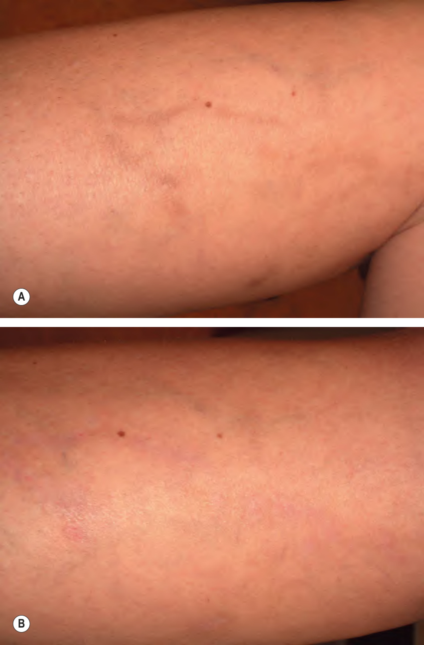
Fig. 155.12 Sclerotherapy-induced hyperpigmentation. A Development of hyperpigmentation along the course of a 3–4 mm diameter reticular vein three months after treatment with sodium tetradecyl sulfate 0.5% foam. B Six months after treatment with the Q-switched ruby laser at 6 J/cm, 5 mm diameter spot. Note complete resolution of hyperpigmentation. The melanocytic nevus serves as a landmark.
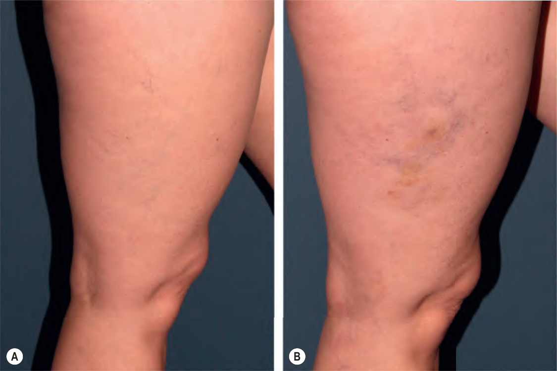
Fig. 155.13 Telangiectatic matting (neoangiogenesis).
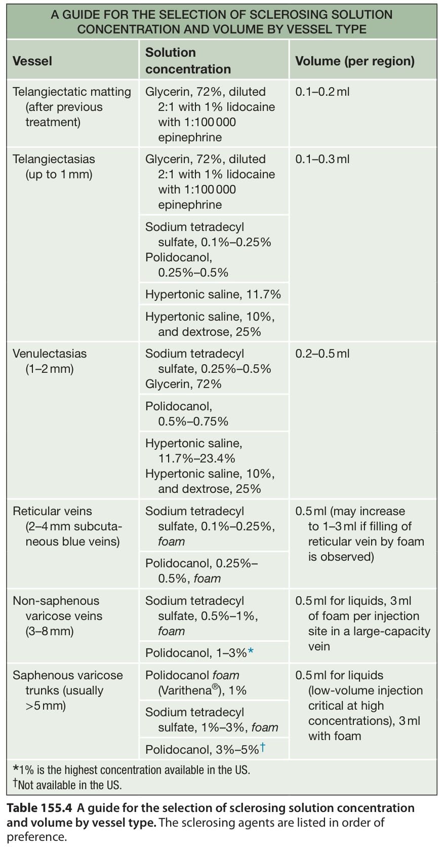
Table 155.4 A guide for the selection of sclerosing solution concentration and volume by vessel type. The sclerosing agents are listed in order of preference.
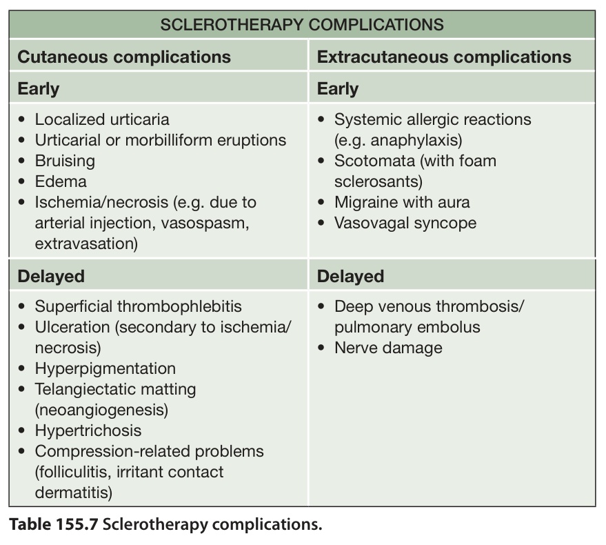
Table 155.7 Sclerotherapy complications.
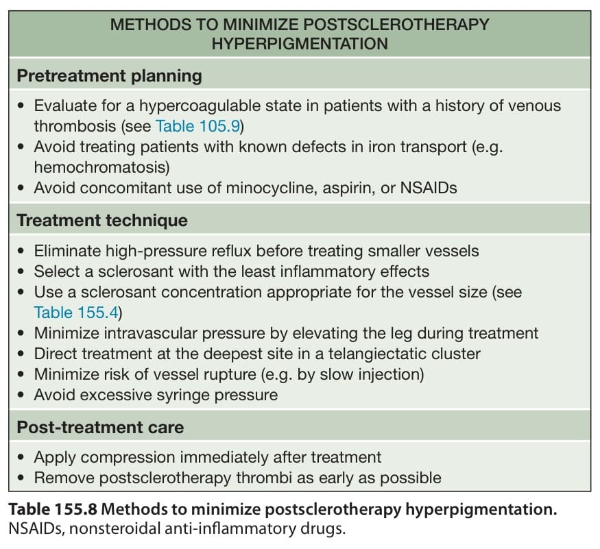
Table 155.8 Methods to minimize postsclerotherapy hyperpigmentation.