HAIR TRANSPLANTATION
Hair transplantation is a procedure performed in an outpatient setting under local anesthesia. It is based on the theory of donor dominance, i.e. terminal hair from the unaffected posterior scalp will continue its growth pattern even when transplanted to the balding frontal scalp. Unfortunately, until the 1990s, hair transplants often had an unnatural appearance due to unsightly and obvious “plugs”, sometimes likened to a “Barbie-doll” appearance. Over the past two decades, there has been a revolutionary movement away from 3–4 mm grafts containing 15–30 hair follicles to grafts containing groupings of one to four hair follicles, referred to as individual follicular units (Fig. 157.5). This has resulted in the transplanted hair having a more natural appearance (Figs. 157.6 & 157.7).
The net perceived density from a hair transplant is equal to the number of successfully transplanted follicles minus ongoing hair loss. In order to maximize long-term results, all patients undergoing this surgical procedure are encouraged to continue topical minoxidil, low-dose oral minoxidil, and/or oral finasteride (see above). Halting or at least slowing the miniaturization process allows for the perception of greater hair density.
Candidate Selection
As with all surgical procedures, the preoperative consultation is key to the success of the procedure. The consultation establishes whether the patient is an appropriate candidate for hair transplantation, both from a medical and a psychological perspective. In addition, it allows for an assessment of areas of greatest concern and whether the patient has realistic expectations of what a hair transplant can achieve.
A complete medical, surgical, and hair loss history is obtained, including previous hair transplants and scalp surgeries as well as scar formation. The etiology of the hair loss is determined, primarily via physical examination of the scalp. Occasionally, additional sites need to be examined and/or clinicopathologic correlation is required. The stage of patterned hair loss also needs to be established (see Figs. 157.2 & 157.3). All medications, prescription, over-the-counter and herbal, are reviewed with regard to their effects on hair growth and hemostasis (see Chs. 21 & 133).
The basic criteria for assessing the appropriateness of a candidate for hair transplantation are outlined in Table 157.1. In particular, the density of donor hair in the occipital scalp and the caliber of the hair need to be appraised, as both have an impact on the perceived density of the transplant (Table 157.2). Obviously, greater donor density and more grafts to transplant as well as thicker hairs rather than fine hairs lead to a fuller-appearing transplant.
Key Concepts
Both men and women should be made aware that AGA represents an ongoing process, i.e. it will progress despite undergoing hair transplantation. Medications (e.g. oral finasteride, minoxidil) or PRP and LLLT can help to maximize hair density from a transplant by minimizing ongoing hair loss. However, in planning for the transplant procedure, the surgeon should always assume that in the future these medications may be discontinued (see Table 157.2). Also, the progressive nature of AGA means that additional hair transplants may be required. The physician should therefore emphasize before the procedure how ongoing hair loss will affect the density and cosmetic appearance of the transplant as well as how this influences the placement of the grafts so as to assure a long-term natural appearance.
The number of expected procedures to accomplish both short- and long-term goals should be reviewed, as well as the limits on available
Courtesy Dowling B. Stough, MD.
The net density from a transplant = number of follicles transplanted minus ongoing hair loss.
donor hairs. Reasons for preferring to transplant primarily the frontal scalp as opposed to the vertex need to be explained – the vertex can consume donor grafts and it exhibits a continued 360° progression of hair loss which can lead to a peripheral rim of bald scalp which will appear unnatural over time. In addition, transplanting the frontal scalp,
not the vertex, produces the maximum cosmetic impact. Therefore, transplanting the vertex should be approached with caution, particularly in younger patients. Concentrating the transplants in the frontal scalp will allow maximum long-term density with minimal long-term cosmetic risk.
Patients have to realize that elliptical donor harvesting will lead to a permanent scar of the posterior scalp (Fig. 157.8). The scar is of no practical concern unless the patient has his or her hair closely cut. If the patient expresses an interest in having closely cropped hair, follicular unit extraction (FUE) should be considered (see below). However, the possibility of small “white dots” following FUE should also be discussed. Lastly, the patient must appreciate the need for a conservative approach to placement of the anterior hairline so that it will have a natural appearance over a lifetime as the temporal and posterior hairlines recede.
Staff Training for Manual Hair Transplantation
Contemporary hair transplantation is a team procedure, and an efficient, high-quality transplant requires a well-trained surgical team. Assistant technicians should be motivated and interested in learning and mastering the procedure. In addition, they need to be able to do repetitive tasks without tiring, losing concentration, or becoming frustrated. Technicians are utilized primarily for cutting and planting. The first task involves sectioning of the donor strip into slivers, followed by further meticulous sectioning into follicular units and prompt placement into chilled saline or other holding solution (Fig. 157.9). After the physician creates recipient sites, technicians can help insert the follicular unit grafts into the scalp. Recipient sites are generally designed at a density of 35–40 follicular units/cm but a lower density may be employed in areas where there is already pre-existing hair growth.
During the procedure, the physician closely monitors the quality of graft creation and placement. It is recommended that physicians learning the procedure have a staff member train at the same time. In order to avoid repetitive strain syndrome, technicians should position their elbows or forearms on a firm, supportive surface while using wrist and finger movements to plant grafts. Rotation of duties can also prove helpful.
Donor Region
The amount of available donor hair is the primary limiting factor in hair transplantation. In general, there are 65–85 follicular groupings/ cm in the occipital donor scalp. If there are <40 follicular units/ cm, the patient is considered a poor candidate (see Table 157.1). The mid-occipital scalp between the upper and lower occipital protuberances is the recommended donor site, given its density of hair and the ability to camouflage the donor scar due to the lack of involvement by AGA. If the harvesting is done too superiorly on the occiput, progressive AGA may reveal the surgical sites. Of note, donor density does not correlate with the extent of current or future hair loss in the frontal scalp or vertex. Harvesting of donor hair is performed via two different techniques: (1) elliptical donor harvesting; and (2) FUE (Table 157.3).
Elliptical donor harvesting (follicular unit transplantation [FUT])
Elliptical donor harvesting is still performed in a large number of patients because it allows for the safe and rapid removal of a large number of hair follicles, with minimal transection of hairs. In addition, if patients
will never wear their hair closely cropped, the resultant linear scar is not a concern and in contrast to follicular unit extractions, there is no need to trim hairs to 1 mm prior to the procedure. Elliptical donor harvesting is also referred to as follicular unit transplantation (FUT) or follicular unit strip surgery (FUSS).
The length and width of the donor ellipse depends on the number of follicular groupings required for the patient. For example, if 1000 follicular groupings are needed for the frontal scalp and a patient has an average donor density of 75 follicular units/cm, a 13.5 cm by 1 cm strip should contain approximately 1000 follicular grafts. It is important to remember that if a greater number of grafts are required, the ellipse should be lengthened rather than widened since increasing the width of a donor ellipse creates more wound tension which may lead to a hypertrophic or wide scar.
Ideal local anesthesia is obtained by infiltrating 1% lidocaine with epinephrine (adrenaline) superficially into the dermis. Once anesthesia has been achieved, bacteriostatic normal saline can be injected to provide further anesthesia, hemostasis, and dermal turgor. Turgor helps to reduce the transection of hair follicles. The blade(s) should be oriented parallel to the exiting follicles to avoid transection of hair follicles. The incision should be into the subcutaneous fat, and not deeper, in order to avoid transecting the occipital arteries (Fig. 157.10). Lateral retraction around the ellipse using fine skin hooks creates good visibility for the surgeon harvesting the ellipse (Fig. 157.11). The ellipse can be removed by scissors or a scalpel, carefully avoiding damage to any follicles in the subcutaneous tissue.
If the incision does not go any deeper than the subcutaneous fat layer, the ellipse can be removed without the use of electrocoagulation or thermal coagulation. Also, if the donor ellipse is <1 cm in width, it can often be primarily repaired without undermining. Some surgeons utilize a two-layer closure with absorbable sutures while others perform a single-layer closure. Staples or sutures can be utilized and then removed 7–10 days postoperatively. In some patients in which there is good surgical technique and optimal wound healing, the donor area from a single strip procedure may be more natural-appearing than with FUE. However, one cannot predict donor site healing following strip harvesting.
Follicular grafts
Hair naturally grows in groupings of 1–4 hair follicles, referred to as follicular units (see Fig. 157.5). During the 1960s to 1990s, 3–4 mm grafts that contained multiple follicular units were employed, leading to an unnatural appearance with obvious bundles of hair. Nowadays, each graft contains one follicular grouping with 1–4 hair follicles. Thus, these grafts mimic the normal grouping of scalp hairs and create a natural appearance.
When an ellipse is harvested, surgical teams carefully separate 500–2000 follicular units from the donor strip over 1–3 hours (see Fig. 157.9). Cutting instruments vary but include #11 and #15 blades as well as #10 prep blades. Good lighting, comfortable chairs, and well-designed instruments are prerequisites for producing follicular units with minimal follicular transection. Most surgeons believe microscopic dissection or magnification reduces transection of follicles during the separation process (Fig. 157.12).
It is important to create intact, minimally traumatized follicular units and place the transplanted hair into the recipient sites as efficiently and quickly as possible. As soon as the follicular unit grafts are separated from the donor ellipse, they must be put into a holding solution until they are placed into the recipient sites. Commercially available holding solutions can enhance survival of these hair follicles, in part by reducing apoptosis. If grafts desiccate for even a few minutes they will die and not grow. One of the advantages of the FUE technique is that following visual inspection, harvested grafts can be transplanted immediately.
The number of follicular units required for each surgery depends on the extent of hair loss, available donor hair, and density of pre-existing terminal hair follicles in the recipient area. This can vary from 100 to 2500 follicular groupings per procedure. Larger cases may be done over two days to avoid technician fatigue and reduce the chance of undesirable sequelae.
Follicular unit excision (FUE)
In the FUE technique, single follicular units consisting of groupings of 1–4 hair follicles are surgically removed from the occipital scalp. Tiny punch devices (~0.75–1.2 mm in diameter) are used to remove
the grafts. The incisions are so small that they usually leave no visible scar after they heal; however sometimes small “white dots” are seen. For patients who like to have closely cropped hair, FUE is generally preferred. It is also an option for patients who have significant tension or extensive scarring from previous elliptical donor harvesting.
Although manual FUE requires more time than elliptical donor harvesting, the introduction of automated mechanical instruments and robotic technology has led to a reduction in extraction times. Important considerations when performing manual FUE are outlined in Table 157.4. Potential disadvantages of FUE include a higher rate of transection of follicular groupings when compared to strip harvesting as well as the risk of “thinning out” the donor area due to repeated extractions. In one small series, the number of lifetime follicles was similar for FUT and FUE.
Numerous devices exist for performing FUE. Earlier devices utilized manual or motorized dull punches to gently extract the grafts in order to avoid damage to bulb anatomy. Subsequently, motorized, sharp punches
were employed to speed up excisions. Punch instruments with a flared or trumpet shape were introduced more recently to try to reduce transection of hair bulbs. Robotic systems such as ARTAS® (Venus Concepts) can be used to minimize physician fatigue and eliminate the need to acquire all the surgical skills needed to dissect grafts.
With FUE, the surgeon can target follicular groups of a specific size or hairs with a specific diameter or pigmentation. Single hairs may be harvested for eyebrow restoration, or larger 3–4 hair groupings may be targeted to add density over the mid scalp. Because of minimal scarring, harvesting from the beard region or other terminal hair-bearing areas is also an option. In addition, harvesting can be done even after multiple strip surgeries have led to a very tight donor area.
For optimal cosmetic results and to preserve symmetry, one should irregularly harvest from the entire safe donor area (SDA) rather than from just a small shaven area. Of note, the surgeon should not be pressured to over-harvest, lest the donor area be depleted. Unfortunately, patients sometimes seek less expensive “black market” hair transplants and then can present with donor and/or recipient site necrosis and soft tissue infections, resulting in few options for repair.
Hairline Design and Recipient Site Creation
In men, the hairline defines the cosmetic success of a hair transplant. Most women have stable frontal, temporal and posterior hairlines, making recreation of a hairline unnecessary. As with hair graft creation, hairline design should mimic, as closely as possible, what occurs in nature. However, patients and physicians often try to mimic the hairline a patient had before their male pattern hair loss began. Even with follicular unit transplants, this approach to hairline design often leads to cosmetic failure. This is due to not considering the slow steady recession of the temporal and posterior hairlines in addition to the frontal hairline.
The design of the frontal hairline should be such that over time it will remain balanced with the temporal and posterior hairlines.
This requires recreating a frontal hairline which is higher and more receded than the one which was present before the process began. Initially, some patients will resist the idea of a higher, more receded hairline, but explaining and demonstrating with a mirror how their temporal and posterior hairlines preclude a low frontal hairline usually changes their opinion.
Rather than considering the hairline to be a fixed boundary, it should be thought of as a natural transition zone of gradually increasing density from skin to terminal hair-bearing skin. This ill-defined “feathering zone” is created by placing, in an irregular pattern, follicular unit grafts (with 1–4 follicles) along the newly created hairline (Fig. 157.13). Dense packing of grafts should not be performed along the frontal hairline because a densely packed hairline has an unnatural appearance. The level at which the hairline is placed varies from individual to individual and it is important to examine each patient in a global, 360° manner before determining placement of the hairline.
While male pattern hair loss is progressive, transplanted hair will have long-term growth. Therefore, when evaluating patients, the surgeon must assume that all patients will progress to the highest grade of involvement, i.e. the worst-case scenario. As noted previously, for several reasons, including the progressive nature of AGA leading to an unnatural appearance, transplanting into the vertex should be approached with caution.
Anesthesia and Recipient Site Creation
A combination of supraorbital/supratrochlear nerve blocks, field blocks, and local infiltration with 1% lidocaine with epinephrine is performed. Hemostasis is essential for good visibility when creating recipient sites and for graft placement. The epinephrine in the local anesthetic (placed into the dermis, not the subcutaneous space) is usually adequate for providing excellent hemostasis. While most surgeons use “off-the-shelf” 1% lidocaine with 1 : 100 000 epinephrine, concentrations of the latter may range from 1 : 50 000 to 1 : 500 000.
Recipient sites should mimic the natural 30–45° angle of hair growth on the scalp (Fig. 157.14). There are a variety of needles, in particular 18, 19, 20 and 21 gauge needles, that are used to make recipient sites for 4, 3, 2, and single hair groupings, respectively. When creating recipient sites, surgeons must be careful not to transect existing hair follicles. Most advocate using magnification to place recipient sites in order to limit the loss of existing hair during surgery. The key to success is to arrange recipient sites in a random, irregular pattern with 10–30 sites/ cm, depending on the density of existing hair on the scalp.
Graft Placement and Postoperative Course
Microvascular forceps are used by surgical assistants to place the grafts. These forceps gently grasp the follicular units by their perifollicular tissue, avoiding trauma to the hair follicles. Placement of the grafts into recipient sites is often the most challenging component of the hair
Correct (A) versus incorrect technique (B). Grafts should not be oriented perpendicular to the scalp surface.
transplant procedure, both for novice and experienced hair transplant teams. The major challenges include hemostasis and “popping” of grafts from recipient sites after they are placed. However, it is difficult to predict which patient will have “popping” grafts. This phenomenon is overcome by applying light pressure over a placed graft for 5–15 seconds with a saline-soaked cotton swab before placing the next graft.
After the procedure, an overnight dressing is placed to protect the grafts. The entire dressing should be non-adherent and removed by the patient the next morning (Table 157.5). Staples or sutures are removed during a 7- to 10-day postoperative visit. The transplanted hair begins to appear 3–6 months after the procedure and is fully grown by 9–14 months. Patients must be warned of the possibility of post-transplant telogen effluvium.
Complications
Medical and surgical complications from a hair transplant procedure are unusual. The extensive vascular supply to the scalp results in rapid wound healing and a low risk of infection. In addition to reactions to medications and vasovagal episodes, complications include excessive swelling (∼5%), postoperative bleeding (<0.5%), folliculitis, a temporary headache, and temporary pruritus or numbness of the scalp. Persistent problems include permanent numbness in the donor or recipient sites, abnormal scarring around the grafts, hypertrophic scarring of the donor site, and poor growth of hair grafts. The latter can be minimized by keeping the grafts constantly moist.
Hypopigmentation often occurs at extraction sites within the donor region but its occurrence is not predicable. It is often larger in diameter than the punch instrument used to obtain the graft. Scalp micropigmentation has been used to successfully camouflage this hypopigmentation. Telogen effluvium of nonreplaced hair, also referred to as “shock” hair loss, is an uncommon complication that in the authors’ experience resolves on its own. It stems from overharvesting of grafts in a specific region. In general, maximal harvesting during a single procedure should be 25% of the total number of follicular units. Because the number of safely harvested grafts depends on the density of follicular groupings and hair caliber, individuals with fine hair and older individuals (number of follicular groupings decreases with age) who undergo harvesting of higher numbers of follicular groupings are at risk for this complication. Lastly, a hair transplant-triggered flare
of LPP has been reported. It is unclear if a hair transplant actually triggered a flare of LPP or subclinical preexisting disease became more active.
Corrective Hair Transplant Surgery
When patients present for corrective surgery due to previous transplantation of 3–4 mm punch grafts that have led to unnatural large “plugs”, i.e. a “pluggy” transplant, three major options exist: (1) add a large number of follicular unit grafts containing one to four hairs between the larger plugs; (2) surgically remove the large grafts; and/or (3) perform laser-assisted hair removal. Which option is best suited for an individual patient is determined during the consultation.
Adding follicular unit grafts
Transplantation of a large number of follicular unit grafts containing one to four hair follicles – in front of, in between, and behind large grafts – will soften the “pluggy” appearance (Fig. 157.15). This option is appealing for many patients because it allows for both cosmetic improvement and increased density (Fig. 157.16). Unfortunately, some patients with an unnatural hairline have a depleted donor supply from previous transplant procedures while others are reluctant to have another surgery following the emotional trauma from the initial transplant.
Surgical removal of grafts
Some hairlines are so cosmetically inappropriate that there is no worthwhile corrective option other than surgically removing the grafts. This option is also indicated for large grafts with perifollicular white scar tissue. The grafts can be removed by either a 2–4 mm punch instrument or an elliptical excision. Another option is to perform FUEs from larger grafts via 1 mm punch instruments. The latter procedure reduces the “pluggy” appearance of the larger grafts while allowing a more natural appearing graft to remain.
In general, scars on the scalp heal well with minimal evidence of the surgical procedure. However, all patients should be warned that cosmetically evident scars develop in a small minority of cases. If unacceptable scars do arise, pulsed dye, ablative, non-ablative, or fractional ablative laser treatments can be used to help improve the cosmetic appearance.
Laser-assisted removal of large grafts
As with other parts of the body, lasers only remove pigmented terminal hair follicles (see Ch. 137). Typically 5–10 treatments are needed to permanently remove the majority of follicles. Similar to FUE of larger
grafts, laser-assisted removal eliminates the majority, but not all, of the hairs, thus providing a substantial cosmetic improvement of the unnatural plugs while retaining some of the transplanted hair for a more natural appearance. Laser therapy is an excellent option for patients who want to improve their cosmetic appearance in a safe, non-invasive manner.
Previous transplantation of 3–4 mm punch grafts can lead to unnatural large “plugs” of hair. B, C Addition of follicular unit grafts between and in front of the larger grafts softens the hairline and the overall appearance.
Hypertrophic or broad scars in the donor region
All patients have a permanent scar from elliptical donor harvesting, but the majority of scars are 1–2 mm wide and are of no practical concern (see Fig. 157.8). That said, patients should be made aware of this permanent donor scar before they have a transplant. If the patient expresses a possible interest in having their hair cut short, FUE should be considered (see above).
When wide donor scars occur, they can be a major cosmetic problem without an easy solution for repair. Scar revision leads to variable improvement. Pulsed dye, non-ablative, or fractional ablative lasers may be used to help reduce the thickness and erythema of hypertrophic scars. The best method for minimizing the risk of a wide scar is to keep the width of the donor strip to ≤1 cm. While this does not guarantee a fine, thin scar, it does reduce the risk of a wide scar. Another option is to transplant a large number of follicular groupings into the scar in an attempt to provide camouflage.
Hair Transplantation in Scarring Alopecias
Hair transplantation can be successfully performed to improve the appearance of scarring due to previous cosmetic or cranial procedures. However, if there is any concern regarding active inflammation, a biopsy specimen should be obtained. Any inflammation, whether from a primary scalp dermatosis, infection or previous surgery, must be resolved completely before hair is transplanted. In the case of inflammatory scalp dermatoses, patients should have no evidence of inflammation for at least 6 months off therapy before the transplant procedure is performed.
Patients undergoing surgery for cicatricial alopecias such as LPP, FFA, or CCCA must understand that even if their disease is quiet at the time of surgery, any subsequent flares may compromise the future growth of the grafts. In other words, when there is any active inflammation, the transplanted hair may not grow and all patients should be informed of this before their first transplant. Of note, in a retrospective review of 51 patients who received hair transplants for FFA, most of them lost grafts over the following 5 years but there was still very high patient satisfaction.
Compared to sites without scarring, the yield is less when hair is transplanted into scar tissue. How large the difference will be varies from patient to patient. In general, more transplant sessions are needed in patients with scarring alopecia.
Hair Transplantation in African-Americans
In addition to pattern hair loss in men and women of African ancestry, traction alopecia and CCCA are also commonly seen in women (see Ch. 69). If there is a question regarding the precise diagnosis, clinico pathologic correlation is recommended prior to transplantation. For CCCA, there should be no evidence of inflammation for 6 months (off therapy) before the transplant procedure is performed. While patients can wear wigs or hair pieces, hair transplantation is the only way to permanently fill in the areas of alopecia (Fig. 157.17A).
A Appearance 1 week following transplantation of 1200 follicular unit grafts in an African- American woman. B Cross-section of Afro hair follicles demonstrating their characteristic C-shaped curve.
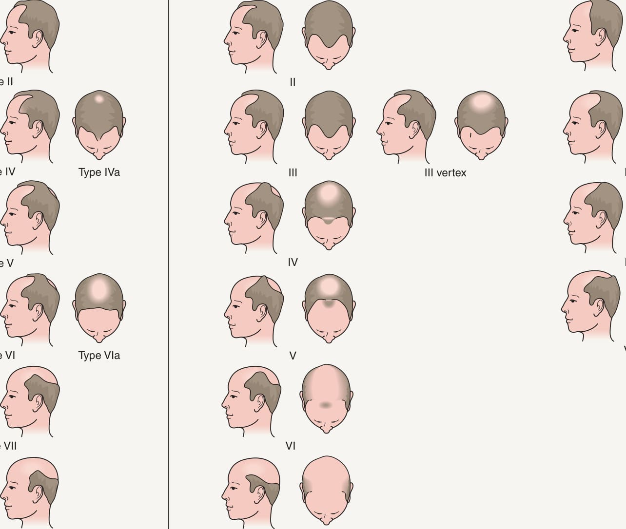
Fig. 157.2 Classification of male pattern baldness (androgenetic alopecia).A Hamilton grading scale. B Norwood grading scale. Adapted from Hamilton JB. Male hormone stimulation is prerequisite and incitant in common baldness. Am J Anat 1942;71:451–80; and Norwood OT. Male pattern baldness: classification and incidence. South Med J 1975;68:1359–65.
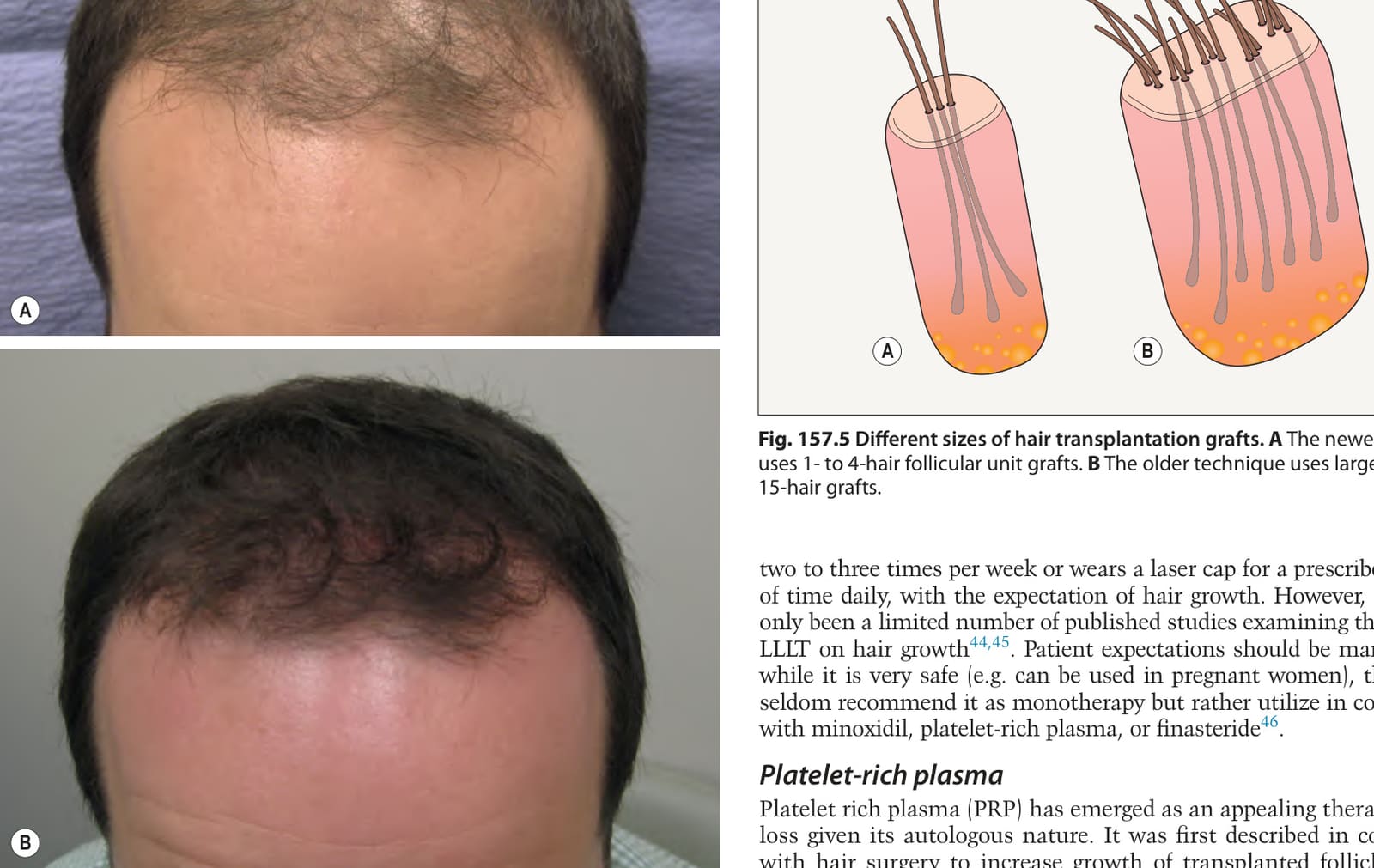
Fig. 157.5 Different sizes of hair transplantation grafts.A The newer technique uses 1- to 4-hair follicular unit grafts. B The older technique uses larger 10- to 15-hair grafts.
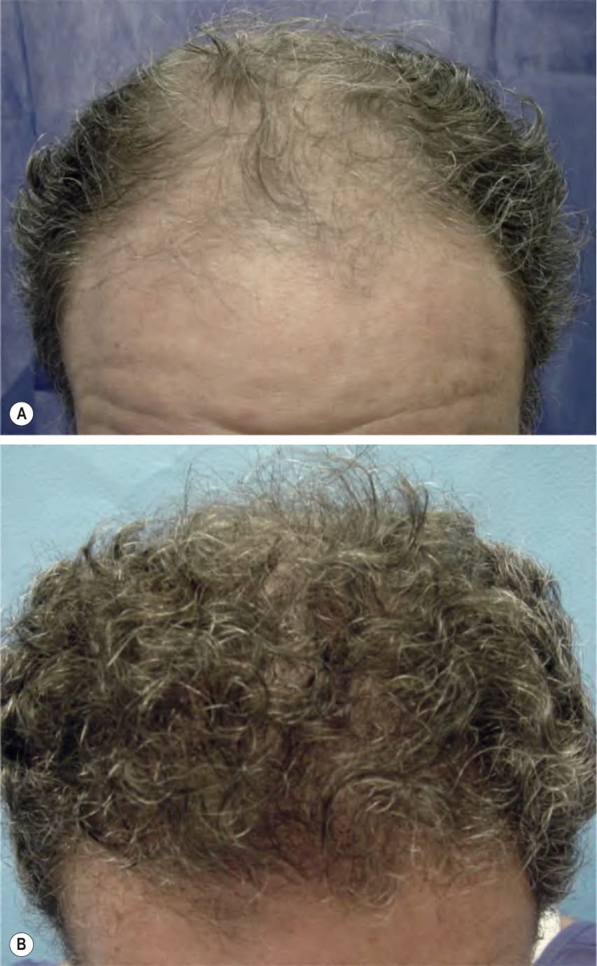
Fig. 157.6 Hair transplantation for male pattern hair loss.A Norwood IV pattern (baseline). B Appearance following transplantation of 900 hair grafts, each with one to three hairs.
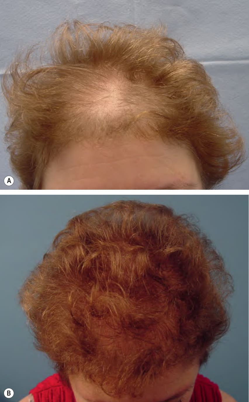
Fig. 157.7 Hair transplantation for female pattern hair loss.A Ludwig II pattern (baseline). B Appearance following transplantation of 750 hair grafts, each with one to three hairs.
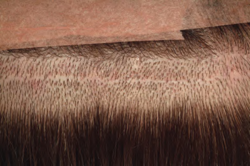
Fig. 157.8 Occipital scalp scar secondary to elliptical donor harvesting.Courtesy Dowling B. Stough, MD.
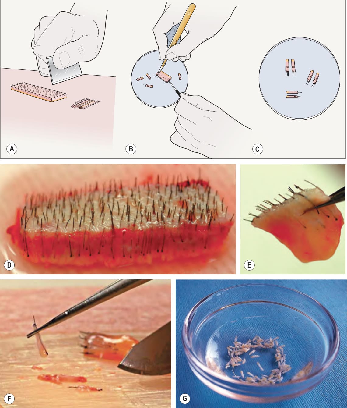
Fig. 157.9 Technique for graft slivering.A Donor slivering which produces slivers 2 mm wide by 10 mm long. B Slivers being sectioned into follicular units. C Follicular unit grafts are placed into dish with saline and separated into one-, two- and three-haired follicular units. D A portion of donor strip prior to slivering. E Appearance of a sliver. F A single follicular unit produced by microscopic dissection of a sliver. G Follicular unit grafts are stored in isotonic saline to maintain their viability. Courtesy Dowling B. Stough, MD.
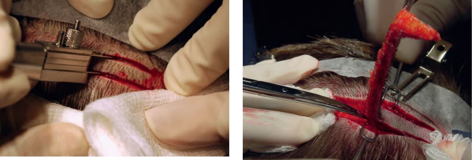
Fig. 157.10 Elliptical donor harvesting. To avoid transection of the hair follicles, the double blades should be oriented parallel to the exiting follicles. The incision should extend into the subcutaneous fat but not deeper (∼5 mm into the scalp). Some surgeons prefer a single blade for performing the ellipse.
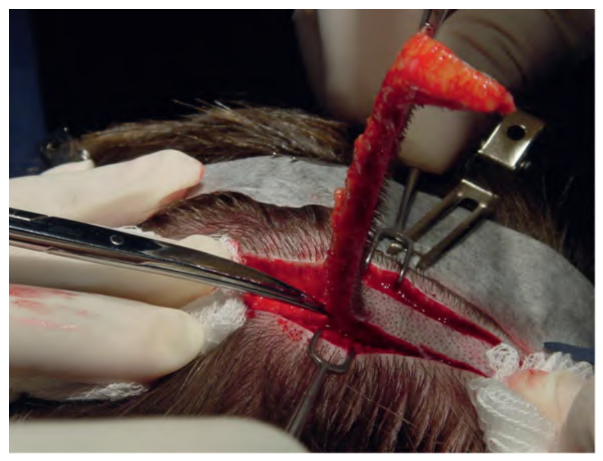
Fig. 157.11 Elliptical donor harvesting. Lateral retraction using fine skin hooks exerts tension away from the excision and creates good visibility. The ellipse can be removed by scissors or a scalpel, being careful to avoid damage to any follicles in the subcutaneous tissue.
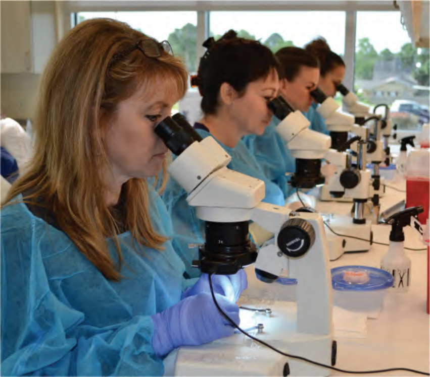
Fig. 157.12 Use of magnification to separate follicular units from the donor strip.
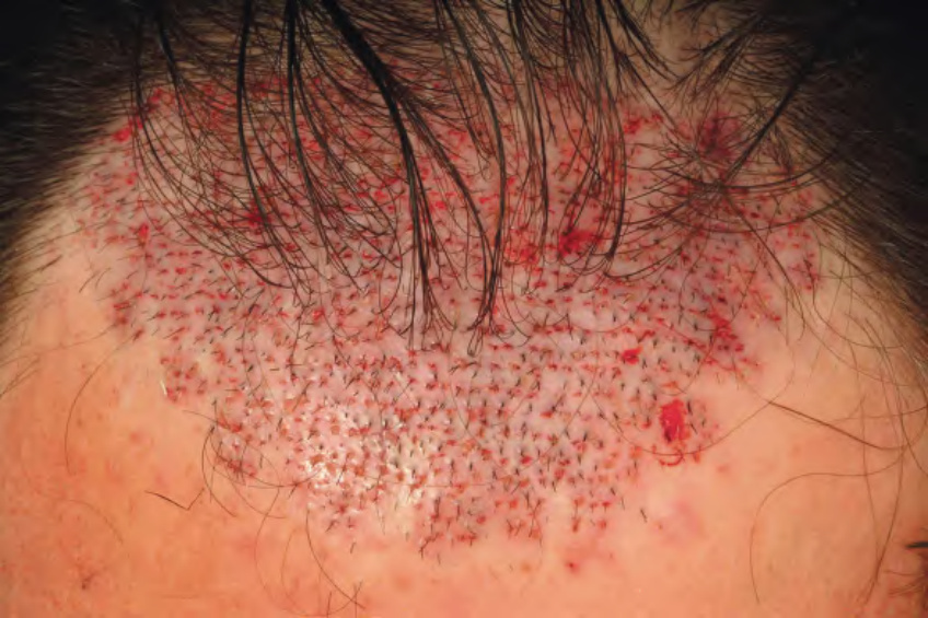
Fig. 157.13 Immediate postoperative appearance with graft placement and hairline design.Courtesy Dowling B. Stough, MD.
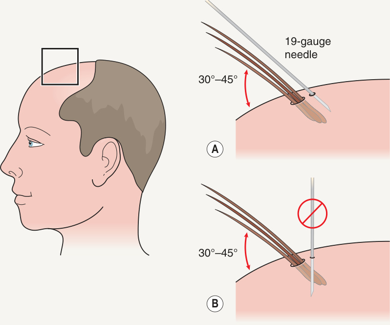
Fig. 157.14 Recreating the 30–45° angle of hair growth on the frontal scalp.
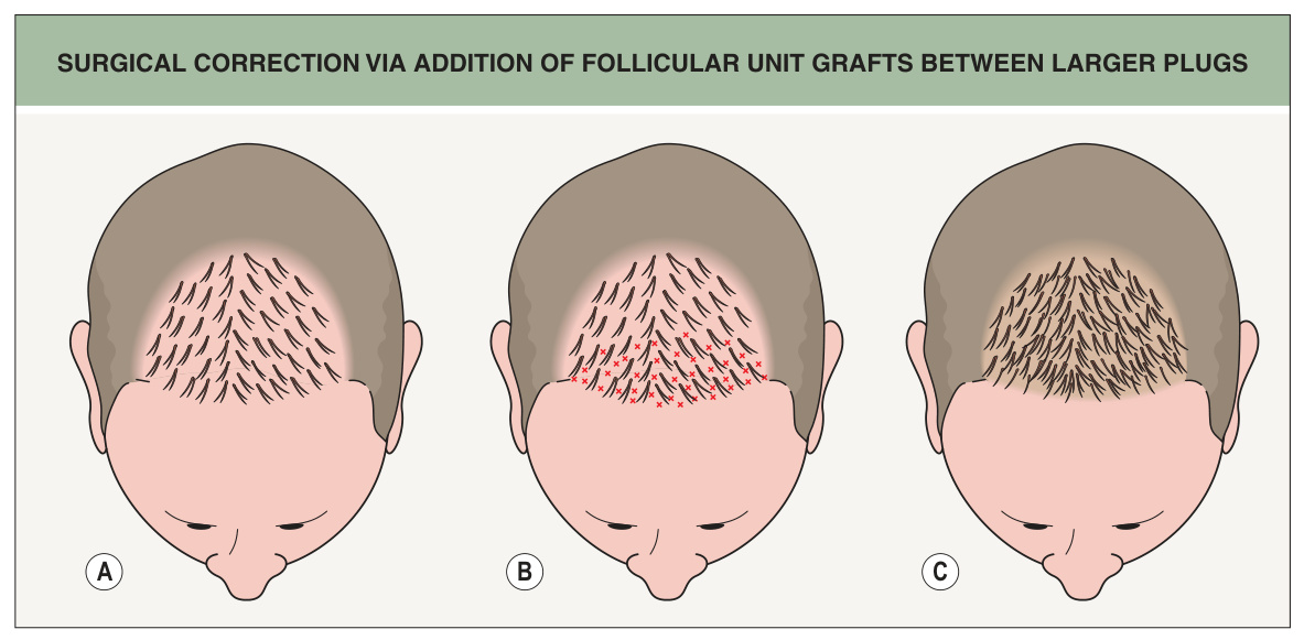
Fig. 157.15 Surgical correction via addition of follicular unit grafts between larger plugs.A
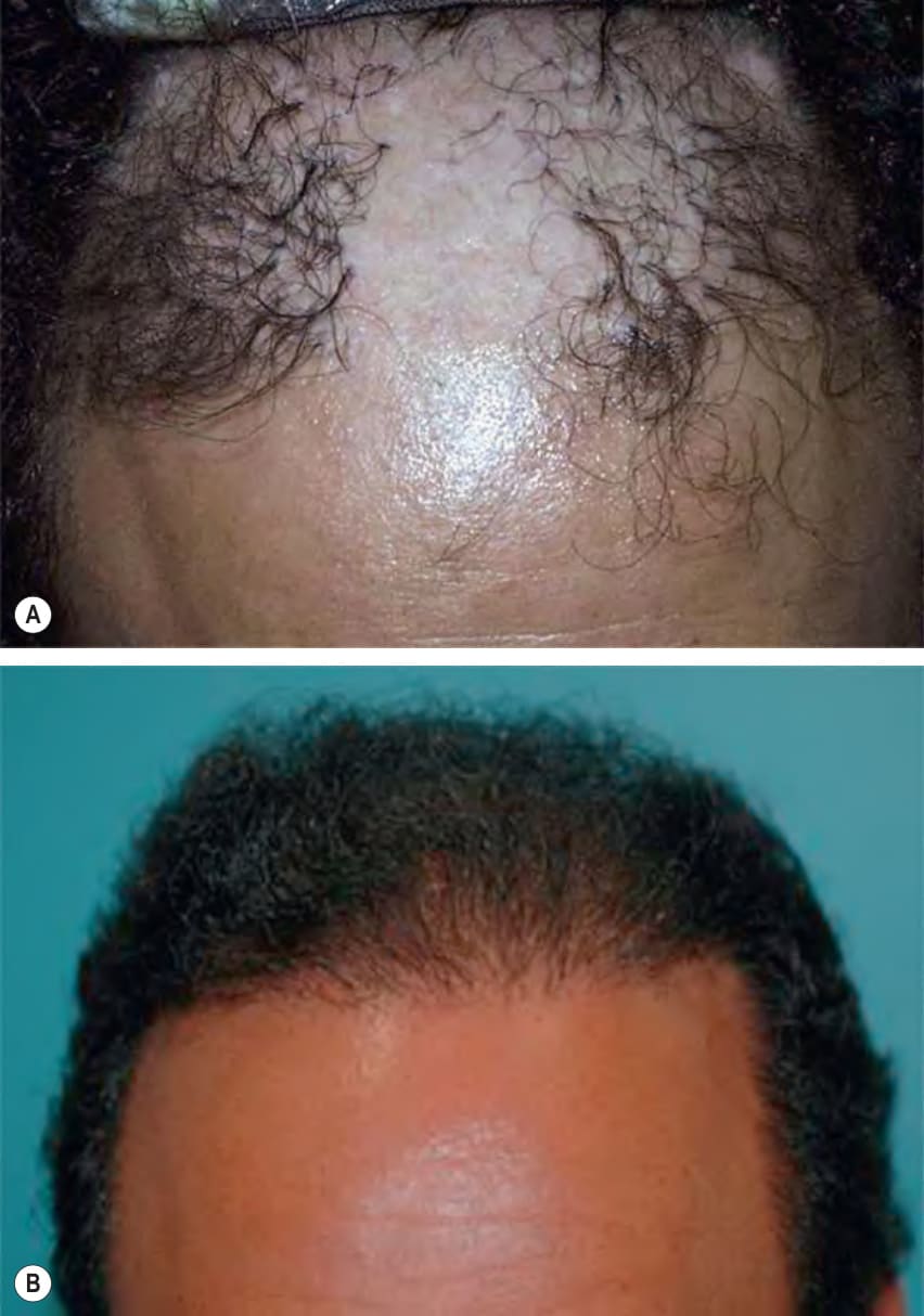
Fig. 157.16 Corrective hair transplant surgery.A Widely scattered large plugs with significant contrast between the lightly pigmented scalp and the black hairs. B Softened and improved appearance after the transplantation of 1000 follicular unit grafts containing one to three hairs.
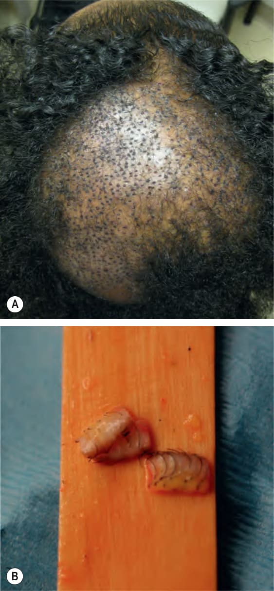
Fig. 157.17 Hair transplantation for central centrifugal cicatricial alopecia.
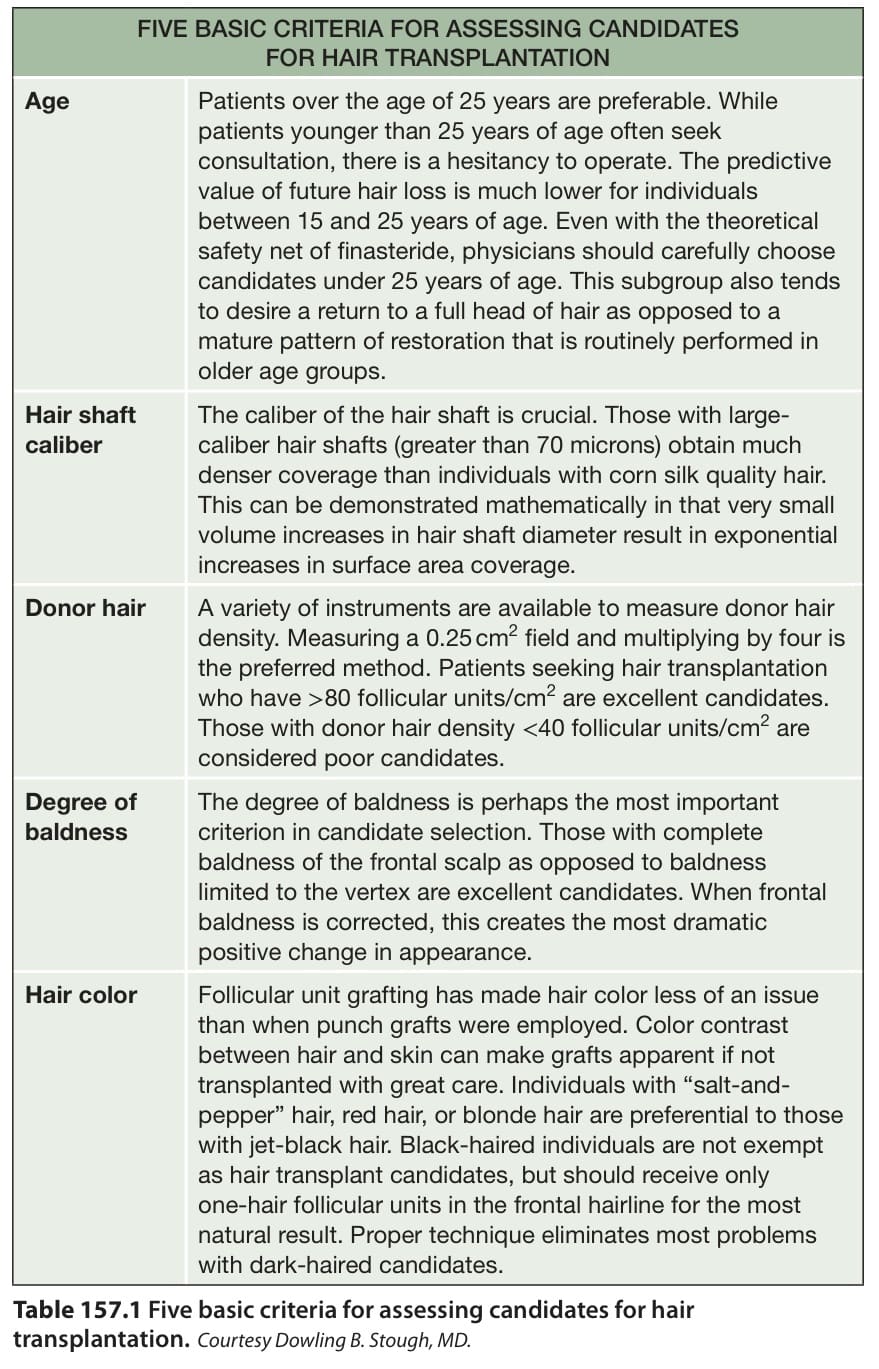
Table 157.1 Five basic criteria for assessing candidates for hair transplantation.
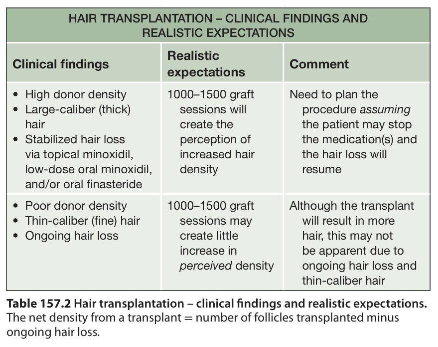
Table 157.2 Hair transplantation – clinical findings and realistic expectations.
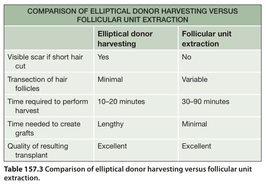
Table 157.3 Comparison of elliptical donor harvesting versus follicular unit extraction.
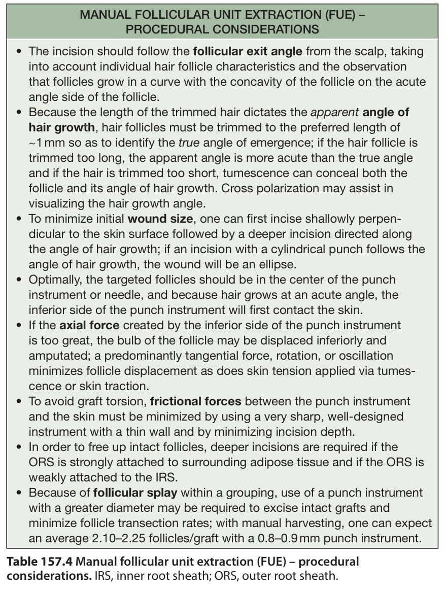
Table 157.4 Manual follicular unit extraction (FUE) – procedural considerations. IRS, inner root sheath; ORS, outer root sheath.
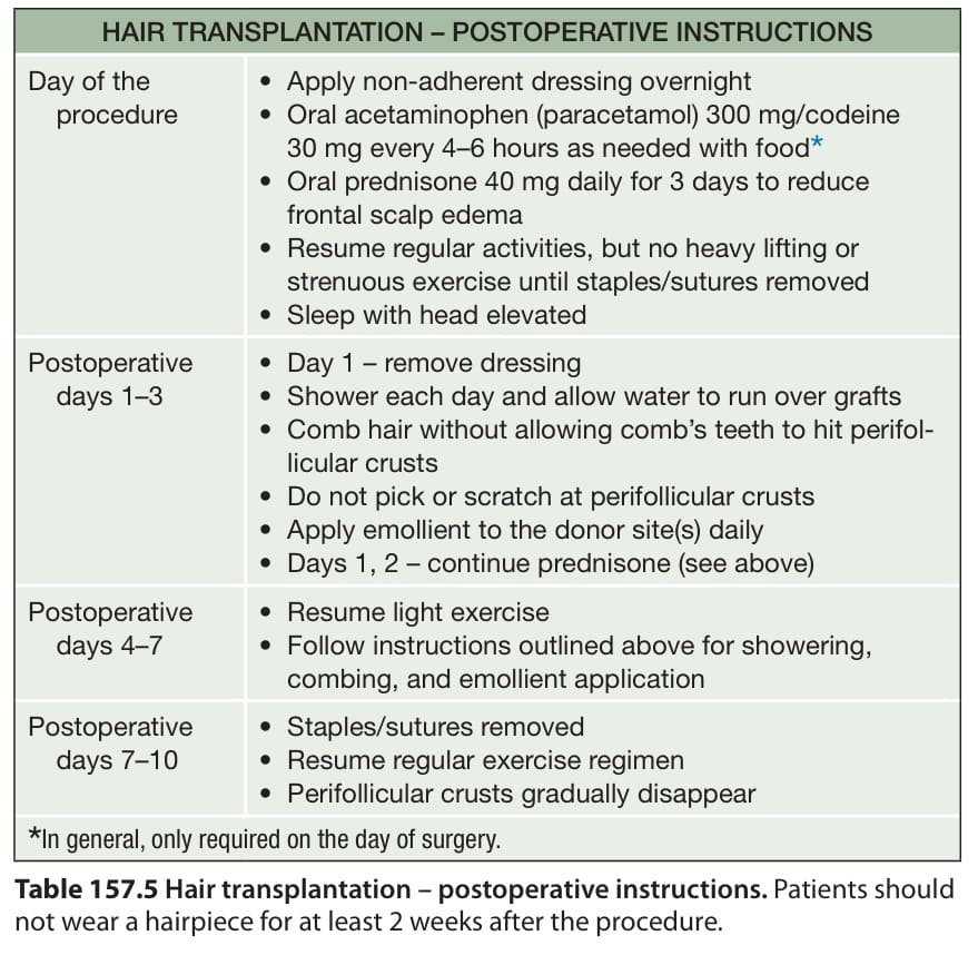
Table 157.5 Hair transplantation – postoperative instructions. Patients should not wear a hairpiece for at least 2 weeks after the procedure.