GENERAL INJECTION PROTOCOLS
Prior to performing any filler procedure, injectors must have a firm grasp of vascular anatomy to ensure safety and help guide optimal injection techniques and product selection. This forms the foundation of all treatments; its importance cannot be overemphasized, since accidental injection into facial arteries can have catastrophic consequences, as outlined by the American Society for Dermatologic Surgery (ASDS) Task Force on evidence-based guidelines for prevention and treatment of adverse events from injectable fillers. While intravascular injection is possible at any location on the face (e.g. lips’ labial arteries), the nose, glabella, forehead, superior nasolabial fold, and medial cheek are considered significant danger zones, from highest to lowest, for vascular complications, visual compromise, and cerebrovascular events.
Studying anatomy and appreciating its variability are the under-pinnings of all injection techniques, which vary by location, and represent a life-long learning endeavor. The facial artery is a branch of the external carotid artery that crosses the jawline periosteally at the antegonial notch just anterior to the border of the masseter muscle (see Fig. 142.3). It takes a circuitous course deeply from the lower lateral cheek to the nasolabial fold, branching off as the inferior and superior labial arteries, then becoming the angular artery near the superior nasolabial fold (see Fig. 142.15). More superficially, the angular artery runs along the medial cheek/lateral nose in variable patterns. It anastomoses with four arteries: (1) dorsal nasal artery; (2) distal ophthalmic artery, which connects to the retinal and cerebral vasculature; (3) supratrochlear artery; and (4) supraorbital artery; the two latter arteries are end branches of the distal ophthalmic artery. The supratrochlear and supraorbital arteries traverse the supraorbital ridge periosteally at the glabella and medial brow, respectively, then run superiorly toward the scalp. These pathways explain the danger zones referenced above.
There is no one completely safe injection protocol only safer strategies based upon location, which are briefly outlined here.
Nose, Glabella, and Forehead
These three regions present the highest risk. Therefore, to minimize the potential for disastrous outcomes, only the most experienced injectors should approach treatment in these areas. Although rare, the chance of blindness is greatest with nasal injections into the dorsal nasal artery. Based on cadaveric studies, the safest plane on the dorsal nose appears to be pre-periosteal or pre-perichondrial. Yet, due to vascular variability, the dorsal nasal artery may lie on the midline periosteum.
The glabella also carries higher risk because of the periosteal exit of the supratrochlear and supraorbital arteries. Injecting deeply within this region is contraindicated. Experts may consider correcting glabellar rhytides that do not respond to neuromodulators with intradermal injections using small needles. However, the vessels are variable, potentially running more superficially, especially in those with thin tissue. These arteries course cephalad to caudal and surface super-ficially within the frontalis muscle superior to the brow. Consequently, forehead correction is safest with cannulas placed in the pre-periosteal, subgaleal plane at least 2 cm superior to the orbital rim and in a horizontal direction, perpendicular to the vasculature.
Nasolabial Fold, Cheek
Two additional danger zones are: (1) the superior nasolabial fold where the angular artery becomes subcutaneous; and (2) the cheek medial to the midpupillary line (medial cheek), where the infraorbital artery runs periosteally and the angular artery subcutaneously. Both needles and cannulas are FDA-approved for injection in these regions, but given the different vascular depths and pathways, a cannula with a minimum size of 25-gauge (G) may be safer (see below). In contrast, the lateral cheek along the zygomatic prominence is considered less risky, although the zygomaticofacial artery does sit on the periosteum. Injection along the periosteum with a needle or cannula is appropriate.
Temple
Periosteal depots, deep to the superficial temporal vessels, are used to correct an atrophic temple. The needle is injected inside the “safe zone” whose border is 1 cm superior to the superior orbital rim, 1 cm lateral
to the temporal fusion line, and 2.5 cm superior to the zygomatic arch where the middle temporal vein lies.
Lips
Injections should be superficial, as the labial arteries usually course more deeply between the mucosa and orbicularis oris at the wet–dry border.
Chin, Jawline
Because the facial artery crosses the mandible just anterior to the border of the masseter muscle, injections along the rami in this region must sit above the bone. Periosteal injection at the gonial angle appears to be safer when sharpening the contour and widening the jaw or the midline mandible to create anterior or inferior chin projection.
Consultation and Evaluation
With a firm knowledge of anatomy, injectors can then approach patient consultation and evaluation. Baseline asymmetry, subjective defects, and realistic expectations should be discussed with the patient (Table 158.5). To fully appreciate the depth and scope of the defect, patients should be placed in a gravity-dependent or seated position with adequate lighting. The latter should be at an acute angle in order to accentuate surface irregularities and deficits. A careful patient selection process will assist in determining ideal candidates for soft tissue augmentation. Contraindications include prior allergy to the filler material or its constituents (e.g. lidocaine). Pre-existing conditions in the planned treatment area such as dermatitis, acne, or infection may require postponement of the procedure. The expected level of improvement and longevity are also key discussion points.
Prior to the procedure, patients should be instructed to abstain from all nonessential medications that can inhibit coagulation and platelet aggregation. For example, aspirin is avoided for 10–14 days and NSAIDs for 5–7 days in order to decrease the risk of bleeding and bruising. In addition, omega-3 fatty acids, fish oil, and a number of over-thecounter supplements should be discontinued (see Table 133.3). Prior to the procedure, written consent is obtained and both make-up and cutaneous debris are removed. Pre- and post-treatment photographs are strongly recommended.
Preparation
Clean technique is critical in preventing contamination during the initial handling, mixing, and injecting of the filler. The skin is cleansed with alcohol, chlorhexidine (avoiding the ear canal and eye), hypochlorous acid or other antimicrobial agent, and gloves are changed after intraoral manipulation. Percutaneous injections of fillers can be painful, necessitating the consideration of adding anesthesia to the treatment protocol. Although the physician can combine fillers with an anesthetic, most commercial products are now premixed with lidocaine (see Table 158.4). Some injectable products may require nerve blocks and/or regional anesthesia (see Ch. 143). More commonly, however, ice or topical anesthetic preparations are sufficient.
Injection Techniques
Proper placement of the filler material is crucial. Common injection techniques include serial puncture, linear threading, fanning, cross-hatching, and depot injection. In the serial puncture technique, small amounts of filler are sequentially deposited along the wrinkle or fold. The injections are placed close to one another so that the filler can blend in a continuous fashion; post-injection massage can also help to blend the filler so that it is evenly distributed. Using the linear threading technique, the full length of the needle/cannula is inserted into the proper dermal or subcutaneous plane, and the filler is injected in a retrograde manner as the needle is withdrawn. Conversely, the material can be injected as the needle is advanced (anterograde), creating blunt dissection within the tissue space. The fanning technique involves multiple passes in different, but evenly spaced, directions without withdrawing the needle/cannula from its original site of insertion. The cross-hatching or radial injection technique involves evenly spaced, linear injections in a grid-like pattern; this technique is used for filling large areas and for the oral commissures. Finally, depot injections are useful when placing volumizing fillers within the subcutaneous fat or along the periosteum. Occasionally, small depot aliquots are also placed along the infraorbital ridge, with a movement toward using cannulas in this region. Massage immediately after depot placement of filler helps to blend the product into the natural contour of the region.
Additional important recommendations:
●Inject small volumes slowly, under low pressure. When there is arterial cannulation or compression from deposition adjacent to arteries, large volume, high pressure boluses may be more detrimental.
●Retract the syringe plunger (reflux test) prior to injection. If positive for blood, then intravascular placement is possible. Stop straight away and reposition. However, a negative reflux test does not exclude possible intravascular injection.
●Continually move the needle or cannula tip to minimize the risk of prolonged intravascular injection of a larger bolus.
Needles and Cannulas
Evolving consensus supports the use of blunt-tipped cannulas, especially those with a larger bore (e.g. 25 G), to reduce the risk of vascular injury. Cadaveric and reflux studies suggest that larger diameter cannulas are less likely to perforate vasculature, when compared to smaller diameter cannulas (e.g. 27 G) or needles. This is of particular importance in areas (e.g. cheeks, lips) where both needles and cannulas are FDA-approved. Blunt-tipped cannulas are best suited for injecting in the subdermal plane and are not appropriate for intradermal injections. One of the authors (DJ) demonstrates high-risk anatomy and the use of cannulas in Video 158.1.
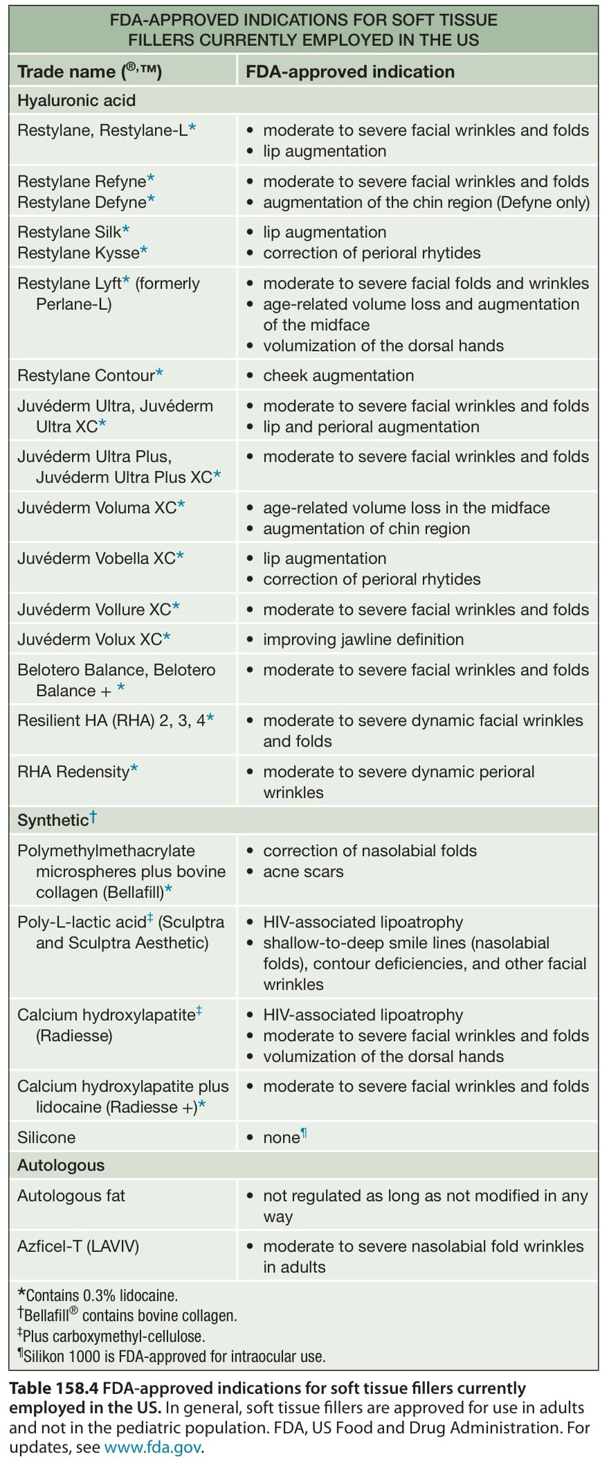
Table 158.4 FDA-approved indications for soft tissue fillers currently employed in the US. In general, soft tissue fillers are approved for use in adults and not in the pediatric population. FDA, US Food and Drug Administration. For updates, see www. fda. gov
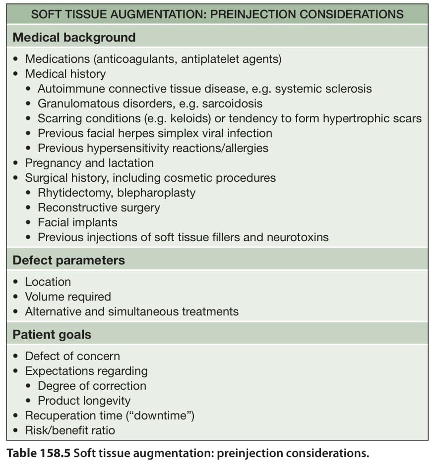
Table 158.5 Soft tissue augmentation: preinjection considerations.