COMPLICATIONS
The popularity of soft tissue fillers is in part due to their favorable side-effect profile. Common injection-related phenomena include erythema, swelling, pain, and bruising. These generally resolve within a week without treatment. The erythema and swelling often appear immediately after injection. Minimizing the number of skin punctures and applying ice may reduce pain, erythema, and swelling. To limit bruising, patients should be instructed to abstain from medications that inhibit platelet aggregation for at least 7–10 days prior to injection. Vigorous exercise with its associated blood pressure elevation should also be avoided for the first 24 hours after the injections. The use of a small-gauge needle, slow injection technique with small aliquots of product as well as blunt cannulas and fewer puncture sites may further minimize bruising. However, bruises are often inevitable and treatment with intense pulsed light or pulsed dye and potassium titanyl phosphate (KTP) lasers may be effective therapies, as they all can target the hemoglobin chromophore contained in the extravasated blood. Superficial bruising from fillers often lightens substantially within a day following laser- or light-based treatment.
More serious complications from soft tissue fillers may occur and include: (1) nodules, beading, and Tyndall effect from superficial placement; (2) infection; (3) hypersensitivity reactions; (4) delayed nodule formation; and (5) vascular compromise (Fig. 158.11A; Table 158.9). Inappropriate placement of fillers may result in the development of papulonodules, which are palpable and often visible. Injecting too superficially with an HA filler may lead to beading as well as blue–gray discoloration due to the Tyndall effect. Hyaluronidase
can be used to treat these complications (see above; Fig. 158.11B). Vitrase® and Hylenex® are the two FDA-approved formulations of hyaluronidase that may be used off-label for dissolving unwanted previously injected HA. Hylenex® is a genetically engineered human recombinant hyaluronidase, whereas Vitrase® is ovine testicle-derived hyaluronidase. In the ophthalmologic literature, the reported incidence of allergic reactions to hyaluronidase is ~1 in 2000. While to date no cases of allergy or anaphylaxis have been reported in the dermatologic literature, a preliminary intradermal skin test with a few units can be considered to exclude hypersensitivity. Because there is a theoretical risk of cross-reactivity in patients who are allergic to honeybees and yellow jackets, patients who have had severe reactions or anaphylaxis to bee stings should be skin tested prior to treatment. Persistent papules can also occur following injection of PMMA microspheres, poly-L-lactic acid, and silicone. Methods to reduce this complication are outlined in Table 158.9.
Any time there is a breach in the integrity of the skin, infection can occur. To minimize the risk, the skin should be cleansed with an antimicrobial agent such as isopropyl alcohol, chlorhexidine (avoiding the ear canal and eye), or hypochlorous acid prior to injection. Patients with an infection may present with tender erythematous fluctuant nodules or abscesses. These skin findings may be accompanied by systemic symptoms such as fever. When an infection is suspected, a microbiological culture of exudate, if present, should be obtained and/or the lesion biopsied for histopathologic examination and tissue culture. Therapeutic interventions include incision and drainage and empiric broad-spectrum antibiotics such as dual therapy with a macrolide (e.g. clarithromycin) and quinolone until the culture results are available. Trauma from injections may also trigger a recurrence of herpes simplex viral infection. Prophylactic antiviral treatment (e.g. valacyclovir) should be considered if a patient has a history of a post-procedure recurrence and when treating the perioral area in a patient with a history of recurrent herpes labialis.
Prior to the introduction of HA fillers, hypersensitivity reactions were more commonly observed (see above). There are a few reported cases of hypersensitivity reactions to HA (Fig. 158.12), and these may be due to residual proteins or impurities from the manufacturing process or they may be due to low-molecular-weight HA fragments. Hypersensitivity reactions to HA fillers often resolve spontaneously after several months, but resolution may be accelerated by application of tacrolimus ointment, intralesional corticosteroids, intralesional hyaluronidase, and/or incision and drainage.
Delayed nodules are variable in their etiology and difficult to precisely diagnose without histopathologic and/or culture results. They may be secondary to a hypersensitivity reaction, soft tissue infection, development of a biofilm, or formation of a foreign body granuloma (see Table 158.9). Although delayed nodules occur more commonly with permanent fillers such as PMMA or silicone (hence our recommendation to avoid their routine use), they can develop following injection of temporary fillers including HA. Hypothetically, delayed nodules secondary to an immune reaction may be triggered by residual proteins in the product or, in the case of HA fillers, by low-molecular-weight fragments of HA. As noted above, these fragments provide a theoretical explanation for the increased incidence of delayed reactions (~1%–4%) seen with the Vycross® fillers, which consist of a mixture of both high-and low-molecular-weight HA.
There are some data to suggest a greater risk of delayed reactions after influenza-like illnesses, following vaccinations, and during cold and flu season. More recently, delayed filler reactions have been observed following administration of SARS-CoV-2 mRNA vaccines. These reactions were typically mild to moderate and resolved within a few days, often without treatment. In a case series, the ACE (angiotensin-converting enzyme) inhibitor lisinopril at a dose of 5–10 mg daily led to a rapid reduction in edema.
Biofilms may also cause delayed nodules. When filler is injected, it can become coated with colonies of bacteria that secrete a protective and adhesive matrix. This results in a low-grade chronic infection resistant to antibiotics and the immune system. Often bacterial cultures will be negative as standard culturing techniques are not sensitive enough to detect this type of infection. Delayed nodules due to formation of biofilms can present as a “sterile” inflammatory nodule, an abscess or other form of soft tissue infection, or a foreign body granuloma. In order to try to prevent biofilm formation, it is important to cleanse the skin with an antimicrobial agent (e.g. chlorhexidine [avoiding the ear canal and eye]) prior to injection. Infectious delayed nodules may be related to biofilms, but can also be due to less common organisms such as atypical mycobacteria.
As all synthetic fillers represent foreign bodies, an intense host response can occur resulting in the formation of foreign body granulomas. Although infrequent, this presentation is more common with permanent fillers and has been well documented histologically (see Fig. 94.11).
While there are several therapeutic interventions for delayed nodules (see Table 158.9), they should be guided by clinical judgment (e.g. inflammatory versus non-inflammatory, sterile versus infected) as well as culture results and/or histopathologic findings when available. Treatment options for removal of as much of the filler as possible include intralesional hyaluronidase (if an HA filler), incision and drainage, and excision. Delayed nodules are often immune-mediated and can be treated with a 1- to 2-week course of oral corticosteroids (e.g. prednisone 30 mg daily). If there is concern regarding an active infection, empiric oral antibiotics are advisable (see above). Additional therapies employed for delayed nodules include topical or intralesional cortico steroids, oral immunosuppressants, and lasers (see Table 158.9).
While the majority of adverse events are reversible, vascular complications can be devastating and often irreversible (Table 158.10). Tissue necrosis may occur if filler is injected directly into the vasculature, resulting in ischemic or embolic phenomena. Of particular concern are the arteries in the mid central face that connect to the ophthalmic circulation, i.e. where the facial artery continues as the angular artery immediately superior to the nasolabial fold (see above). The angular artery then branches into the lateral nasal artery and other collateral vessels, which connect the internal and external carotid artery systems (see Fig. 142.15).
Signs and symptoms of vascular compromise include pain, arborizing blanching, duskiness, and reticulated erythema that may extend along the artery and its branches (Fig. 158.13). Over time, this can progress to a necrotic eschar, ulceration, and scarring. Treatment should be instituted immediately at the first sign of vascular compromise. If blanching occurs while injecting, immediately discontinue. If the complication occurs with an HA filler, injection of hyaluronidase is recommended.
Different formulations of hyaluronidase are available, which makes it difficult to establish standardized dosing. Variable doses have been reported in the literature, from 10–30 units per 2 × 2 cm2 area along the artery and its branches up to a total of 1500 units injected within the affected region. Larger doses may be required for higher cross-linked fillers such as Voluma®. As outlined in Tables 158.9 and 158.10, additional conservative measures can also be initiated. A thorough assessment and detailed treatment plan should be instituted for each patient. Immediate and ongoing care with frequent monitoring ensures optimal outcomes and decreases the risk of permanent complications.
Although very rare, more serious complications can occur secondary to embolism of the filler into the ocular circulation, leading to blindness and/or ophthalmoplegia, or into the brain, causing a cerebrovascular accident. Blindness may occur when there is sufficient pressure and retrograde arterial displacement of injected material from the peripheral vessels into the ocular circulation.
The anatomic sites most commonly associated with visual complications include the nose, glabella, forehead, and nasolabial fold, with the first three sites accounting for 85% of reported cases in a recent review. Here, the filler can travel through the branches of the internal carotid system (e.g. supratrochlear, supraorbital, dorsal nasal artery) to the ophthalmic artery, which further branches into the retinal artery. Alternatively, at sites such as the nasolabial fold, the filler can enter the ocular circulation via rich anastomoses between the external and internal carotid systems (see Ch. 142).
The signature feature of ocular embolism is immediate visual impairment, which may be accompanied by ocular pain, nausea, and vomiting. Such events represent an ophthalmologic emergency. If any of these signs or symptoms occur, the patient should be sent urgently for ophthalmologic consultation, since treatment aimed at recanalizing the affected vessel(s) and restoring tissue perfusion needs to be performed within a short window of time. The most frequently cited interval is 90 minutes, but more recent investigations have suggested an interval as short as 10–15 min. Therefore, a predetermined and detailed action plan is required. In an attempt to restore vision, prebulbar and retrobulbar injections of hyaluronidase can be performed immediately, but this intervention remains controversial (see Table 159.10). Because treatment of embolic blindness is usually unsuccessful, prevention is of critical importance.
When performed with care, soft tissue augmentation is a highly effective and safe procedure. To ensure the best outcomes while minimizing any adverse events, a thorough understanding of facial anatomy as well as proper injection technique are imperative. Injectors should be aware of both prevention and management strategies to minimize complications and improve patient outcomes (see Tables 158.9 and 158.10).
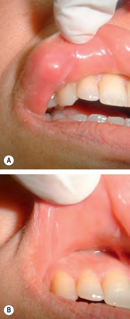
Fig. 158.11 Foreign body reaction to hyaluronic acid.A Nodule due to granulomatous inflammation arose six weeks after injection. B Resolution following intralesional hyaluronidase and triamcinolone. Courtesy Mahmoud Abdallah, MD.
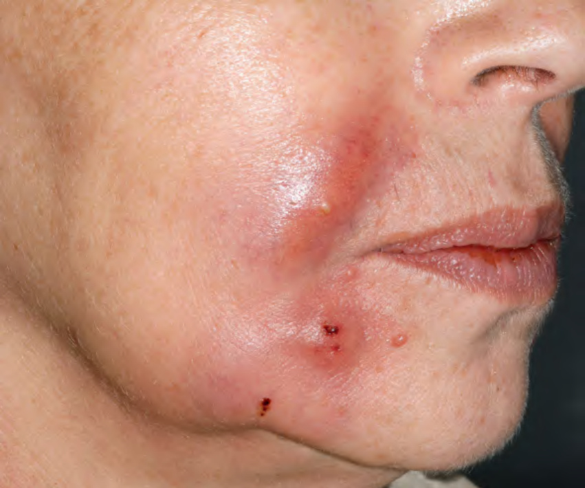
Fig. 158.12 Pustular and granulomatous reaction to injections of hyaluronic acid (Restylane®).Courtesy Julie V. Schaffer, MD.
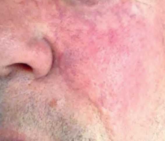
Fig. 158.13 Evolving vascular occlusion following injection of Juvéderm® Ultra XC into the cheek. Note the arborizing blanching, duskiness, and reticulated erythema that is observed in addition to pain.
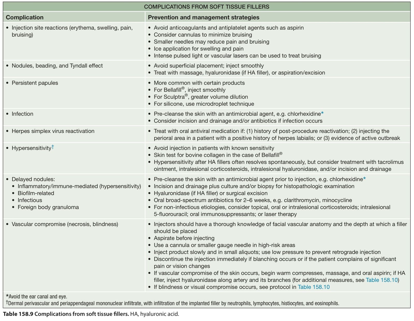
Table 158.9 Complications from soft tissue fillers. HA, hyaluronic acid.
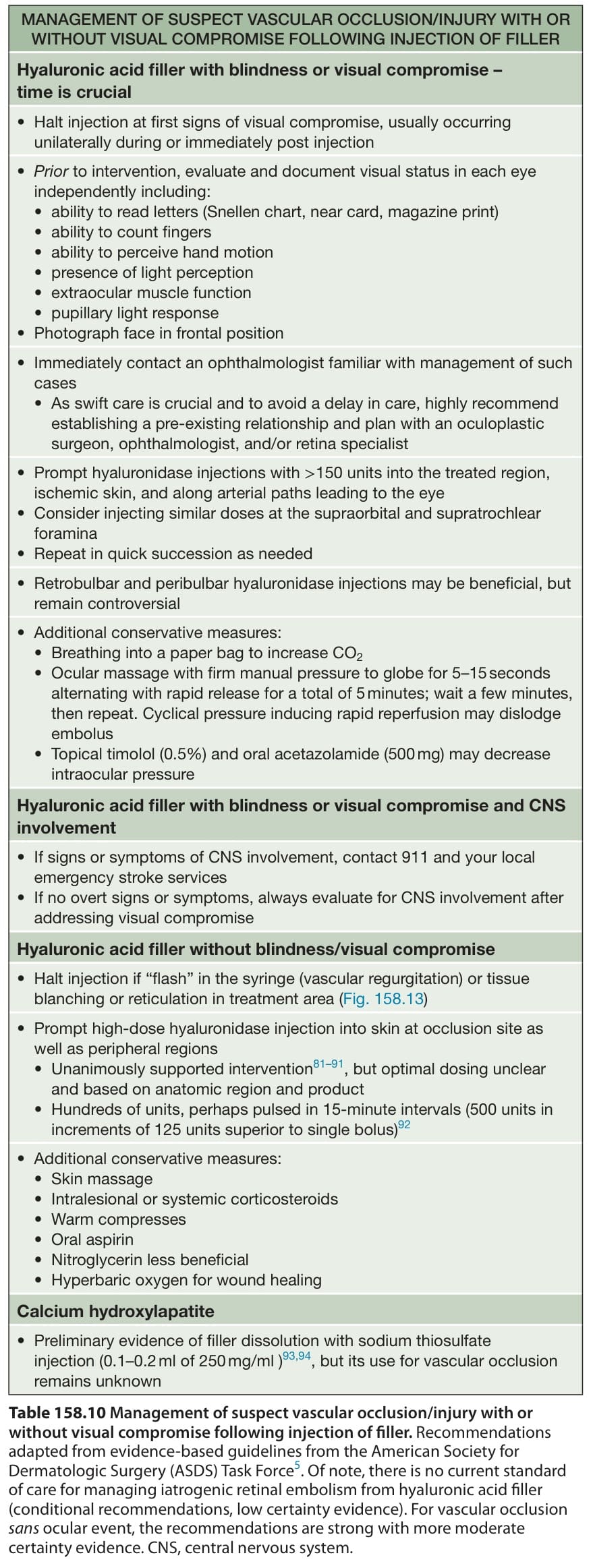
Table 158.10 Management of suspect vascular occlusion/injury with or without visual compromise following injection of filler. Recommendations adapted from evidence-based guidelines from the American Society for Dermatologic Surgery (ASDS) Task Force. Of note, there is no current standard of care for managing iatrogenic retinal embolism from hyaluronic acid filler (conditional recommendations, low certainty evidence). For vascular occlusion sans ocular event, the recommendations are strong with more moderate certainty evidence. CNS, central nervous system.