THE OCCUPATIONAL DERMATOSES
Contact Dermatitis
Synonym: Contact eczema
Key features
The most common causes of occupational allergic contact dermatitis are (in order of decreasing frequency): rubber, nickel, epoxy and other resins, and aromatic amines The most common causes of occupational irritant contact dermatitis are (in order of decreasing frequency): soaps, wet work, petroleum products, solvents, and cutting oils and coolants
Introduction
Contact dermatitis accounts for the majority of occupational skin disease that predominantly affects the hands. The prevalence of hand dermatitis in the population has been estimated at ~0.5%, rising in some studies to 10% when an industrial workforce has been examined. While most individuals will remain at work, over half will change jobs and a large proportion will be absent from work for a month or more.
History
In the first century CE, Pliny the Younger recorded dermatitis in individuals after they cut down pine trees, presumably representing allergic contact dermatitis to colophony in pine resin. Ramazzini in his 1700 treatise described irritant contact dermatitis of the hands and forearms in soap and laundry workers.
Epidemiology
An age-related increase in rates of occupational dermatitis is seen in men, but in women the peak age of incidence is 16–29 years with a decline thereafter. However, a comparison of rates by age within specific occupational groups suggests that the sexes follow a similar pattern. Among chefs and cleaners of either sex, higher rates are seen in young workers, whereas rates among machine tool operators increase with age, reflecting the type of occupational exposure. Materials giving rise to contact dermatitis in the United Kingdom (UK) are shown in Fig. 16.2, and the highest risk UK occupations in Fig. 16.3. In contrast to the UK, in Finland and the US, the highest rate of skin disease is among agricultural workers.
Pathogenesis
The initial events of both irritant and allergic contact dermatitis (see Chs. 14 & 15) are similar. Depending upon the severity of the insult, the tissue response can reach a level at which it becomes clinically apparent as cutaneous inflammation (Fig. 16.4). If tissue perturbation is maintained via repeated insults, then chronic dermatitis ensues, while an early withdrawal of the insult may result in healing.
Clinical features
Contact dermatitis, both allergic and irritant, is morphologically indistinguishable from endogenous eczema (Fig. 16.5). Although most cases of dermatitis will result from direct exposure that leads to localized
dermatitis, there are other routes of absorption that can lead to dermatitis. In the case of solvents, inhalation may result in widespread dermatitis, often with systemic involvement.
Diagnosis of an occupational cause is dependent on a thorough dermatologic and occupational history (Table 16.2), combined with
examination of the entire skin. Material safety data sheets (MSDS) provide a guide to the nature of materials handled at work. They list the chemical composition and the hazards associated with the product, e.g. irritant, corrosive, contains a sensitizer. However, total reliance should not be placed on the MSDS as they may not list all ingredients of relevance, and communication with the manufacturer may prove helpful. When occupational dermatitis is suspected but a cause cannot be established from the history, a workplace visit may prove invaluable and may also elucidate the source of an allergen previously detected via patch testing.
Although the primary site is usually the hands, spread to adjacent areas of skin (even without primary contact) is relatively common. Spread to distant sites, such as the face and feet, is more frequently seen in allergic contact dermatitis than in irritant contact dermatitis. Notably, airborne allergens, e.g. from paint sprayers, can lead to a pattern of dermatitis that suggests this type of exposure. Covered areas such as the trunk and feet are unusual sites of onset of disease. Improvement of the dermatitis during periods away from work is an important clue to an occupational cause, but it should be remembered that endogenous eczema may also be exacerbated by workplace exposures. As the dermatitis becomes chronic, the relationship between work and exacerbations becomes less clear-cut.
Occupational contact dermatitis can be either irritant or allergic. Personal protective equipment (PPE) is defined as equipment that protects the wearer’s body against health/safety risks at work. Although it is designed to help reduce the incidence of contact dermatitis at work, in some cases PPE itself can cause contact dermatitis. Hand dermatitis due to gloves is the most common PPE-related dermatitis, but non-glove exposures also occur. Dermatitis associated with the use of face masks during the recent SARS-CoV-2 pandemic would be a prime example.
Patch testing is essential for establishing the diagnosis and should be performed in any individual with a dermatitis that may be workrelated or for whom a change of job is being contemplated. Testing with work materials is essential (Table 16.3) if contact allergy is not to be missed, as not all allergens are commercially available and the MSDS may not disclose all relevant ingredients. A guide to testing 5200 chemicals has been published. When a reaction occurs to an unknown substance, the use of a dilutional series and the testing of ~20 controls can confirm that the reaction is allergic rather than irritant.
In an extensive surveillance report, the most common causes of occupational allergic contact dermatitis were rubber (23.4% of cases), nickel (18.2%), epoxy and other resins (15.6%), aromatic amines (8.6%), chromate (8.1%), fragrances and cosmetics (8.0%), and preservatives (7.3%). Recently, the more frequent use of long-lasting artificial nails has led to an increase in allergic contact dermatitis due to methacrylates in occupationally exposed, and sensitized, nail technicians.
pattern of eczema. Investigation demonstrated allergic contact dermatitis to chromate found in cement and contact urticaria to latex in the gloves he was using for hand protection.
Most occupational dermatitis results from repeated exposures to weak irritants which cause cumulative damage to the skin (see Fig. 16.4). On the other hand, strong irritants are usually recognized and protection provided. Employees with previous atopic dermatitis, especially with hand involvement, are particularly at risk, as are those with mutations in the filaggrin gene.
Soaps (22% of cases), wet work (20%), petroleum products (9%), solvents (8%), and cutting oils and coolants (8%) are the most frequently cited causes of occupational irritant dermatitis.
A chemical burn is an acute irritant reaction in which the injury to the skin is irreversible and cell death occurs. It can occur following a single exposure. Initial symptoms consist of burning and stinging with progressive development of erythema, blisters, erosions, and ulceration (see Fig. 15.2). Symptoms usually develop in close association with the exposure, but some chemicals, such as phenols and weak hydrofluoric acid, can have a delayed onset. Common occupational causes include:
●strong acids: e.g. sulfuric, nitric, hydrochloric, chromic. Most coagulate skin proteins and as a result form a barrier that impedes further penetration. Hydrofluoric acid differs in that it causes a liquefactive necrosis: penetration, even down to bone, can continue for several days after exposure. Pain, which can last several days, is typical of hydrofluoric acid and other fluorides. If more than 1% of the body surface area is affected, systemic toxicity can develop
●strong alkalis: e.g. sodium, calcium, and potassium hydroxides; wet cement (Fig. 16.6); sodium and potassium cyanides. Degradation of lipids and saponification of the resulting fatty acids form soaps which aid the penetration of alkalis deeper into the skin. As a consequence, damage is more severe than with most acids (apart from hydrofluoric acid) and pain is also a feature
●organic and inorganic chemicals: e.g. dichromates; arsenates; phenolic compounds. Phenols and unhardened phenolic resins easily penetrate the skin. Nerve damage may cause anesthesia, but rarely in the absence of visible skin damage. Vasoconstriction may contribute to the necrosis that develops and following systemic absorption, shock and renal damage may ensue
●solvents and gases: e.g. acrylonitrile; ethylene oxide; carbon disulfide; mustine. Ethylene oxide gas is used to sterilize medical instruments, textiles, and plastic materials, and it can remain on these items for several days if not allowed to evaporate off prior to use. The possibility of exposure may therefore not be obvious.
Glass fibers are subdivided into various types, depending on fiber diameter, and are used for their insulating (thermal, acoustic, and electrical), strengthening, and filtering abilities. They are chemically inert and the mechanism of skin injury is via direct penetration, which is directly proportional to the diameter (>3.5 microns) of the fiber and inversely proportional to its length. Histologically, eczematous changes including spongiosis are seen. Occasionally, granulomatous dermatitis is seen with birefringent fibers visible in polarized light.
Pruritus and tingling are the usual initial symptoms of fiberglass dermatitis. Subsequently, erythematous papules develop (often with follicular accentuation), either on exposed areas when there is airborne exposure or on the forearms when there is contamination of a work surface (Fig. 16.7). Contamination of clothing leads to involvement of sites where there is close contact with the skin, particularly flexural areas. Paronychia is common, and airborne exposure may also cause burning eyes, sore throat, and cough.
The diagnosis is often based on clinical findings, but can be confirmed by detecting the glass fibers either by tape stripping affected skin or by examining skin scrapings in 20% potassium hydroxide. The dermatitis resolves rapidly after cessation of exposure. In most individuals, hardening occurs and symptoms resolve over a few weeks despite continued
exposure; of note, the use of resins to bind glass fibers can result in an allergic contact dermatitis.
Most occupational photosensitivity is phototoxic in nature and resolves with hyperpigmentation. The most common scenario consists of outdoor workers exposed to plant-derived psoralens. This may result from direct contact or be airborne as in “strimmer” dermatitis when plant sap splatters the body of a worker inadequately protected when cutting down plants (see Ch. 17).
Coal tar and its products, including creosote, can cause a reaction known as tar/pitch smarts. A burning or stinging sensation develops after as little as 15 minutes of sun exposure.
Occupational marks are usually areas of lichenification or calluses and corns that develop at sites of friction and are specific to a particular job (Fig. 16.8). Mechanical injury to the skin is also thought to cause dermatitis analogous to irritant chemical damage. It may result in a keratotic hand dermatitis or post-traumatic eczema (Koebner phenomenon).
Pathology
The histopathologic features of allergic contact dermatitis and irritant contact dermatitis are discussed in Chapters 14 and 15, respectively.
Differential diagnosis
The etiology of occupational dermatitis is frequently multifactorial and coexisting endogenous eczema may contribute to the clinical appearance. Tinea manuum may resemble hand dermatitis, especially following treatment with topical corticosteroids, and scabies infestation of the interdigital spaces can simulate irritant dermatitis. Rarely, blistering of the dorsal hands due to porphyria cutanea tarda is misconstrued as contact dermatitis.
Psoriasis frequently affects the palms, resulting in keratotic plaques, sometimes with fissures. This can be difficult to distinguish from dermatitis when there are no lesions elsewhere. Furthermore, psoriasis may be exacerbated by repeated, work-related trauma leading to Koebner phenomena. Nail involvement and plaques over the interphalangeal joints are helpful clues.
Treatment
Occupational disease prevention is divided into primary, secondary, and tertiary measures (Table 16.4). During pre-employment screening, a history of severe childhood atopic dermatitis, particularly with hand involvement, indicates an individual at risk of developing dermatitis from exposure to irritants. Employment in a “dry” job should be recommended. In the workplace, the use of PPE and skin care preparations should be encouraged (Table 16.5). Use of protective creams prior to exposure may aid in the subsequent removal of irritants and application
of emollients throughout the day may prevent the development of dermatitis. Education of the workforce in skin care has been shown to reduce the development of skin disease.
Treatment is discussed in Chapters 14 and 15. The most important aspect, however, is avoidance of the cause. Ideally, a change in the production process may avoid the need for exposure, but this may not be feasible. A practical compromise is the use of PPE and/or sub-stitution of a particular chemical. Advice on appropriate glove type may be found on the MSDS or from glove manufacturers (e.g. www. ansellpro.com). Gloves need to be replaced regularly and each particular type of glove will have a penetration time for any given chemical. For example, acrylate glues (orthopedic surgeons, dentists), the hair dye para-phenylenediamine (hairdressers), and “acid perm” solutions containing glycerol monothioglycolate (hairdressers) rapidly penetrate latex gloves.
Initial treatment of chemical burns requires irrigation with large volumes of water. When the chemical is insoluble in water, a soap solution may be used instead. High pressures should not be used, in order to avoid splashing other areas of the body or bystanders with the corrosive material. For some chemicals, specific antidotes can then be used, e.g. 2.5% calcium gluconate gel for hydrofluoric acid; reduction of pain is a sign of successful treatment. When there is a risk of toxicity from systemic absorption, as with chromic acid, early debridement of necrotic areas reduces blood levels.
Several chemicals (e.g. hydrofluoric acid, phenolic compounds, chromic acid, gasoline) carry a significant risk of systemic toxicity even when the area of skin involved is small (~1% of body surface area). In these instances, regular monitoring of blood, liver, and kidney function plus appropriate supportive treatment (e.g. dialysis) is required. When the chemical is also a sensitizer, allergic contact dermatitis may subsequently appear on re-exposure to non-irritant concentrations, as burns and irritant dermatitis promote sensitization.
Once it has developed, the outlook for occupational contact dermatitis is poor, and patients frequently have persistent disease despite interventions. While a change of occupation is associated with a better prognosis, the possibility exists that the new workplace will have the same or similar chemical exposures. Prognosis is also worse when there is a contact allergy (delayed or immediate) to a chemical that is present in the domestic as well as the industrial environment. Some individuals (~10%) have persistent disease in the absence of any obvious cause, for which the term “persistent post-occupational dermatitis” has been coined.
Contact Urticaria
Synonyms: Contact urticaria syndrome Immediate contact reaction Variant: protein contact dermatitis
Key features
Pruritus and wheal-and-flare reaction Develops within 60 minutes of exposure and resolves within 24 hours The protein content of latex rubber is responsible for the associated contact urticaria In suspected cases of latex-induced contact urticaria, the specific
IgE test may be negative, requiring prick testing with a commercial latex extract and a usage test
Introduction
Contact urticaria has been reported following exposure to a wide range of substances. With regard to occupational skin disease, plant- and animal-derived proteins are recognized causes, especially among food handlers and agricultural, animal laboratory, and veterinary workers. In extreme circumstances, systemic symptoms (i.e. rhinoconjunctivitis, bronchospasm, and anaphylaxis) may be seen. With the introduction of universal precautions in the 1980s and increased use of natural rubber gloves, latex protein emerged as an important cause of contact urticaria, particularly in the healthcare setting.
History
Contact urticaria was first defined as a clinical entity during the 1970s but it had long been recognized.
Epidemiology
Based on official statistics, occupational causes of contact urticaria have been well classified in Finland. The relative frequency of various urticants is shown in Fig. 16.9 and reflects the high prevalence of reactions in the farming community. The reactions to cow dander are probably the result of high exposure, as cattle are kept indoors from September to May/June. The prevalence varied from 140/100 000 for bakers to 2.1/100 000 for shop assistants and was higher in women, irrespective of occupation. Relative risk by occupation is shown in Fig. 16.10. The incidence of contact urticaria declines with age, and ~30% of individuals have coexistent contact dermatitis.
Pathogenesis
Contact urticaria is classified as either irritant/nonimmune-mediated or allergic/immune-mediated. An additional category includes those cases in which the mechanism is uncertain, exemplified by ammonium persulfate in hairdressing.
The mechanism of nonimmune contact urticaria is not well defined but involves the release of vasoactive mediators. It is inhibited by non-steroidal anti-inflammatory drugs but not by antihistamines, suggesting a role for prostaglandins. Symptoms develop in the majority of those exposed and are most often due to simple chemicals, e.g. sorbic acid.
Immunologic contact urticaria is mediated by allergen-specific IgE (see Ch. 18). Binding of antigen, usually protein, to mast cells in a previously sensitized individual results in degranulation and release of mast cell mediators, including histamine. Atopic individuals are at greater risk of this type of reaction.
Clinical features
Signs and symptoms range from nonspecific, i.e. pruritus, tingling and burning, to more typical urticaria with a wheal and flare, usually noted within 30 minutes of exposure at the site of contact. Resolution occurs within hours. Individuals with immunologically mediated urticaria may also experience systemic symptoms with generalized urticaria, rhinoconjunctivitis, orolaryngeal and gastrointestinal symptoms, bronchospasm, and even anaphylaxis.
One of the more common causes of contact urticaria is foodstuffs, which can provoke either orolaryngeal symptoms when ingested or hand symptoms when handled (e.g. fish processors, slaughterhouse workers, individuals in the catering industry). There is a diverse range of responsible foods, including meats, fish, eggs, fruits, vegetables and flour, as well as associated enzymes such as α-amylase (found as an additive in flour). In Scandinavia, a strong association is seen between the incidence of birch pollen allergy and contact urticaria to fruits and vegetables, due to the presence of similar peptides. Birch pollen is a common aeroallergen in Scandinavia, whereas in the UK the most common aeroallergens are the house dust mite and grass pollen; this may explain why there are fewer reports of contact urticaria in food handlers in the UK. In the US, incidences of birch pollen allergy vary depending upon geographic region. If contact urticaria is confirmed, there are recognized cross-reactions between the various foodstuffs (Table 16.6).
Contact urticaria to latex was first described in 1979. The term “latex” defines an aqueous dispersion of a rubber. The rubber obtained from latex by drying or coagulation is termed “latex rubber”. Natural latex is derived from the sap of the tree Hevea brasiliensis. Natural latex contains polyisoprene (30%–40%) together with a variety of other plant chemicals, including proteins (2%).
After 1985, the demand for latex gloves for medical and dental use to prevent the transmission of infectious agents more than doubled. Paralleling this rise in usage were reports of type I allergy to latex gloves. Occupational latex contact urticaria occurs more frequently in women, atopic patients (particularly those with hand eczema), and workers frequently exposed to latex gloves (e.g. hairdressers). Rates ranging from 3% to 16% have been reported among healthcare workers. Most problems arise from items made by coating a mold with concentrated liquid latex, e.g. gloves, balloons, condoms. Nowadays, however, many gloves are manufactured by methods designed to leave lower levels of protein allergen in the final product. Latex that is allowed to dry before processing by compression molding or extrusion (e.g. syringe plungers, vial stoppers) causes fewer problems, due to degradation of the protein allergen during the manufacturing process. When latex protein is absorbed onto starch particles in gloves, it can become airborne and cause conjunctivitis, rhinitis, and asthma. Ideally, powder-free gloves should be used to avoid this risk.
In a subgroup of latex-sensitive patients, hypersensitivity reactions to bananas, avocados, chestnuts, kiwis and other fruits may also occur (see Table 16.6). Radioallergosorbent inhibition studies have shown that they contain a similar antigen. In some individuals, the primary
sensitization is to the fruit, with latex sensitivity developing as a secondary phenomenon.
Common household sources of exposure to latex are gloves, balloons, latex contraceptives, latex mattresses and pillows, rubber bands, swimming caps, and baby pacifiers. The term “hypoallergenic glove” refers to latex gloves with reduced levels of accelerators and antioxidants (causes of allergic contact dermatitis); these gloves are not suitable for individuals allergic to latex (with immediate-type hypersensitivity).
Non-latex alternatives should be sought. Vinyl (PVC) gloves are suitable for home use. For those in the medical and dental professions, alternative gloves, typically nitrile (both sterile and non-sterile), are available from the major glove suppliers. Affected individuals should warn any doctor or dentist that they visit of their sensitivity so that measures can be taken to prevent a reaction. The most severe reactions have followed mucosal and parenteral exposures. Death has occurred following the use of a natural rubber latex cuff on a barium enema device and anaphylaxis after intraoperative, oral, or vaginal exposure to latex gloves. Use of a bracelet or necklace to alert medical professionals in the event of an emergency has been advocated.
Although rare, these chemicals can be important causes of occupational contact urticaria in the industrial setting. Some are also potential causes of occupational asthma. The diagnosis may prove difficult to establish because skin testing may require conjugation of the low-molecular-weight chemical with protein to form the allergen. Chemicals (and industries) associated with contact urticaria include: antibiotics (pharmaceutical industry); ammonium persulfate and para-phenylenediamine (hairdressing); phthalic anhydrides, epoxy resin systems, and polyfunctional aziridines (plastics and glue industry); and reactive dyes (textile workers).
The term “protein contact dermatitis” was originally used to describe an eczematous reaction to protein-containing material in food handlers (see Table 12.5). The reactions were both allergic and non-allergic, although many had a positive prick test or the presence of specific IgE antibodies, implying an IgE-mediated mechanism. In some, positive patch tests pointed to the coexistence of delayed-type hypersensitivity. The clinical picture is usually that of a chronic eczema with episodic exacerbations following contact with the allergen.
Pathology
The histologic findings of contact urticaria are described in Chapter 18.
Differential diagnosis
After a detailed history and clinical examination, skin testing may be performed to confirm the diagnosis of contact urticaria. When the patient has experienced anaphylactic symptoms and a specific IgE test is available, the blood test may confirm the diagnosis, avoiding the risk of anaphylaxis as a result of skin testing. Skin testing should be performed with appropriate positive and negative controls. With an unknown allergen, exposure should be graded; an initial application test (open and subsequently occluded) is followed by a prick test and, if necessary, an intradermal test. If the patient has a positive test, control individuals should be tested; a positive response in the latter group points to the presence of a nonimmune contact urticant. While commercial allergen extracts are available, it should be remembered that, unless adequately standardized, they may not contain the relevant protein allergens, resulting in a false-negative test result. The gold standard for testing is a sample of fresh material. Skin tests should only be performed where resuscitation facilities are available.
In the case of latex, the specific IgE blood test is not sensitive and a negative test does not exclude the diagnosis (Fig. 16.11). While skin tests with glove extracts have been recommended, many gloves now contain low levels of latex protein and prick testing with these homemade extracts frequently leads to false-negative results. Prick test solutions of latex are commercially available, some of which claim >98% sensitivity and 100% specificity (Fig. 16.12). If a particular glove type appears to elicit symptoms, a so-called “prick to prick” test can be used: the suspected item itself is pricked with the lancet, after which the patient’s skin is pricked with the antigen-“contaminated” lancet. The
IgE-mediated immediate hypersensitivity is detected by prick tests, whereas delayed-type hypersensitivity, which manifests as allergic contact dermatitis, is detected by patch testing. In the open usage test, the patient wears the suspect glove on a moistened hand.
final arbiter is a usage test in which a patient wears the suspect glove on a moistened hand and is observed for any reaction.
In the absence of a latex allergy, localized symptomatic dermographism is a common cause of urticaria to gloves. Lastly, a proportion of individuals who have reactions to gloves have symptoms caused by irritation.
Treatment
Management consists of avoidance, as desensitization for the majority of involved allergens is not available. Avoidance may be achieved by improved occupational hygiene and the use of PPE, but in extreme circumstances may necessitate a change of occupation. Treatment of the acute episode includes the use of systemic antihistamines and epinephrine (adrenaline), depending on the severity of the attack.
In the case of latex, the use of powder-free gloves containing low levels of protein has been shown to prevent the development of latex hypersensitivity by reducing the level of exposure in the at-risk population. There has also been a movement in healthcare facilities to use non-latex gloves.
Occupational Skin Cancer
Key features
A skin cancer in which occupation has played a major etiological role In the workplace, the most important exposures are UV radiation, ionizing radiation, and carcinogenic chemicals (e.g. polycyclic hydrocarbons)
Introduction
Occupational skin cancers have been estimated to account for <1% of all skin cancers. In a study of workers exposed to coal and diesel combustion products, the relative risk for developing non-melanoma skin cancer was 1.5%. The tumor most frequently associated with occupational chemical exposure is squamous cell carcinoma (SCC).
History
The first association between occupation and cancer was made in 1775. Sir Percivall Pott, a surgeon at St Bartholomew’s Hospital in London, described the occurrence of scrotal cancer in chimney sweeps. The carcinoma was often preceded by keratotic lesions known as soot warts. In 1873, von Volkmann described skin cancers from exposure to the distillation products of tar and pitch.
In the early 1800s, inorganic arsenic was associated with skin cancers, especially of the scrotum in smelter workers, but also from exposure in the mining industry and in users of end products (e.g. sheep dip). In the early twentieth century, ionizing radiation was recognized as a cause of radiodermatitis followed by skin cancer, particularly of the hands of medical workers administering radiotherapy.
Epidemiology
As carcinogens have become recognized and limits on occupational exposure introduced, solar UV exposure has risen to account for ~95% of cases of occupational skin cancer reported in the UK. In Australia, the most common cause of compensated occupational cancer (of all types) was UV exposure (22%), followed by asbestos (21%). It was estimated that 34 000 cases of non-melanoma skin cancer per year and 4% of cases of cutaneous melanoma in men were attributable to occupation. Recently, in Germany, actinic keratoses (AKs) and cutaneous SCCs were recognized and compensated as occupational diseases, provided in the case of AKs they cover at least 4 cm2 or ≥5 separate AKs develop each year. The SCCs must arise in sun-exposed skin and the worker must have been occupationally exposed to an additional 40% more UV than that of an indoor worker, e.g. at age 50 the worker would have to have spent 15 years in an outdoor occupation.
Occupations at high risk of occupational skin cancer include:
●outdoor workers, especially in agriculture and construction industries, and welders (UV exposure). Squamous cell carcinoma occurs with an odds ratio (OR) of 1.77 (95% confidence interval [CI] 1.40–2.22; p<0.001) and basal cell carcinoma with an OR of 1.43 (95% CI 1.23–1.66; p = 0.0001) amongst outdoor workers
●workers exposed to polycyclic hydrocarbons (e.g. tars, pitches, oil fractionation products); in addition to direct skin contact, fumes may be inhaled
●workers exposed to arsenic in glass production, copper, zinc or lead smelting, and the production of pesticides, herbicides, and semiconductors
●radiologists, dentists, X-ray technicians, and those who work with radioactive materials (ionizing radiation).
Pathogenesis
The major carcinogenic hazards include UV radiation, ionizing radiation, and carcinogenic chemicals. Occupationally induced cancers are not pathologically different from those which arise spontaneously. However, the former tend to occur at an earlier age than spontaneous tumors at the same anatomic site. They arise following repeated or continuous exposure to the carcinogen, and there is a long latency period between the exposure to the carcinogen and appearance of the tumor which can make it difficult to attribute causation.
There are two major classes of chemical carcinogens: (1) polycyclic aromatic hydrocarbons including benzo(a)pyrene; and (2) aromatic amines such as dichlorobenzidine, an intermediate in the dyestuffs industry. For many compounds, carcinogenic mechanisms are not well understood.
The molecular pathogenesis of UV radiation is discussed in Chapters 86 and 107.
Clinical features and diagnosis
In evaluating a patient with suspected UV-related occupational skin cancer, a careful history is required to establish the relative contributions of work and leisure exposure. The final judgment is essentially clinical.
With polycyclic aromatic hydrocarbons, a variety of skin changes may precede the development of cancer and aid in the clinical diagnosis. Associated changes include (in order of development) erythema and burning, folliculitis, poikiloderma, and keratotic papillomas (tar warts) within the poikilodermatous skin. The verrucous tumors may then develop into SCC, although basal cell carcinomas and keratoacanthomas also occur.
The clinical features and diagnosis of these cutaneous malignancies are discussed in detail in Chapters 108 and 113.
Pathology and treatment
The pathology and treatment of skin cancers are discussed in detail in Chapters 108 and 113.
Occupational Acne
Key features
Open and closed comedones Non-inflamed nodules and cysts Inciting factors include exposures to oils, halogenated polycyclic hydrocarbons, and repeated frictional trauma
Introduction
Occupational acne is a form of acne resulting from various chemical exposures as well as environmental and physical factors. Chloracne is a rare acne-like skin condition caused by certain toxic chemicals and is a cutaneous sign of systemic exposure.
History
Chloracne was first reported in 1897. In 1949, the first descriptions of human exposure to 2,3,7,8-tetrachlorodibenzo-p-dioxin (2,3,7,8-TCDD)-contaminated chemicals were reported after a trichlorophenol reactor explosion in Nitro, West Virginia, USA. The majority of health effects have been reported among groups with high exposure, including occupational populations such as chemical production workers, pesticide applicators, and individuals who handled or were exposed to materials treated with 2,3,7,8-TCDD-contaminated pesticides. In addition, residents of communities contaminated with tainted waste oil (Missouri, USA) and industrial effluent (Seveso, Italy) have been affected. During the Vietnam War, chloracne in US troops was ascribed to exposure to the defoliant Agent Orange. More recently, dioxin poisoning was implicated in the development of chloracne in the Ukrainian President Viktor Yushchenko.
Epidemiology
Oil acne is the most common form of occupational acne.
Pathogenesis
Continued exposure to oils results in reactive hyperkeratosis and subsequently follicular occlusion with typical manifestations of acne vulgaris.
Chloracne is caused by exposure to certain halogenated polycyclic hydrocarbons such as polychlorinated dibenzodioxins (PCDDs) and dibenzofurans (PCDFs), which are most often found in fungicides, insecticides, herbicides, and wood preservatives. It occurs within 2 months of exposure to chloracnegens and can persist for years despite cessation of exposure. The condition is always a sign of systemic exposure to the causal agent(s), but entry may occur via percutaneous absorption as well as from inhalation or ingestion of the chemicals. After exposure of workers in a large chemical factory to hexachlorobenzene, blood levels in those with chloracne were compared to the levels in workers without chloracne. In the chloracne group, blood levels ranged from 1168 to 22 308 pg/g blood lipid. In the exposed but chloracne-free group, the range was from 424 to 662 pg/g. It was concluded that chloracne developed at blood levels between 650 and 1200 pg/g.
Clinical features
Contact with acnegenic substances, such as creosote, oils and greases, can worsen existing acne. Oils used in industry, such as cutting oils (paraffin/oil mixtures), tars (pitch and creosote), and crude petroleum oils such as diesel oil, can all cause acne. Lesions may develop both in the usual sites for acne and in unusual sites, including beneath clothing that is saturated with oils (e.g. arms, abdomen). Oils mists can also induce acneiform eruptions.
Acne mechanica is a form of acne, usually inflammatory, that is exacerbated by repeated frictional trauma that occludes the follicle. It is localized to sites of pressure, e.g. the back in long-distance drivers, the neck in violinists, the chin in football players who wear helmets with chin straps, the face from mask-wearing during the SARS-CoV-2 pandemic.
Certain cosmetic ingredients used by models and thespians, e.g. lanolin, petrolatum, some vegetable oils, can induce comedo formation.
Chloracne is distinguished from ordinary acne vulgaris by the predominance of open (over closed) comedones and a concentration of lesions over the malar crescent and behind the ears (Fig. 16.13). The first sign of chloracne may be excessive oiliness of the skin. This is accompanied or followed by the appearance of numerous open comedones. In mild cases, open comedones may be limited to the area around the eyes, extending across the temples to the ears. In more severe cases, they may appear more extensively on the body, especially in the malar region, other facial areas, behind the ears, and on the arms. The open comedones are usually accompanied by fluid-filled cysts and by an increased or darker growth of body hair.
As the disease progresses, milia form together with distinctive yellow-ish epidermoid inclusion cysts (some of which may become inflamed) on the face, behind the ears, and on the neck, buttocks, scrotum, and thighs. Some authors have proposed using the term “metabolizing acquired dioxin-induced skin hamartomas” for these dioxin-induced cysts. The skin may become thicker and hyperpigmented and it may desquamate or peel. In severe cases, ulcerations and permanent scars appear. Chloracne fades slowly after exposure. Minor involvement may disappear altogether, but more severe disease may persist for years. This disorder is difficult to treat and can last for long periods without known additional exposure to chloracnegens.
In addition to conjunctivitis and “ophthalmic acne”, there may be systemic manifestations including altered liver function (elevated transaminases), headaches, and peripheral neuropathy.
Chemicals that contain iodides, bromides, and other halogens can induce an acneiform eruption similar to that due to corticosteroids (see Chs. 36 and 125); however, the iodide-induced eruptions
(i.e. iododerma) may be more severe. Patients with bromoderma may also have lesions that resemble pyoderma gangrenosum.
Pathology
Histopathologic features of comedones are reviewed in Chapter 36. Small infundibular cysts and dilated infundibula filled with orthokeratotic plugs are seen in chloracne. A disappearance of sebaceous glands has been described.
Treatment
Treatment consists primarily of avoidance of the cause, although some forms can be persistent and require treatments designed for acne vulgaris, e.g. topical or oral retinoids (see Ch. 36). However, chloracne is particularly recalcitrant to therapy.
Vibration White Finger
Synonyms: Hand–arm vibration syndrome Dead finger
Key features
Development of a white finger or fingers in response to cold Associated transient loss of sensation Possible permanent finger neuropathy and pain in the affected limb Associated with exposures to vibrations between 30 and 300 Hz Workers at risk include operators of chainsaws and pneumatic tools
Introduction
Vibration white finger is a relatively frequent disorder among operators of chainsaws, pneumatic tools, and hand grinders who work in cold climates. Up to half of the exposed workforce can be affected.
History
Loriga first described the disorder in Italian marble-quarry workers in
1911. Six years later, this was followed by a description in sandstone cutters in Indiana, USA.
Epidemiology
Prevalence studies have suggested a relationship between symptoms and degree of exposure. This varies, however, with the type of tool and the work process involved. From 1972 to 1990, the prevalence amongst Finnish lumberjacks fell from 40% to 5%. This was attributed to a reduction in the weight and acceleration-induced vibration of their chainsaws.
It has been suggested that vibration white finger does not occur in countries with a warm climate. However, this may reflect the absence of cold temperatures that serve as a trigger for symptoms rather than the absence of vibration-induced vascular changes. In tropical countries, neurological symptoms predominate.
Pathogenesis
Vibrations between 30 and 300 Hz are most strongly associated with development of this disorder. While increased activity of the sympathetic nervous system is viewed as the major cause of vibration white finger, local factors are likely also important. Vibration-induced damage to endothelial vasoregulatory mechanisms with alteration in α-adrenoceptors and impaired release of nitric oxide have been described.
Smoking is known to contribute to the development of the disorder, presumably because of the vasoconstrictive effect of nicotine.
Clinical features
Onset may occur within 3 months of beginning work but more frequently it takes 2–3 years to develop. Typically, a white patch of skin develops in response to cold-induced vasospasm. It is well demarcated from surrounding areas. Reduction in sensation occurs because the touch receptors are unable to function without adequate blood supply and this results in reduced dexterity. Episodes cease upon rewarming, often with the development of reactive hyperemia. Although caused by vibrating tools, attacks are not usually triggered by work with the vibrating tool.
The condition is graded according to the severity and frequency of symptoms, starting on the tips of the fingers (stage 1) and spreading to affect the whole digit (stage 3). In the most severe form, there are trophic changes and ulceration (stage 4). To aid in the diagnosis, finger skin temperature and finger systolic blood pressure can be measured in response to cold exposure, but the results are not discriminatory.
Pathology
The histopathologic features of vibration white finger include a significant reduction in the number of unmyelinated nerve fibers containing the neuropeptide calcitonin gene-related peptide (CGRP). There also may be hypertrophy of arterial smooth muscle. However, histology is not routinely utilized in the diagnosis of vibration white finger.
Differential diagnosis
In contrast to the symmetric distribution seen in Raynaud disease, in vibration white finger the blanching is asymmetric and occurs only on those digits most exposed to vibration; the thumbs are usually spared.
However, Raynaud phenomenon can also result from repeated occupational trauma, particularly in those who use tools that require a squeezing action, e.g. farmers, mechanics.
The hypothenar hammer syndrome results from occlusion of the ulnar artery as a consequence of repeated trauma to the palms and may be misdiagnosed as Raynaud phenomenon.
Treatment
The prognosis for affected individuals is good: the disease regresses upon cessation of exposure. This is particularly true in the early stages, provided there are no other environmental or constitutional factors contributing to the disorder. As a consequence, no specific treatment may be required other than avoidance. Therapeutically, topical nitroglycerin ointment or sustained-release glyceryl trinitrate patches are effective, but the condition relapses upon withdrawal. Various systemic agents used for Raynaud disease, e.g. calcium channel blockers, have also been used for symptom control (see Ch. 43).
Infections
Many occupations, particularly those involving contact with animals, are associated with exposure to infectious agents. For example, individuals with occupations that involve wet work combined with rough
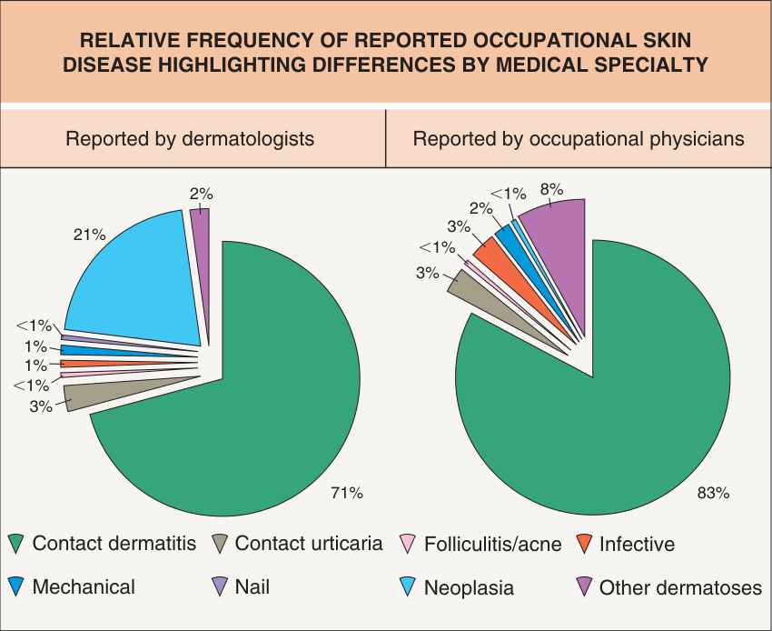
Fig. 16.1 Relative frequency of reported occupational skin disease highlighting differences by medical specialty.Courtesy The Health and Occupation Research (THOR) network of the Centre for Occupational and Environmental Health (COEH) of the University of Manchester, UK.
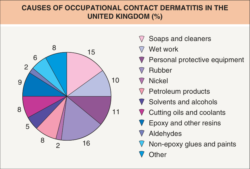
Fig. 16.2 Causes of occupational contact dermatitis in the United Kingdom (%).

Fig. 16.3 Reported cases and incidence rates of contact dermatitis by occupation in the United Kingdom.A Actual and estimated cases of contact dermatitis, with crude data adjusted for sampling bias. B Contact dermatitis incidence rates (per 100 000 employed); crude data were adjusted for the size of the labor force by utilizing the labor force survey data (2002–2009) as the denominator. Courtesy The Health and Occupation Research (THOR) network of the Centre for Occupational and Environmental Health (COEH) of the University of Manchester, UK.
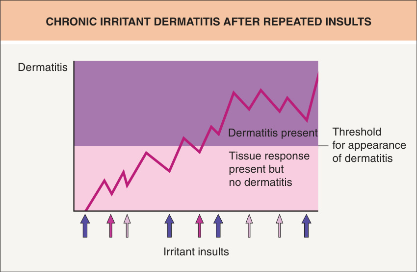
Fig. 16.4 Chronic irritant dermatitis after repeated insults. Initial subclinical response to tissue perturbation by an irritant becomes manifest as chronic irritant dermatitis after repeated insults. Each arrow represents an irritant insult of different intensity.
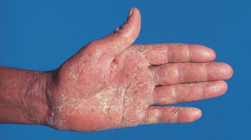
Fig. 16.5 Hand of a builder who presented with a dyshidrotic (pompholyx)
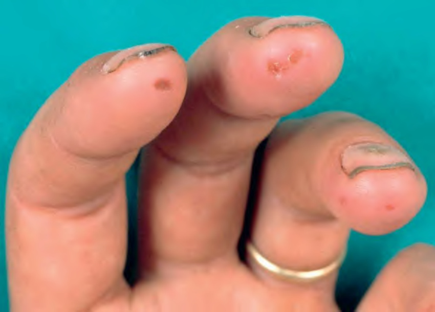
Fig. 16.6 Cement burns. Ulcerations on the fingertips of a construction worker exposed to wet cement. Courtesy P. J. Coenraads, MD.
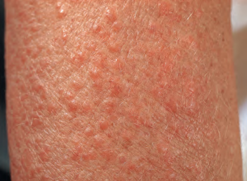
Fig. 16.7 Fiberglass dermatitis. Multiple pruritic pink papules at the site of exposure.
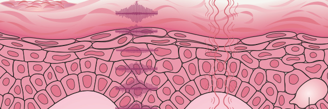
Fig. 16.8 The effects of mechanical injury to the skin. Both friction and pressure lead to callus formation. Adapted from Adams RM. Occupational Skin Disease, 3rd ed. Philadelphia: WB Saunders, 1999:36.
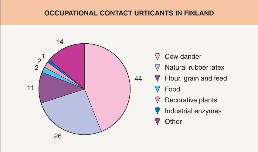
Fig. 16.9 Occupational contact urticants in Finland. Relative frequencies are represented. Reproduced from Kanerva L, Elsner P, Wahlberg JE, et al. Handbook of Occupational Dermatology. Berlin: Springer Verlag, 2000.
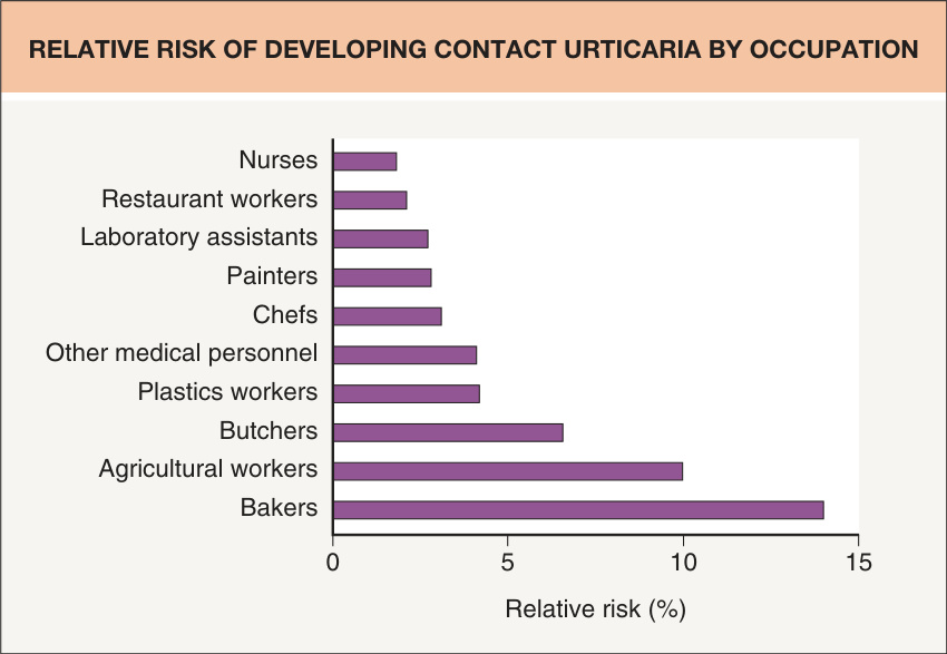
Fig. 16.10 Relative risk of developing contact urticaria by occupation. Based upon statistics from Finland. Reproduced from Kanerva L, Elsner P, Wahlberg JE, et al. Handbook of Occupational Dermatology. Berlin: Springer Verlag, 2000.
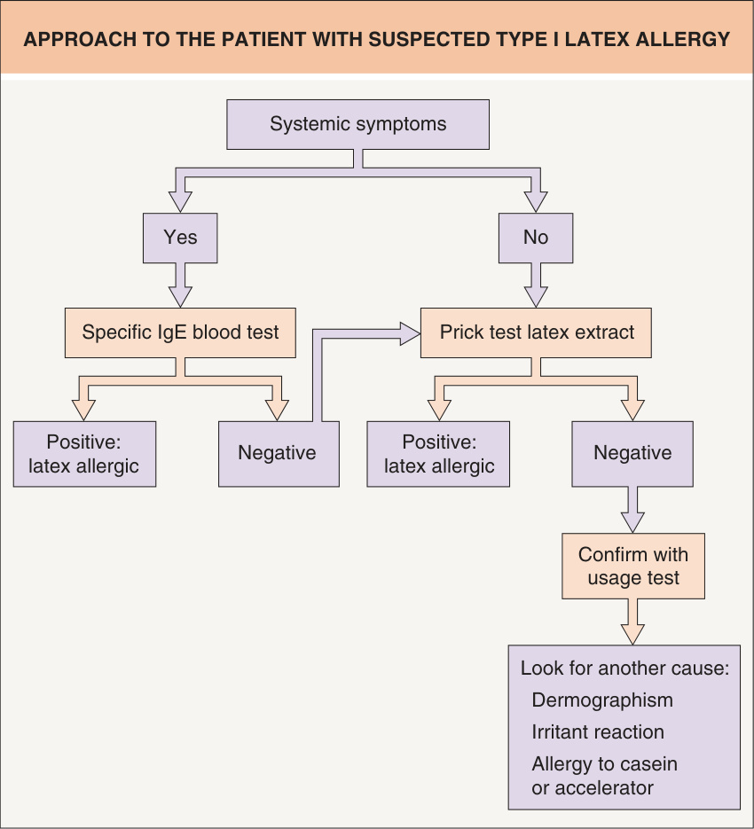
Fig. 16.11 Approach to the patient with suspected type I latex allergy.
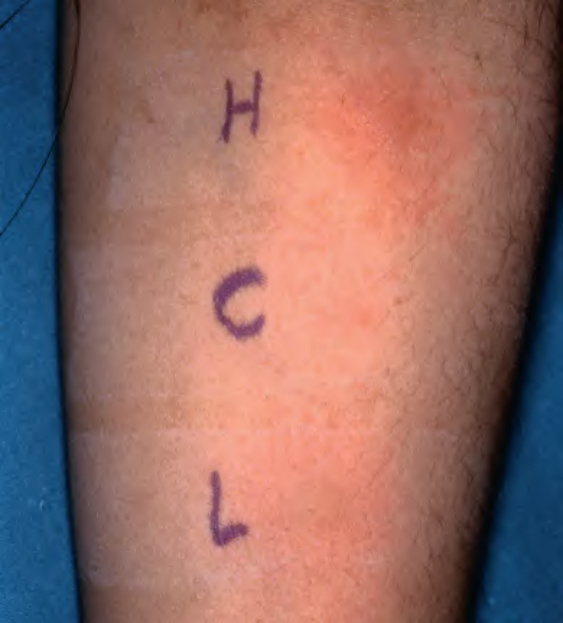
Fig. 16.12 Positive prick test with a commercial latex extract (L). Histamine (H; 10 mg/ml) and saline (C) controls are appropriately positive and negative. The histamine-positive control confirms that the effects of previous medications (including antihistamines) are no longer an issue and the negative control assesses for dermographism. When a standardized allergen is available for a prick test, a small drop is placed on the skin and the skin is pierced with a special lancet with a 1 mm tip. Surplus allergen is blotted off and a fresh lancet is used for each test substance to prevent cross-contamination. Reactions develop over 15 minutes and a wheal 3 mm larger than the negative control is considered positive.
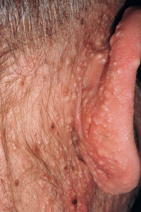
Fig. 16.13 Chloracne. Note involvement of retroauricular skin with numerous comedones (primarily closed) and cysts. The clinical differential diagnosis includes folliculotropic mycosis fungoides.
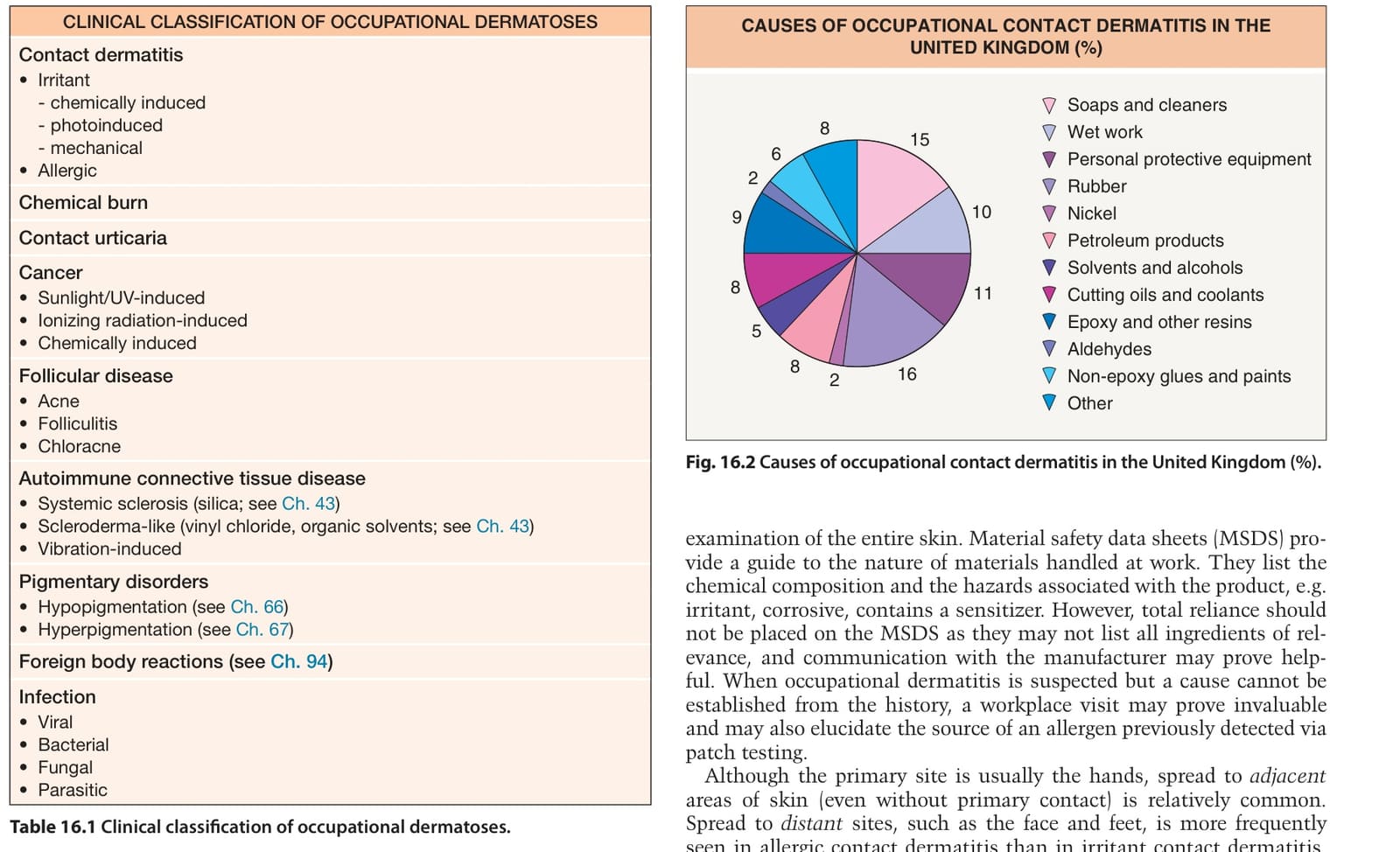
Table 16.1 Clinical classification of occupational dermatoses.
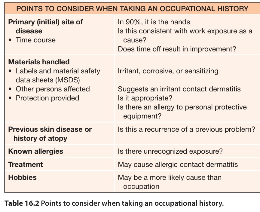
Table 16.2 Points to consider when taking an occupational history.
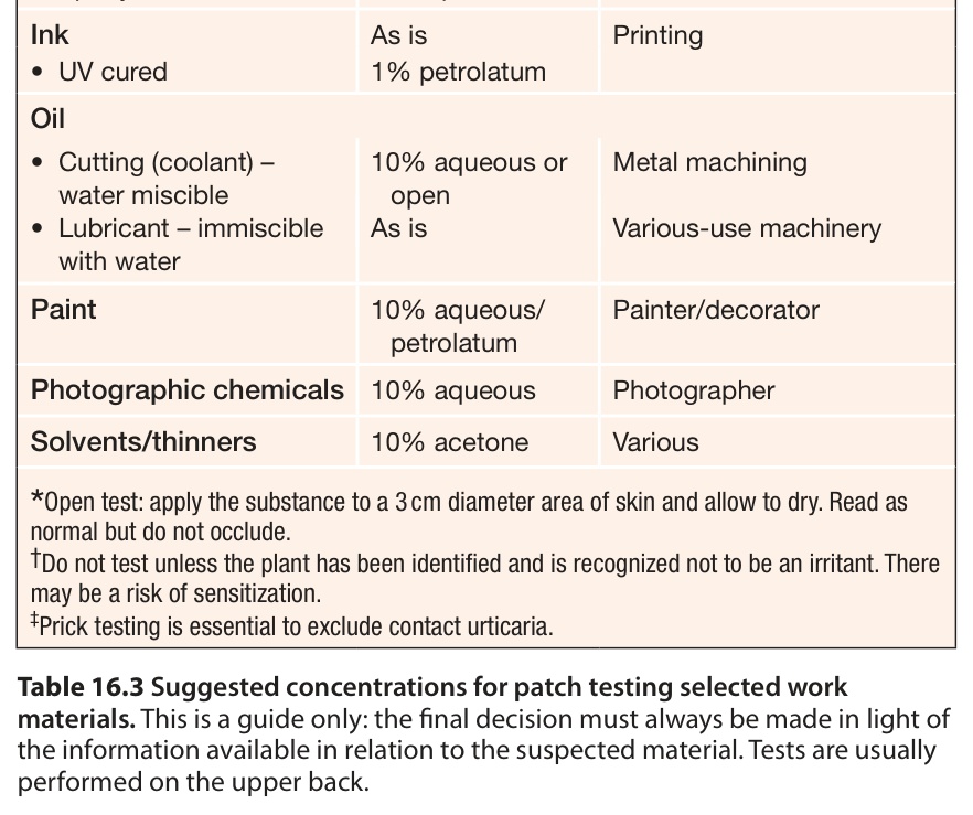
Table 16.3 Suggested concentrations for patch testing selected work materials. This is a guide only: the final decision must always be made in light of the information available in relation to the suspected material. Tests are usually performed on the upper back.
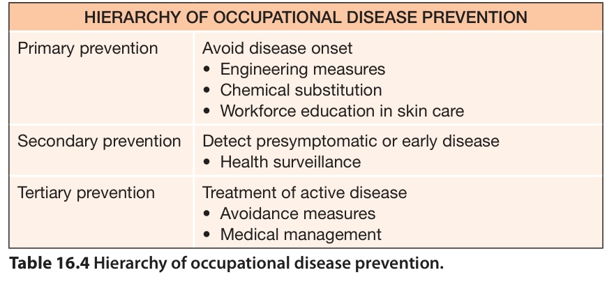
Table 16.4 Hierarchy of occupational disease prevention.
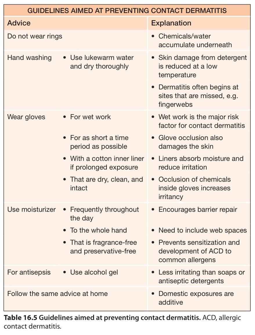
Table 16.5 Guidelines aimed at preventing contact dermatitis. ACD, allergic contact dermatitis.
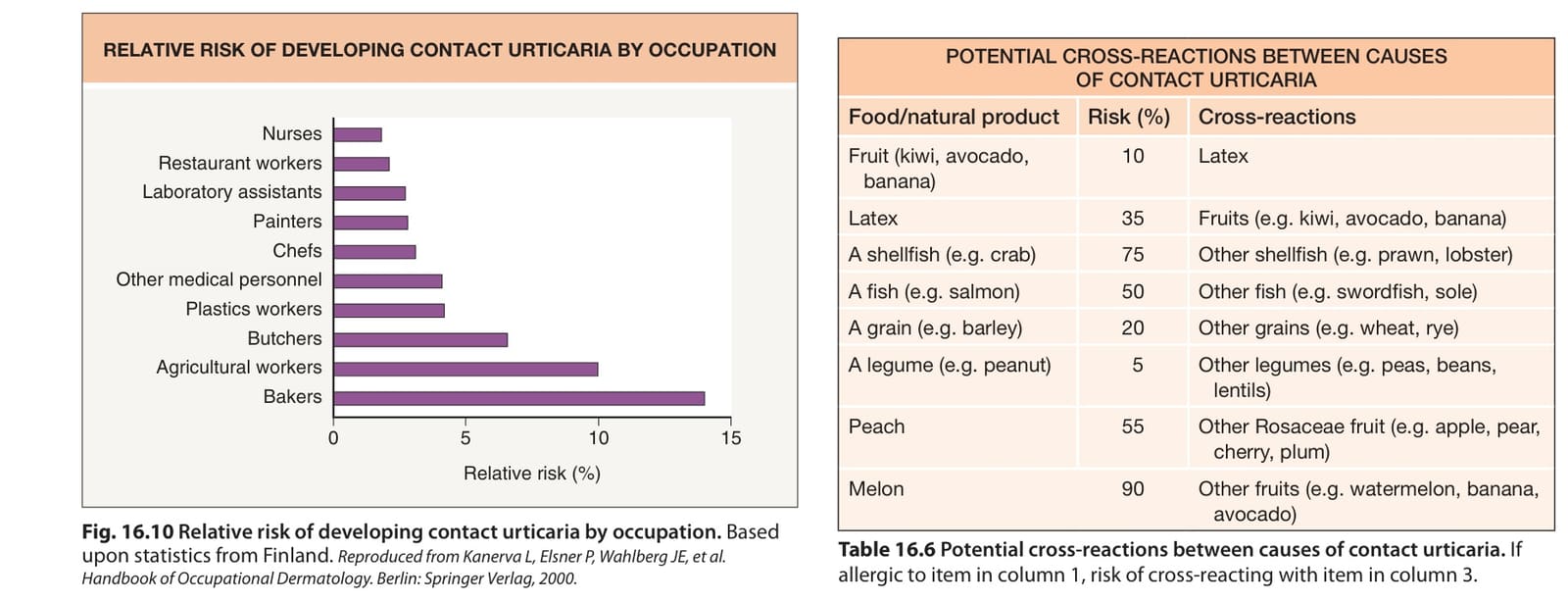
Table 16.6 Potential cross-reactions between causes of contact urticaria. If allergic to item in column 1, risk of cross-reacting with item in column 3.
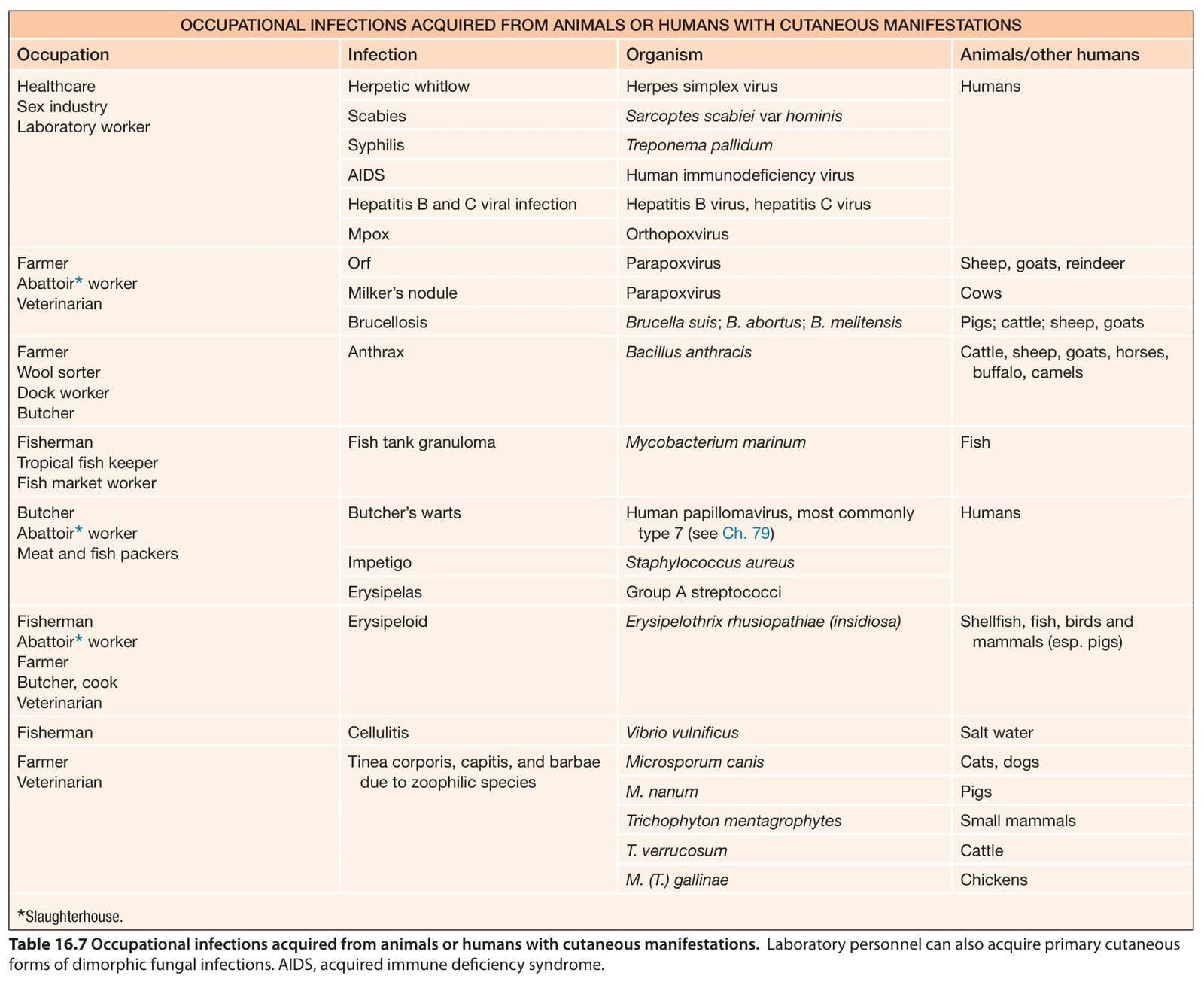
Table 16.7 Occupational infections acquired from animals or humans with cutaneous manifestations. Laboratory personnel can also acquire primary cutaneous forms of dimorphic fungal infections.AIDS, acquired immune deficiency syndrome.
physical conditions, such as butchers, slaughterhouse workers, and meat and fish packers, commonly develop infections with pyogenic bacteria and human papillomavirus (butcher’s warts) through cuts and abrasions. Epidemics of methicillin-resistant Staphylococcus aureus infection have also occurred in professional football players in the US.
In the metal working industry, staphylococcal folliculitis can develop as a result of exposure to contaminated work materials such as coolant fluid. In addition, outdoor workers are exposed to environmental pathogens either by direct contact or implantation with soil or plants (e.g. mycetoma, chromomycosis, sporotrichosis), as well as following inoculation, most commonly via insect bites (e.g. Lyme disease, tularemia, spotted fevers).
Common infections may also occur more frequently as a result of environmental factors in the workplace. Increased humidity may lead to bacterial and fungal infections (due to dermatophytes or Candida spp.) in flexural body sites along with maceration of the skin. Pitted keratolysis caused by Corynebacterium spp. or Micrococcus sedentarius occurs on the soles in association with the use of occlusive footwear.
Infectious organisms related to specific occupations are summarized in Table 16.7. Further information on these infections can be obtained by reference to the relevant chapter in this book (see Section 12).
Occupational Skin. Diseases. 3rd ed: Springer; 2020.2. Health and Safety Executive. Health and Safety Statistics
Vehicles for 5200 Chemicals. 5th ed. Wapserveen: AC deGroot; 2022.11. Gonçalo M, Pinho A, Agner T, et al. Allergic contact
Management of Individuals Exposed to Hand Transmitted Vibration. An Evidence Review. London: Faculty of Occupa-tional Medicine; 2004.36. Kazakova SV, Hageman JC, Matava M, et al. A clone
Table 16.7 Occupational infections acquired from animals or humans with cutaneous manifestations. Laboratory personnel can also acquire primary cutaneous forms of dimorphic fungal infections.AIDS, acquired immune deficiency syndrome.