ALLERGIC CONTACT DERMATITIS (SEE CH. 14)
Members of the Anacardiaceae and Asteraceae families represent the most common causes of allergic contact dermatitis due to plants.
Epidemiology
Anacardiaceae and related families
Key features
Poison ivy/poison oak plant identification
Compound leaves with three leaflets
Flowers or fruit arise from axillary position
Black dots of urushiol often present on leaves and fruit
Common poison ivy climbs as a vine with hairy aerial roots
Allergens are long-chain catechols and resorcinols found in the urushiol oleoresin
Members of the Anacardiaceae family cause more allergic contact dermatitis than all other plant families combined. Most allergenic family members belong to the genus Toxicodendron (meaning “poison tree”). In the US, over a third of patients present during June or July.
Compound Toxicodendron leaves possess three or more odd-numbered leaflets. Flowers and fruit arise in an axillary position in the angle between the leaf and the twig from which it arises (Fig. 17.7A,B). The leaf stalk is enlarged at its origin from the supporting twig and leaves a “U”- or “V”-shaped scar after it falls off. Green fruit turn off-white when mature. Plants cling to trees via “hairy” aerial rootlets (Fig. 17.7C,D).
Toxicodendron oleoresin contains urushiol and the enzyme laccase which oxidizes urushiol to form an insoluble, black, plastic-like polymer with great tensile strength. Black spots are commonly found on the leaves of plants (Fig. 17.7E), and the “black-spot test” helps identify toxic Anacardiaceae, but performing it also increases one’s likelihood of contracting a nasty dermatitis! A stone is used to thoroughly crush plant contents, especially the leaf stalks, between folds of white paper. Urushiol turns dark brown within 10 minutes and black by 24 hours.
As noted in Table 17.10, there are two species each of poison ivy and poison oak and one species of poison sumac that are common to the US
(Fig. 17.8). Some have multiple subspecies. Poison oak and poison ivy are weeds that grow along roads, trails, or streams; they possess three (or occasionally five) leaflets per leaf. Poison sumac contains 7–13 leaflets per leaf (see Fig. 17.7A). Young leaves are frequently red in color, and the mature fruit (drupes) are tan or cream-colored and hairless (young fruit has hair). Classically, poison ivy leaves have pointed tips and are ovate (widest point below the center). Poison oak leaves usually have rounded ends. Western poison oak has oval leaves, whereas Eastern poison oak has variable leaf appearances that can mimic white oak leaves.
Virtually all portions of the plant can induce dermatitis, even in the winter. The “winter” shape of poison ivy on fence posts resembles “Medusa heads” that are identifiable while driving down the highway. Strongly suspect any vine climbing by hairy aerial rootlets that is connected at the bottom to a tree or log. In snow, Western poison ivy grows so low to the ground that the upward, tine-like branches may be the only clue to its identification.
The cashew nut tree (Anacardium occidentale) grows in the tropics worldwide. The nut contains an oily, brown juice between the two layers of its shell. Contact with concentrated phenols in the nutshell and bark leads to immediate vesication. Any part of the tree, except the nut, can cause dermatitis.
Mango (Mangifera indica) is the most popular fruit tree in tropical and subtropical America, and some 35 species grow throughout Southeast Asia. The leaves, bark, stems, and fruit skin contain sensitizing resorcinols. Peeling the fruit before eating it typically prevents allergic contact dermatitis, although allergy to mango pulp has been demonstrated; the pulp allergens appear to be distinct from the resorcinols. Hawaiian natives rarely react to mango, perhaps because early oral exposure induces immunologic tolerance.
The Florida holly (Schinus terebinthifolius, Brazilian pepper tree) is probably the most common cause of allergic contact dermatitis in southern Florida. The sap and crushed berries possess a variety of sensitizing catechols and resorcinols.
Although the mango, cashew, and Brazilian pepper trees are incredibly common throughout Latin America, dermatitis to them there is rare, in contrast to the frequent reactions seen in US residents. The main allergens in toxicodendrons are catechols, whereas those in the listed trees are resorcinols, and catechols possess greater allergenicity than resorcinols. Early cutaneous exposure to catechols may allow
cross-reactions to other Anacardiaceae, whereas early oral exposure to resorcinols may induce a state of tolerance.
The 15- to 20-meter tall Japanese lacquer tree (Toxicodendron verniciflua; also referred to as the Chinese lacquer tree) produces a thick, self-melanizing, viscous bark sap used for varnishing wood. Because polymerized urushiol remains in the lacquer, it maintains its allergenicity for many years. Patients allergic to poison ivy usually react to Japanese lacquer tree catechols. In a survey of 232 lacquer craftsmen, 189 (81%) developed dermatitis from the lacquer, but 83% of these reactions resolved with continued lacquer exposure (natural hyposensitization).
Other common urushiol-containing Anacardiaceae family members include Smodingium argutum (African poison ivy) in South Africa, Toxicodendron striatum (Manzanillo tree) in Colombia, and Lithraea caustica (Litre tree) in central Chile.
The seed of the Ginkgo tree (Ginkgo biloba), the only living member of the family Ginkgoaceae, possesses the allergenic catechol ginkgolic acid. Because of the tree’s large size, beauty, and resistance to air pollution, male ginkgo trees are widely grown in suburban North America and Europe. The yellowish seeds of female trees disintegrate and release rancidsmelling butyric acid. Contact with the soft outer layer (pulp) of the seed causes most allergic reactions. Dermatitis following contact with the seed kernel, seed shell, and leaves has been reported, but these parts likely only cause irritant reactions (Fig. 17.9). Handling the intact seed (without the seed pulp) does not cause dermatitis in sensitized individuals. The unique, fan-shaped leaf makes the ginkgo easy to recognize.
The genus Grevillea (Proteaceae) includes 250 plant species common to Australia that contain pentadecylresorcinol. Having no side-chain double bonds, it is less allergenic than poison ivy and poison oak allergens. The flower of the Hawaiian kahili tree (Grevillea banksii) is a significant cause of allergic contact dermatitis in Hawaii.
Asteraceae and related families
Key features
Asteraceae (Compositae) identification
Flower head with all strap-like florets (e. g. dandelion)
Flower head with small, central tubular florets and peripheral strap-like florets (e. g. daisy, sunflower)
Leaf-like bracts surround undersurfaces of flowers
Allergens are sesquiterpene lactones (SQLs)
The compound flower heads in this largest of all plant families (~24 000 species) are composed of clusters of tiny flowers (florets) subtended by a whorl of leaf-like bracts (Fig. 17.10). In some plants, an inner “disc” of short, tubular florets is surrounded by long, strap-like “ray” florets (see Fig. 17.10A). In some plants, all the florets are similar (see Fig. 17.10C).
The daisy family includes many troublesome weeds, ornamentals, herbaceous perennials, and vegetables (Table 17.11).
In 1956, Parthenium hysterophorus (“congress grass”), a native of northeastern Mexico, accidentally traveled in a consignment of US wheat to India, where it found an inviting ecologic niche. It now infests approximately two million hectares within India as well as large areas of the Americas, South Africa, Madagascar, eastern Australia, and the Pacific Islands. Unlike South American P. hysterophorus, the Indian plant contains high concentrations of SQLs and is likely the most common cause of Asteraceae dermatitis in the world.
Only the fresh seed covering contains the allergen, ginkgolic acid, which typically cross-reacts with urushiol.
Asteraceae (Compositae). Daisy (Leucanthemum spp. ), demonstrating a composite flower head of numerous, tiny, yellow tubular florets centrally and white ray florets peripherally (A) as well as green leaf-like bracts beneath the flower head (B), both of which are characteristic of flower heads of the family Asteraceae. C A cushion chrysanthemum (X Dendranthema cvs), demonstrating all ray, or strap-like, florets. D A composite chrysanthemum (X Dendranthema cvs), demonstrating central yellow tubular florets and peripheral ray florets.
Allergic reactions to SQLs occur following contact with tulip poplar bark (Liriodendron tulipifera) and Magnolia trees (Magnoliaceae); liverworts (Frullania spp. [Jubulaceae]) in the Pacific Northwest; and leaves of the bay laurel tree (Laurus nobilis [Lauraceae]), a Mediterranean basin native.
Pathogenesis
Anacardiaceae allergens
The allergenic substance urushiol derives its name from the Japanese word for the sap (kiurushi) of the Japanese lacquer tree (T. verniciflua). It contains a mixture of catechols (1,2-dihydroxybenzenes) and resorcinols (1,3-dihydroxybenzenes) that avidly binds to skin.
Catechols and their alkyl side chains are immunologically inert. However, combining them produces potent sensitizers. The
immunologic activity of alkyl side chains results from weak van der Waals forces. Longer side chains increase irritancy and allergenicity; location of the catechol ring at position 3 increases antigenicity, but placement at position 6 induces tolerance. Although virtually all individuals who are allergic to poison ivy react to the C15 catechol with two double bonds (a diolefin), only 35% respond to the C15 catechol with an unsaturated side chain (pentadecylcatechol). Therefore, pentadecylcatechol, which is easily synthesized, is an inappropriate patch test allergen for Anacardiaceae sensitivity, because the diolefin is the major component in poison ivy and poison oak urushiols.
Asteraceae allergens
The major sensitizers, sesquiterpene lactones (SQLs), are found in the leaves, stems, and flowers, and they are composed of a sesquiterpene (CH) and a lactone ring (cyclic ester). Direct plant contact, botanical extracts in skin care products and cosmetics, and dry airborne plant parts including pollen can transfer SQLs to individuals. Over 5000 SQLs have been described, of which about 4000 are potentially allergenic. The composition of SQLs produced by any species varies according to location and weather.
Patch testing for SQL allergy has traditionally relied on the SQL mix consisting of three SQLs – costunolide, alantolactone, and dehydrocostus lactone – or the “Compositae mix” of non-standardized extracts from five species of Asteraceae. The SQL mix has a false-negative rate of 35%–65% in SQL-allergic patients, while the Compositae mix has a false-negative rate of at least 15%. The SQLs present in these mixtures poorly represent SQL structural diversity. A combination of three commercially available patch tests – SQL mix, Compositae II mix, and parthenolide (also contained in Compositae II mix) – can be used as this combination detects 96% of known SQL allergic individuals. Ideally, patients should be tested to portions of the suspect plant.
Clinical Features
Anacardiaceae dermatitis
Key features
Urushiol is a partially water-soluble allergen that must be washed off quickly
Treat for at least 2 weeks, otherwise rebound dermatitis commonly occurs
Eruption “progresses” to “new areas” because of variability in antigen concentration and stratum corneum thickness
Lightly brushing against uninjured leaves is innocuous, since plants must be damaged to release urushiol (see Fig. 17.7E). However, in the late fall, plants spontaneously release urushiol. In addition, contaminated clothing, pets, lacquered furniture, sawdust, and smoke (potentially causing severe respiratory tract inflammation, dermatitis, and even temporary blindness) can transfer urushiol.
After urushiol contact, a sensitized person typically develops an erythematous, pruritic eruption within 2 days (range, 4–96 hours) that peaks within 1–14 days . However, dermatitis may occur up to 3 weeks after primary contact or within hours of secondary contact. Streaks of erythema and edematous papules typically precede vesicles and bullae (Fig. 17.11A,B,E). If the antigen load is lower, only erythematous, edematous reactions may be seen (Fig. 17.11C,D). Although allergic contact dermatitis is the most common cause of such a streaky, vesicular dermatitis, plants may cause this picture by other means (e.g. chemical irritant dermatitis or the initial phase of phytophotodermatitis). Patch tests of bulla fluid are routinely negative. The illusion that this fluid propagates the eruption results from variations in the time it takes for a clinical reaction to occur at different sites that have received differing antigen loads and have variable stratum corneum thicknesses.
Erythema multiforme appears to be an underreported sequela of severe poison ivy reactions that may appear 2 weeks following onset of the dermatitis (Table 17.12). Uncommonly, nephritis or eruptions resembling measles, scarlatina, or urticaria develop and are ascribed to immune complex deposition. Without treatment, poison ivy dermatitis lasts about 2–3 weeks. Prolonged postinflammatory hyperpigmentation may occur in individuals with darker skin phototypes.
Over 70% of the US population react to poison ivy allergens after patch testing, but only 50% react to plants in nature. Interestingly, only 15% of atopic persons are sensitive, and studies suggest that sensitivity is hereditary. Urushiol-sensitive AIDS patients with CD4+ counts <200/mcl maintain sensitivity to poison ivy allergens because the predominant effector T cells against poison ivy catechols are CD8+.
In “black-spot” poison ivy dermatitis, urushiol acts as both irritant and allergen; an irritant contact dermatitis is superimposed on an allergic contact dermatitis25,25a. Oxidized resin creates a black discoloration on the skin (Fig. 17.11F). If urushiol stains clothing black, it cannot be washed off and will retain its allergenicity indefinitely. A more widespread spotted pattern in non-covered sites is seen if a patient has been weed-whacking areas of poison ivy (Fig. 17.11G).
Asteraceae dermatitis
Key features
Caused by sesquiterpene lactones (SQLs)
Most common cause for home gardeners is chrysanthemums
Most common worldwide cause is Parthenium hysterophorus
Often presents with an airborne contact pattern
Patients frequently become photosensitive
Chrysanthemums probably cause more allergic contact dermatitis than any other home garden plant (Fig. 17.12). The flowers and leaves are more potent sensitizers than the stems, and contact is common when removing dead flowers to encourage further blooming (“dead-heading”).
Middle-aged men with a history of outdoor exposure may develop Asteraceae allergy that resembles airborne contact dermatitis (ABCD) (Fig. 17.13). SQLs, though not volatile, are bound to airborne trichomes and dried leaves in sufficient concentrations to cause ABCD. Typically, a single region of the body is involved for several years, flaring in the summer during the plant’s growing season and disappearing during the winter. Later, all exposed areas develop a chronic pruritic and lichenified dermatitis. However, the eyelids, melolabial folds, retroauricular sulci and antecubital fossae are generally involved, unlike the situation in photosensitive dermatitis (see Fig. 87.15).
Ragweed (Ambrosia artemisiifolia) commonly causes ocular and airway immediate hypersensitivity reactions via pollen allergens, with the major allergen identified as Amb a 1. Although pollen can rapidly penetrate the skin, it contains no SQLs and has not been demonstrated to cause contact dermatitis. SQLs found in other parts of ragweed plants can induce ABCD (see above) and dyshidrotic eczema.
Parthenium hysterophorus allergic contact dermatitis accounts for 40% of all visits to contact dermatitis clinics in India and most commonly presents with an ABCD pattern (45%); 15% of patients have erythroderma while 10% develop chronic actinic dermatitis. The remainder display a mixture of these three patterns. Parthenium-allergic patients sometimes demonstrate positive prick tests suggesting the
coexistence of type I and type IV hypersensitivity reactions. Allergens found in Asteraceae plants have been shown to cause systemic allergic dermatitis which can involve both skin and mucosae. Although patients are initially sensitized via skin contact, subsequent exposure to the allergen by other routes (e.g. intravenous, intramuscular, subcutaneous, inhalation, oral, mucosal) results in dermatitis in the absence of contact of the allergen with the skin.
Photosensitivity and Asteraceae allergy
SQLs possess neither phototoxic nor photoallergic properties, yet allergic patients often develop abnormally low minimal erythema and minimal phototoxic doses to wavelengths of 300–350 nm, and conversely, up to 85% of patients with chronic photosensitivity dermatoses react to Asteraceae allergens. Chronic actinic dermatitis (CAD) that develops in some patients who are allergic to SQLs is thought to be an autoimmune photodermatosis mediated by sunlight-induced antigens in the skin (see Ch. 87). For example, the SQL isoalantolactone has been shown to be highly reactive with DNA in solution during UVA irradiation, and the UV action spectrum for CAD corresponds with the UV absorption peak for DNA. An interaction between SQLs and DNA in the presence of UV light may alter DNA to render it antigenic and trigger a CAD response.
Although α-terthienyl and various polyacetylenes with phototoxic properties have been found in some Asteraceae, no link has been established between their presence and dermatitis.
Treatment (Anacardiaceae and Asteraceae Dermatitis)
The primary goal of treatment is to relieve pruritus. As soon as exposure to poison ivy or related plants is recognized, one approach is to thoroughly wash the entire body three times with liquid dishwashing soap and a damp washcloth – stroked in one direction, not back and forth – under very warm to hot running water. If no dishwashing soap is available, washing with just water will still remove some resin.
Weepy lesions are best treated with tepid baths, wet-to-dry soaks, or bland shake lotions (calamine) that can dry the lesions. An astringent such as Burow’s solution (aluminum acetate) works well to cool and dry lesions when applied as a wet-to-dry dressing or open wet dressing. Topical antihistamines, anesthetics containing benzocaine, and antibiotics should be avoided to prevent sensitization34,34a.
Superpotent topical corticosteroids only help if applied during the earliest stages of the outbreak when vesicles and bullae are not yet present, and their abrupt discontinuation can lead to rebound inflammation. Topical calcineurin inhibitors are ineffective in treating urushiol-induced dermatitis. One surfactant product (Zanfel® wash) reportedly relieves redness and itching.
Systemic corticosteroids are extremely effective when indicated. Oral prednisone should be dosed at 1–2 mg/kg/day based upon ideal weight and gradually tapered over 2–3 weeks. Six-day tapering courses of methylprednisolone (e.g. Medrol® Dosepak) are too short and too low dose to be effective; rebound commonly follows completion. The author routinely uses intramuscular long-acting triamcinolone acetonide (1 mg/kg) mixed with fast-acting betamethasone (0.1 mg/kg). Patients experience rapid relief and fewer side effects than with oral prednisone. Such systemic treatment is also less expensive than application of high potency topical corticosteroids. Soporific oral antihistamines often induce poor-quality sleep while doing nothing for the itch, since histamine is not responsible for the pruritus.
Hyposensitization programs for Anacardiaceae- and Asteraceae-allergic individuals have generally failed. Patients develop pruritus ani, generalized pruritus, or urticaria and comment that the “treatment is worse than the disease”. It appears that hyposensitization after becoming cutaneously sensitized to the potent allergens in poison ivy and poison oak is difficult, but not impossible, as demonstrated by Japanese lacquer craftsmen (see above) and one study of Parthenium-allergic individuals in which 70% of patients who underwent oral hyposensitization improved symptomatically.
A recent study in guinea pigs suggests that vaccination may be possible. Intramuscular administration of water-soluble derivatives of certain urushiol components not only desensitized previously sensitized animals, but also prevented sensitization of naive animals.
Applying 5% quaternium-18 bentonite lotion before touching poison ivy reduces allergic reactions, but currently the once commercially available product is no longer available. Of note, toxicodendron allergens pass through rubber gloves, but not heavy-duty vinyl gloves.
In chronic actinic dermatitis or other widespread Asteraceae-induced dermatitis, potent topical corticosteroids and oral prednisone are relatively ineffective unless employed early (before year-round dermatitis occurs). Very-low-dose PUVA with initial oral corticosteroid coverage, azathioprine, or mycophenolate mofetil may be necessary to control the disease (see Ch. 87 for details).
Other Plant Families Causing Allergic Contact Dermatitis
Alliaceae
The genus Allium includes onions, garlic, and chives. Garlic is the most frequent cause of fingertip dermatitis in cooks. It typically presents on the thumb, index, and middle fingertips of the non-dominant hand with hyperkeratosis, desquamation, and fissuring. Patch testing to whole garlic should never be done because of its irritant properties. The most important irritant and allergen is diallyl disulfide. Onion (Allium cepa) dermatitis is rare. Common disposable gloves, such as vinyl, latex, polyethylene, and nitrile, do not protect sensitized individuals who handle garlic or onions. Reusable domestic rubber gloves are protective.
Alstroemeriaceae and Liliaceae
“Tulip fingers” is a combined allergic and irritant contact dermatitis caused by tulip bulbs (Tulipa spp.). Erythematous, scaly plaques appear on the fingertips and periungual skin, particularly on the first and second fingers of the dominant hand, but a diffuse, xerotic hand dermatitis often develops after prolonged exposure. Pulpitis, paronychia, and dissemination to the face, neck, arms, and anogenital area may ensue. Tuliposide A, a glycoside, is found in the white epidermis of bulbs. Acidic hydrolysis converts it to tulipalin A, the allergen. Tuliposide B is also found in tulips, but its hydrolytic product, tulipalin B, is a much weaker sensitizer.
Since their popularization in Holland in 1963, Peruvian lilies (Alstroemeria aurantiaca and A. ligtu) have become incredibly popular in floral arrangements. Handlers remove individual flowers, thread wire through the stems, and/or cut off leaves (Fig. 17.14A). These workers can develop erythema, fissuring, vesicles, hyperkeratosis, and exfoliation of the fingertips, greater on the dominant hand (Fig. 17.14B). Most often, allergic reactions present with non-inflamed distal finger fissuring and hyperkeratosis. Unfortunately, the allergen passes through vinyl (polyvinyl chloride) gloves. Although nitrile (synthetic rubber) gloves are protective, few florists will wear them. Tuliposide A and B are found in virtually all portions of the plant. The flowers have more allergen than the stems, and the leaves have the least amount.
Myrtaceae
Since the early 1990s, oil distilled from the leaves of Melaleuca alternifolia (the Australian tea tree) has been increasingly reported as a cause of allergic contact dermatitis. At least two-thirds of Australians have been exposed to it, and between 2% and 7% of them are allergic to it. While tea tree oil possesses broad-spectrum antimicrobial, anti-inflammatory, and antihistaminic effects, at least 16 different constituents are strong sensitizers. Because most sensitizers are degradation products formed after exposure of tea tree oil to light, warmth, and moisture, fresh tea tree oil is a weak sensitizer.
Botanical products
Hundreds of botanical extracts are added to skin care products for fragrance and purported healing properties. Fifty to 60% of patients with a presumed contact allergy to botanical extracts display at least one relevant positive reaction. In a 2004 report, the most common culprit was tea tree oil, followed by feverfew (Asteraceae family) and lichen acid mix (found in deodorants). A more recent analysis of allergic contact dermatitis due to the use of herbal remedies found that Asteraceae plants were the most common source of botanical allergens.
Balsam of Peru (Myroxylon spp.), fragrance mix, Compositae mix, and SQL mix are poor screens for botanical allergy in that they uncover only 33%, 30%, 20%, and 7% of relevant allergic reactions, respectively. A repeat open application test performed by applying the suspected allergenic product to the antecubital fossa twice daily for seven days will help diagnose an allergy to botanical extracts.
Table 17.13 lists plant dermatoses most commonly observed by dermatologists in various countries from around the world.
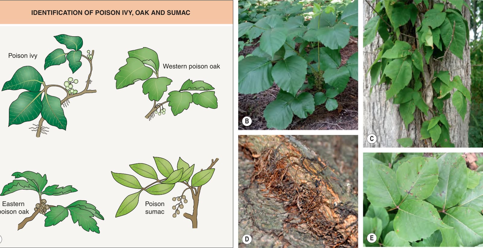
Fig. 17.7 Characteristic features useful for identifying poison ivy, poison oak and poison sumac.A Shapes and numbers of leaflets and appearance of fruit (drupes). B Shrub type poison ivy (T. radicans) with flowers arising from an axillary position just below point where leaf attaches to stem. Leaves possess three leaflets (“Leaves of three, let them be” or “One, two, three, don’t touch me”). C, D Climbing vine of poison ivy (T. radicans) on an oak tree demonstrating aerial roots that anchor the vine to the tree; close-up of vine anchored to tree bark by fine hair-like aerial roots (“Hairy rope, don’t be a dope”). E Shrub type poison ivy (T. radicans) with black spots on leaflets. Leaf trauma (e. g. wind, rain, trampling by animals or people) allows urushiol to reach the surface, oxidize, and turn black.
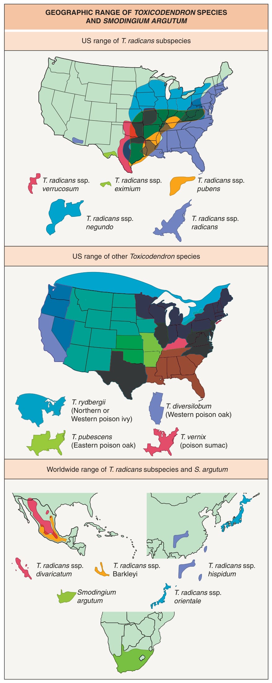
Fig. 17.8 Geographic range of Toxicodendron spp.andSmodingium argutum (African poison ivy).
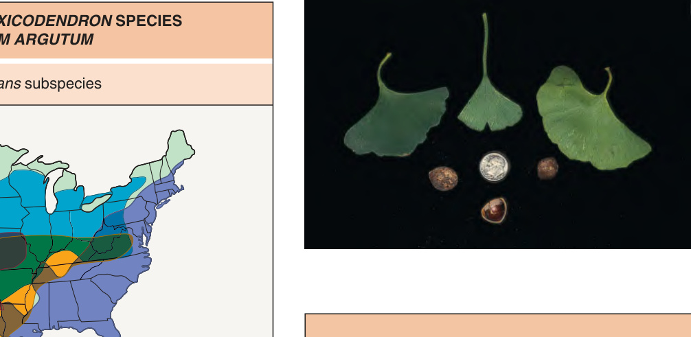
Fig. 17.9 Leaves and seeds of Ginkgo biloba.
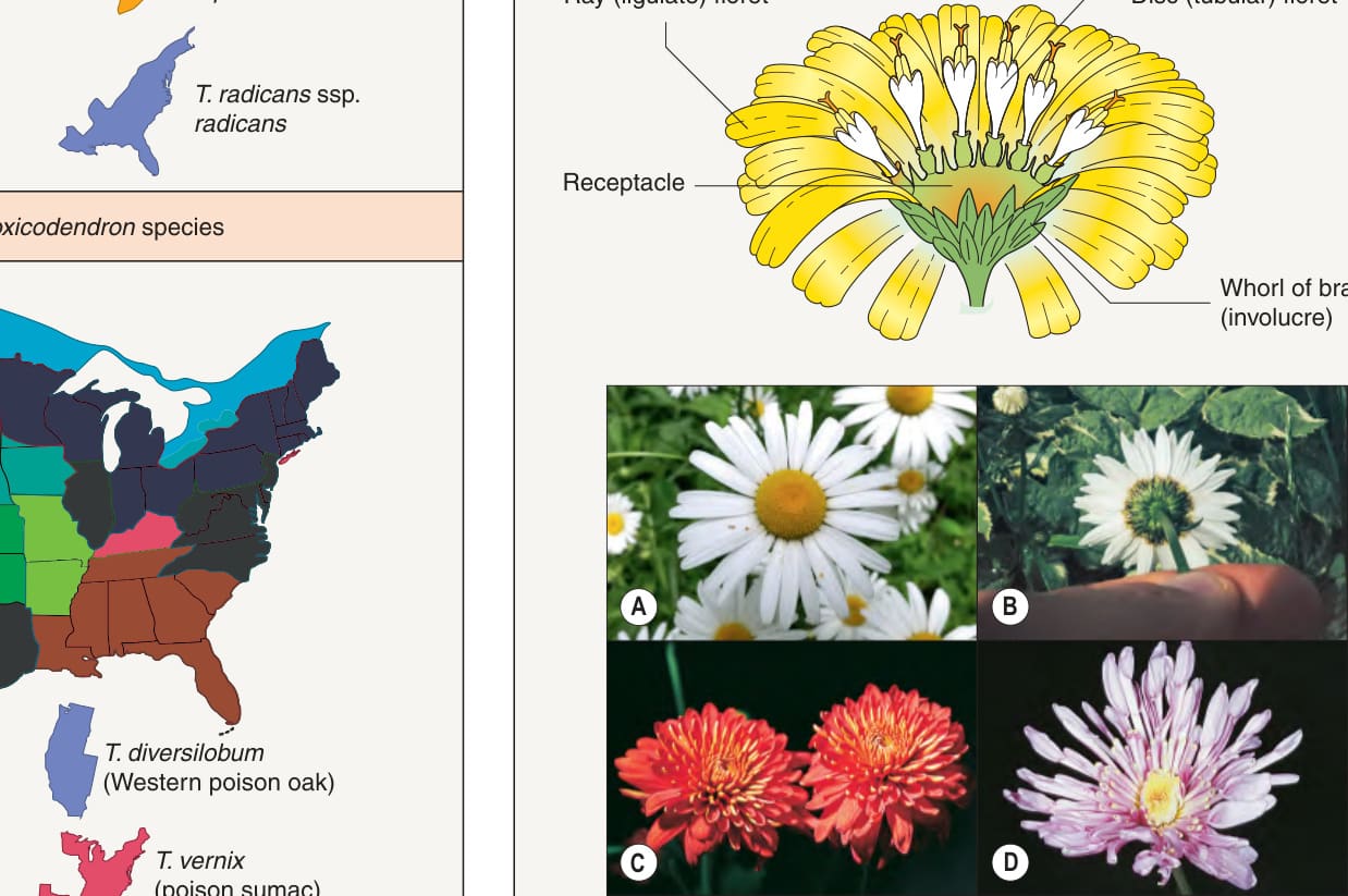
Fig. 17.10 Typical composite flower heads of members of the family
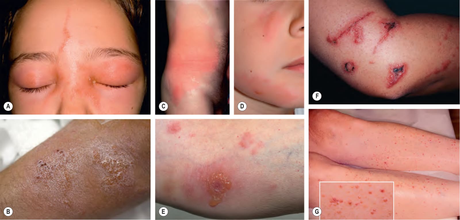
Fig. 17.11 Clinical manifestations of Anacardiaceae dermatitis.A Poison ivy (Toxicodendron radicans) dermatitis: acute streaks of erythema with superimposed vesicles and eyelid edema with serous crusting. B Poison ivy dermatitis: multiple vesicles and a few hemorrhagic crusts with superimposed zinc oxide from application of calamine lotion. C Widespread erythema and edema associated with intense pruritus after carrying logs of the poisonwood tree (Metopium toxiferum) of the family Anacardiaceae. D Pink edematous streaks on the face. E Compared to B, more obvious erythema in association with edema and multiloculated vesicles. F “Black-spot” poison ivy dermatitis: note the black discoloration in the central portion of the edematous plaques due to polymerized plant oleoresin. G Weed whacker dermatitis with widespread spotted pattern. A, G, Courtesy Louis A. Fragola, Jr, MD; B, Courtesy Julia Pettersen Neckman, MD; D, Courtesy Julie V. Schaffer, MD; E, F, Courtesy Kalman Watsky, MD.
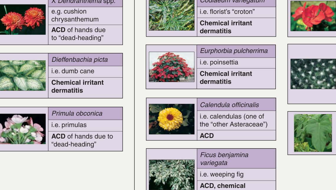
Fig. 17.12 House and garden plants that commonly cause dermatoses. ACD, allergic contact dermatitis. Photograph of poinsettia courtesy Denver Botanical Gardens.
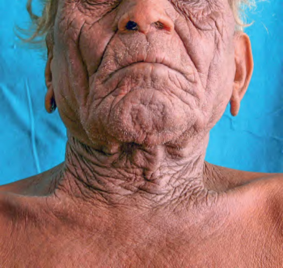
Fig. 17.13 Airborne contact dermatitis. This farmer from Gujarat, India was allergic to the sesquiterpene lactones in Parthenium hysterophorus. Courtesy Shyam Verma, MBBS, DVD, FRCP.
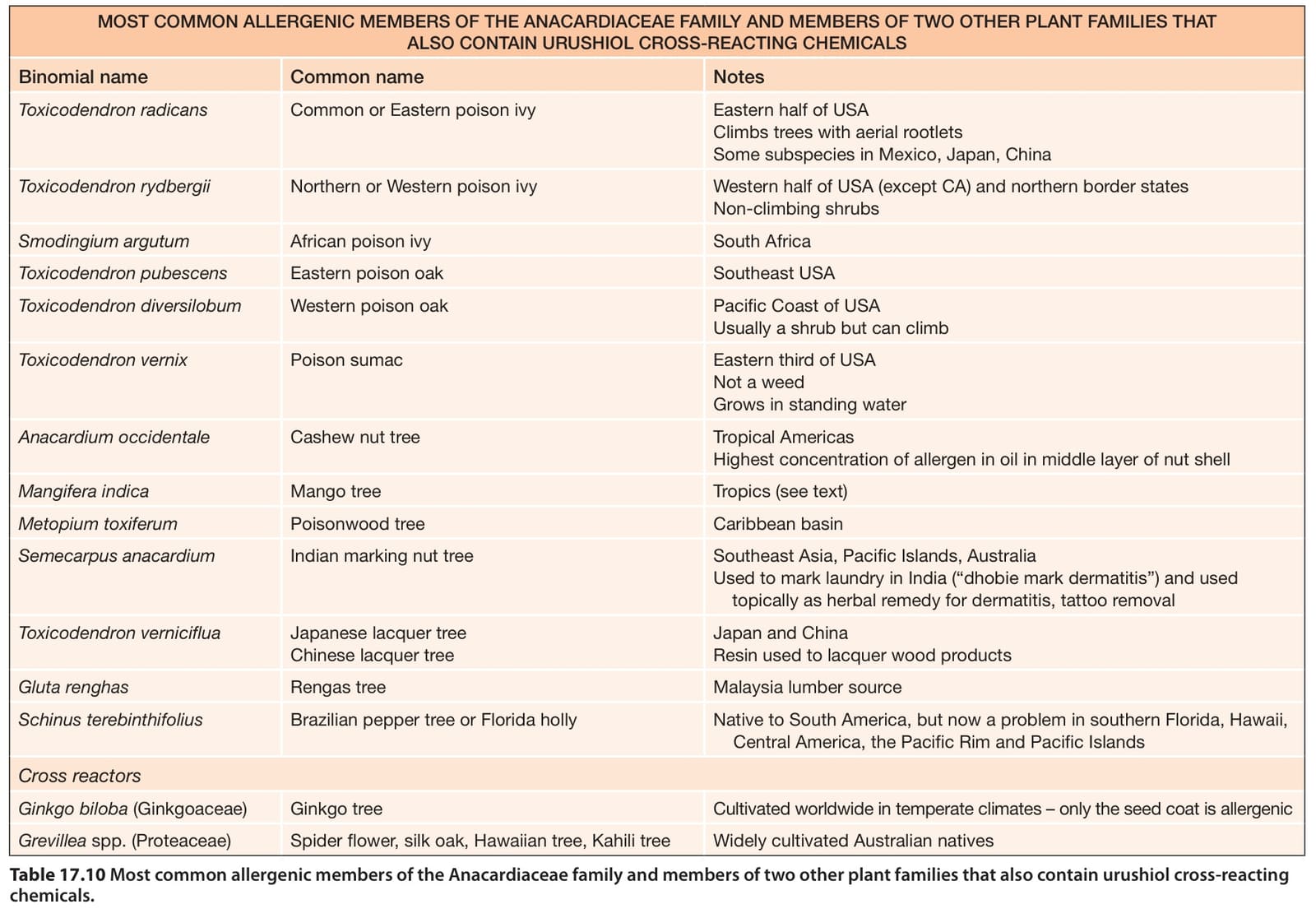
Table 17.10 Most common allergenic members of the Anacardiaceae family and members of two other plant families that also contain urushiol cross-reacting chemicals.
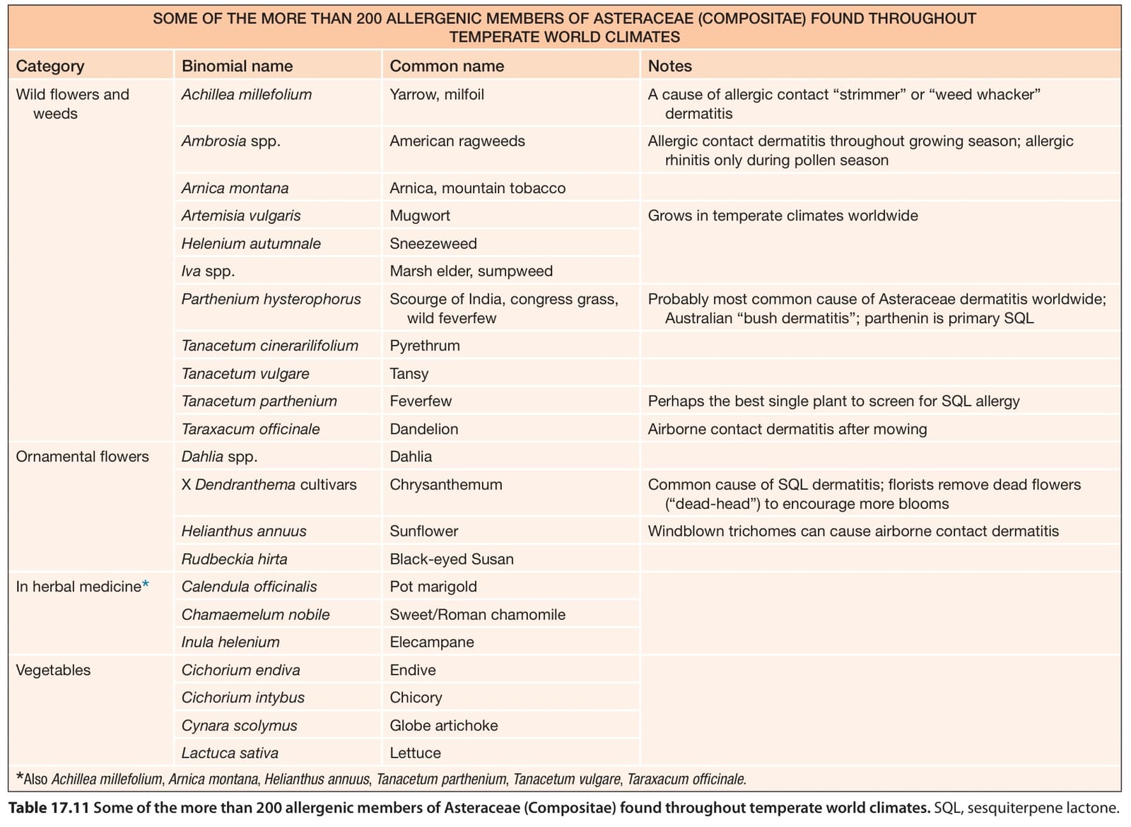
Table 17.11 Some of the more than 200 allergenic members of Asteraceae (Compositae) found throughout temperate world climates. SQL, sesquiterpene lactone.
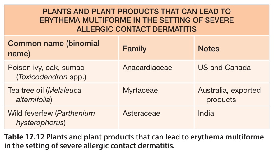
Table 17.12 Plants and plant products that can lead to erythema multiforme in the setting of severe allergic contact dermatitis.
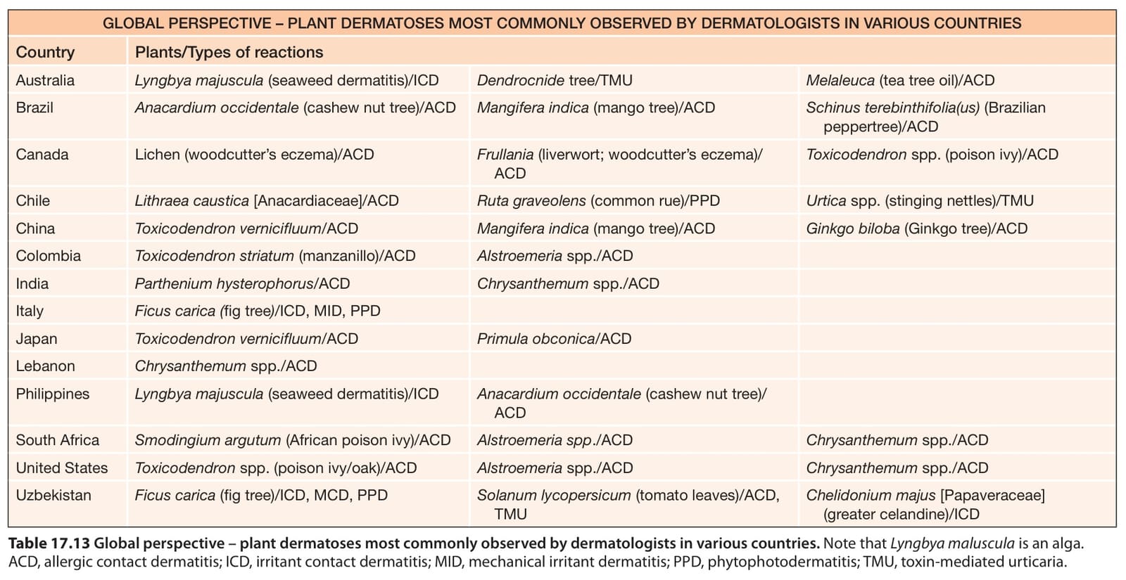
Table 17.13 Global perspective – plant dermatoses most commonly observed by dermatologists in various countries. Note that Lyngbya maluscula is an alga. ACD, allergic contact dermatitis; ICD, irritant contact dermatitis; MID, mechanical irritant dermatitis; PPD, phytophotodermatitis; TMU, toxin-mediated urticaria.