PATHOGENESIS
The Mast Cell
Distribution and diversity
The mast cell is the primary effector cell of urticaria. Mast cells are widely distributed throughout the body but vary in their phenotype and response to stimulation. This may explain why systemic features, such as those seen in anaphylaxis, do not accompany the activation of cutaneous mast cells in urticaria. The majority of mast cells in the skin and intestinal submucosa contain the neutral proteases tryptase and chymase (MCTC), whereas those in the bowel mucosa, alveolar wall, and nasal mucosa contain only tryptase (MCT). Both types, however, express high-affinity IgE receptors (FcεRI) and are therefore capable of participating in IgE-dependent allergic reactions. There is conflicting evidence on the number of cutaneous mast cells in chronic urticaria, but there is agreement that they may be more likely to degranulate in response to certain stimuli, such as intradermal codeine injection, and in this sense may be in general more “releasable”. Little is known about the beneficial effects of mast cells, but there is evidence that they are involved in the innate immune response to infection, wound healing, and the neuroendocrine system. They have also been shown to help initiate the extracellular matrix formation and angiogenesis required for neurofibroma development.
The swelling is deeper than wheals and may affect mucosal surfaces. Note the swelling of the lips and periorbital region and the lack of erythema.
Degranulating stimuli
Cross-linking of two or more adjacent FcεRI on the mast cell membrane will initiate a chain of calcium- and energy-dependent steps leading to fusion of storage granules with the cell membrane and externalization of their contents. This is known as degranulation. Classic immediate hypersensitivity reactions involve binding of receptor-bound specific IgE by allergen. There are also several recognized immunologic degranulating stimuli that act through the IgE receptor, such as IgG anti-IgE and anti-FcεRI antibodies (Fig. 18.3).
Autoantibodies against FcεRI (IgG > IgM, IgA) are associated with disease markers of CSU, but the degree to which they mediate release of histamine from mast cells or basophils in vitro can vary. In addition, because anti-IgE and anti-FcεRI autoantibodies have been identified by immunoassays (but not functional assays) in patients with other diseases such as systemic lupus erythematosus and occasionally in healthy controls, the role of autoantibodies in the pathogenesis of urticaria has been debated. Stimuli that can lead to mast cell degranulation independent of FcεRI activation include C5a, stem cell factor, and agents that bind MRGPRX2 (MAS-related G-protein coupled receptor X2; see Fig. 18.3); the latter include substance P and drugs such as opioids.
Proinflammatory mediators
Mast cell granules contain preformed mediators of inflammation, the most important of which is histamine (Fig. 18.4). A wide range of cytokines has been identified in human mast cells from different tissues, including tumor necrosis factor (TNF), interleukins (IL)-3,
-5, -6, -8, and -13, and granulocyte–macrophage colony-stimulating factor (GM-CSF). Synthesis and secretion are upregulated following FcεRI stimulation. TNF is expressed constitutively in resting human cutaneous mast cells. Prostaglandins and leukotrienes are synthesized from arachidonic acid derived from cell membrane phospholipids. The most important proinflammatory eicosanoids are prostaglandin (PG) D and the leukotrienes (LT) C, D, and E (slow releasing substance of anaphylaxis). PGE has inhibitory effects on immunologic mast cell degranulation and may therefore have a protective role in urticaria. Increased levels of TNF, IL-1β, -6, -10, -12p70, -13, and B cell activating factor (BAFF) have been detected in the sera of urticaria patients.
Blood Vessels
Histamine and other proinflammatory mediators released upon degranulation bind receptors on postcapillary venules in the skin, leading to vasodilation and increased permeability to large plasma proteins, including albumin and immunoglobulins. Furthermore, histamine, TNF, and IL-8 upregulate the expression of adhesion molecules on endothelial cells, thereby promoting the migration of circulating inflammatory cells including eosinophils, basophils, neutrophils, and Th0 cells from the blood into the urticarial lesion.
Blood
Autoantibodies
Based upon in vitro assays, functional IgG autoantibodies that release histamine (and other mediators) from mast cells and basophils have been detected in the serum of 30%–50% of patients with CSU. The majority of these autoantibodies bind the extracellular α subunit of FcεRI. Those recognizing the α domain compete with IgE for the binding site, whereas non-competitive autoantibodies directed against the terminal α domain are able to bind the receptor in the presence of IgE (Fig. 18.5). Increased serum levels of IgM anti-FcεRIα autoantibodies have also been found in patients with CSU and have been linked to features of autoimmune CSU. Approximately 10% of chronic urticaria sera contain functional autoantibodies directed against the Fc portion of IgE itself (see Fig. 18.3). Binding of the autoantibodies to mast cells may initiate complement activation with the generation of C5a anaphylatoxin, which in turn facilitates or augments degranulation.
Circulating IgE autoantibodies directed against a range of autoantigens such as thyroid peroxidase (TPO), thyroglobulin, tissue factor, and IL-24 have been detected in patients with CSU. However, healthy individuals and patients with disorders such as atopic dermatitis also have these IgE autoantibodies. In patients with CSU and IgE
anti-TPO antibodies, skin testing with recombinant TPO can elicit a wheal and flare reaction, indicating potential functionality, but it is still unclear as to why such patients develop only local wheals rather than systemic anaphylaxis.
Other mast cell activating factors may also exist in urticaria sera, e.g. a non-IgG “mast cell-specific factor” has been described, although its identity remains unknown. There is currently no evidence that characterized cytokines cause mast cell degranulation in urticaria. Evidence from small series of CSU patients treated with plasmapheresis or cyclosporine indicates that functional autoantibody levels correspond to disease severity.
Leukocytes
The importance of peripheral blood leukocytes in the pathogenesis of urticaria is becoming clearer. In general, blood basophils from CSU patients are less responsive in vitro to anti-IgE antibodies as an immunologic stimulus, possibly through desensitization, and these cells are reduced in number. CSU patients can be further classified into responders and non-responders based upon the release of histamine by their basophils in response to anti-IgE antibodies or the flow cytometry-based basophil activation test (BAT). This basophil functional phenotype appears to remain stable during the course of the active illness, but then, during disease remission, the basophils become more responsive to anti-IgE antibodies. Expression of the negative regulator SHIP (src homology 2-containing inositol phosphatase) is increased in the basophils from anti-IgE non-responders, although the significance of this to the pathogenesis of chronic urticaria is unknown.
Evidence has emerged that basophils are recruited into urticaria wheals and may sustain the inflammatory response by releasing histamine and other mediators, analogous to the delayed phase of immediate hypersensitivity reactions. A correlation between disease severity and the degree of reduction in circulating basophil counts has been observed. This basopenia can normalize following successful treatment with omalizumab, providing support for the role of basophil migration into lesional skin during periods of disease activity.
In addition to basopenia, circulating eosinopenia may be present in those CSU patients who have greater disease activity and a lack of response to H1 antihistamines. Eosinophils can contribute to the persistence of wheals by generating LTC, LTD, and LTE and by releasing toxic granule proteins, including major basic protein (MBP); the latter can induce release of histamine from basophils and trigger mast cells via MRGPRX2. Interactions between eosinophils and mast cells also include expression of tissue factor by eosinophils which then leads to activation of mast cells via protease activated receptors (PARs).
While lymphocyte numbers are normal in the peripheral blood of CSU patients, these cells are often present in biopsy specimens from spontaneous wheals. In one study, ~50% of the cells in the perivascular cellular infiltrates were T cells and ~20% were monocytes. Additional investigations in CSU patients noted the dermal infiltration of CD4+ helper T cells, the majority of which co-expressed HLA-DR antigen; T cell activation in the peripheral blood as well as lesional and nonlesional skin; and increased expression of Th2-initiating cytokines in lesional skin. Lastly, autoreactive CD4+ T cells specific for FcεRIα were detected in the peripheral blood of patients with CSU.
Nerves
Substance P and other neuropeptides release histamine from mast cells in vitro and can induce a wheal and flare reaction in human skin when injected intradermally. Vasoactive intestinal polypeptide (VIP) caused a greater wheal reaction in those with chronic urticaria than did other skintested neuropeptides, but the relevance of this to urticaria is still uncertain. Elevated levels of brain-derived neurotrophic factor, β-endorphin, and substance P have been detected in patients with chronic urticaria. Because several of these neuropeptides can bind MRGPRX2 on mast cells, the roles of neuro-immuno-cutaneous factors and neurotransmission in chronic urticaria require further clarification.
Mechanisms of Urticaria Formation
Mast cell-dependent urticaria
Potential mechanisms for mast cell-dependent urticaria are included in Table 18.1. Cross-linking of the Fab portion of specific IgE on mast
cells by percutaneous or circulating allergen undoubtedly accounts for some cases of acute or episodic urticaria, especially in children (see Fig. 18.3), but this is probably never the cause of adult chronic continuous urticaria. Examples of the former would be contact urticaria from natural rubber latex and acute urticaria from foods, including nuts, fish, and fruit. However, the majority of acute urticaria cases do not relate to allergen exposure.
IgE has been implicated in the pathogenesis of symptomatic dermographism, cold urticaria, and solar urticaria, but the mechanism by which it renders skin mast cells more sensitive to physical stimulation is not certain. It is proposed that the physical stimulus in these patients induces a neoantigen that reacts with specific IgE antibody bound to mast cells. An additional mechanism, such as neuropeptide release, could initiate or potentiate mast cell activation (see above). Using electron microscopy, localized platelet clumping has been demonstrated in cold urticaria, and the release of platelet mediators, including plateletactivating factor (PAF) and platelet factor 4/CXCL4, could contribute to wheal formation. A mutation in F12, which encodes the coagulation protein factor XII, has been identified in familial cold urticaria and a bradykinin-dependent, rather than mast cell-dependent, pathomechanism has been proposed (see below). Cold-induced urticarial lesions also occur in familial cold autoinflammatory syndromes (FCASs) due to mutations in NLRP3, NLRP12, or PLCG2, thus implicating a role for the cryopyrin inflammasome and its production of IL-1β (see Tables 45.2 & 45.7).
Cholinergic urticaria develops in response to stimulation of cholinergic sympathetic innervation of the sweat glands. How release of acetylcholine from the nerve endings leads to mast cell activation and histamine release is unknown. An allergy to sweat has been demonstrated by one group of investigators. It has been proposed that pressure-induced wheals may be due to a late-phase reaction, but an antigen has never been identified. In three Lebanese families with autosomal dominant vibratory urticaria, a missense gain-of-function variant was detected in ADGRE2 which encodes adhesion G protein-coupled receptor E2. This was thought to lead to destabilization of the inhibitory interaction between the alpha and beta subunits of ADGRE2, leading to sensitization of mast cells to vibration-induced degranulation.
The initiating event for spontaneous urticaria wheals is unclear but may involve plasma leakage due to local factors such as heat or pressure, which allows the extravasation of autoantibodies or IgE-directed antigens that then activate the IgE receptor, thus leading to mast cell degranulation and a subsequent urticarial response. As functional autoantibodies cannot be detected in ~70% of chronic urticaria sera by currently available tests, other mechanisms may operate in “non-autoantibody” urticaria, which, nevertheless, has a similar clinical presentation. Increased plasma levels of prothrombin fragment 1 + 2 (F 1 + 2) and D-dimer (a measure of fibrinolysis) have been demonstrated in CSU and relate to disease severity, but the contribution of coagulation abnormalities to the pathogenesis remains unclear. There are other serum factors in CSU patients that can activate mast cell lines in vitro and lead to endothelial activation, independent of IgE receptors on mast cells and the presence of IgG.
A popular hypothesis is that dietary food additives and natural salicylates as well as nonsteroidal anti-inflammatory drugs (NSAIDs) may cause urticaria via the diversion of arachidonic acid metabolism from prostaglandin to leukotriene formation. How this leads to urticaria is not clear, but it is known that intradermal injections of LTC, LTD, and LTE cause whealing by a direct action on small blood vessels. There is some evidence from studies of rat peritoneal mast cells that PGE can have inhibitory effects on immunologic mast cell degranulation, so a reduction in their formation may facilitate the latter. Aspirin can aggravate urticaria in up to 30% of patients with chronic disease, and while some clinical studies of dietary pseudoallergen avoidance have given encouraging results, the proportion of complete responders confirmed by rechallenge is small. Aspirin allergy as a cause of urticaria is much less common, and the proportion of patients with urticaria due solely to pseudoallergens is probably low. Tolerance to NSAIDs is thought to be achievable in many CSU patients who are well controlled by H1 antihistamines.
Understanding “idiopathic” urticaria remains an important challenge. From a clinical perspective, it should be regarded as a multifactorial problem, and searching for aggravating factors is just as important as looking for causes.
Mast cell-independent urticaria
There are several recognized circumstances where angioedema or wheals are due to mechanisms that do not involve mast cells. These need special consideration because their management and prognosis are different. For example, prostaglandins are involved in the pathogenesis of some patterns of non-immunologic contact urticaria (e.g. to benzoic acid), and the latter can be suppressed by NSAIDs. In the cryopyrinassociated periodic syndromes (CAPS; see below), patients often develop urticarial lesions. Systemic symptoms, such as fever, help to distinguish patients with autoinflammatory syndromes from those with CSU (see Ch. 45). The significant improvement that results from the administration of anakinra, an IL-1 receptor antagonist, rilonacept, a fusion protein that contains the extracellular domain of the IL-1 receptor and functions as an IL-1 trap, or canakinumab, a human anti-IL-1β monoclonal antibody, points to the role of the cryopyrin inflammasome and its production of IL-1β (see Figs. 4.2 & 45.13).
C1 inh deficiency is usually hereditary but may be acquired. Two major forms of hereditary angioedema (HAE), types I and II, are primarily caused by mutations in one allele of the structural gene for C1 inh (C1NH/SERPING1) and are inherited in an autosomal dominant pattern. These mutations result in reduced levels of C1 inh (85% of cases; type I) or reduced C1 inh function (15% of cases; type II). Because these heterozygous mutations lead to levels that are 5%–30% of normal (rather than the expected 50%) in patients with type I HAE, it is thought that there is trans inhibition of the normal allele or increased catabolism of C1 inh. Rare families with autosomal recessive severe HAE and homozygous mutations have been reported.
Deficiency of C1 inh leads to loss of inhibition of factor XII (FXII; Hageman factor), resulting in the generation of bradykinin by the action of kallikrein on high-molecular-weight kininogen (Fig. 18.6). Activation of the C1 component of complement by proteolytic enzymes, including plasmin and FXIIa, leads to low levels of C4 in the serum, which is an almost constant feature between and during attacks in untreated patients.
HAE with normal C1 inh activity, also known as nC1-INH-HAE and previously referred to as type III HAE, is currently subclassified as HAE3 to HAE8 based upon underlying genetic mutations. HAE3 is due to gain-of-function mutations in one allele of the gene that encodes FXII, leading to increased formation of bradykinin (see Fig. 18.6). A positive feedback loop between kallikrein and FXII also contributes to the pathogenesis. One possible explanation for the preponderance of women with HAE3 is enhanced transcription of F12 by estrogens. Mutations in PLG, ANGPT1, KNG1, MYOF, and HS3St6 which encode plasminogen, angiopoietin-1, kininogen 1, myoferlin, and heparan sulfate 3-O-sulfotransferase 6 have also been described in HAE with normal C1 inh activity.
Acquired deficiency of C1 inh may result from activation of C1q and the complement cascade in patients with B cell lymphoproliferative disorders and plasma cell dyscrasias, or in those with autoimmune connective tissue diseases. This leads to consumption of C1 inh plus low serum levels of C1q as well as C4 (acquired, type I) or the formation of inhibitory autoantibodies directed against the C1 inh (acquired, type II).
Angiotensin-converting enzyme (ACE) inhibitor-induced urticaria is believed to result from the inhibition of endogenous kininase II (also known as ACE), which leads to increased production of bradykinin via inhibition of its metabolism (see Fig. 18.6). It usually presents with angioedema, which is usually orofacial and may be life-threatening.
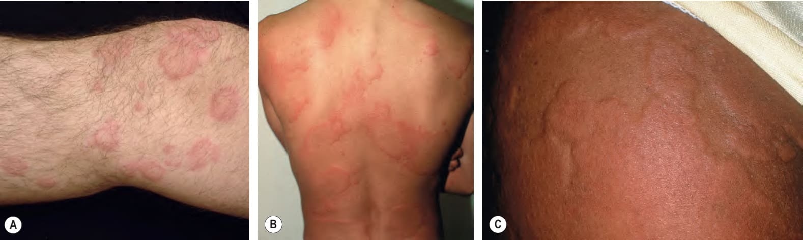
Fig. 18.1 Wheals.A, B Wheals can vary in size from small to large as well as be annular, but they still retain the classic central pallor and erythematous flare. C Occasionally, more uniform edematous plaques are seen.
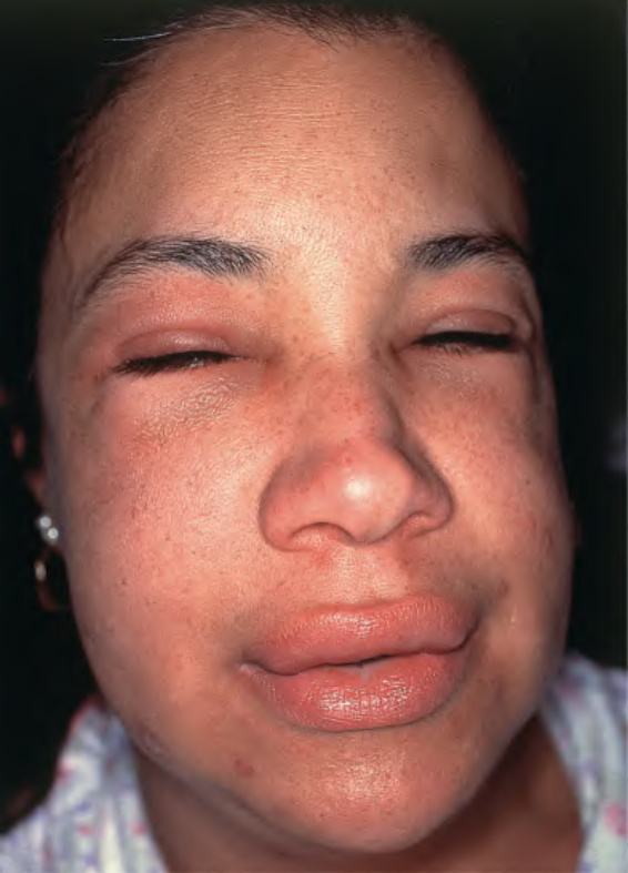
Fig. 18.2 Angioedema.
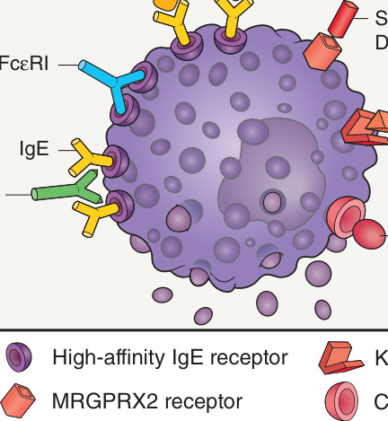
Fig. 18.3 Mast cell degranulating stimuli. Both immunologic and non-immunologic stimuli can lead to release of mediators. Substance P also binds to neurokinin 1 receptor; stem cell factor is also known as KIT ligand. MRGPRX2, MAS-related G-protein coupled receptor X2.
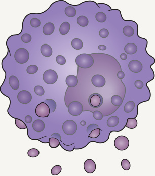
Fig. 18.4 Mediators released by human dermal mast cell degranulation. Both preformed and newly synthesized proinflammatory mediators are released from mast cells.
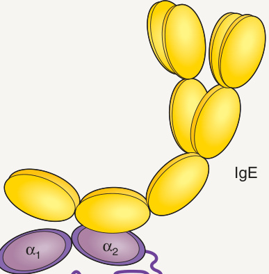
Fig. 18.5 IgE antibody binding to the extracellular α subunit of the high- affinity IgE receptor (FcεRI). Histamine-releasing autoantibodies directed against the terminal α domain are able to bind the receptor in the presence of IgE and are therefore non-competitive, whereas those recognizing the α domain compete with IgE for the binding site.
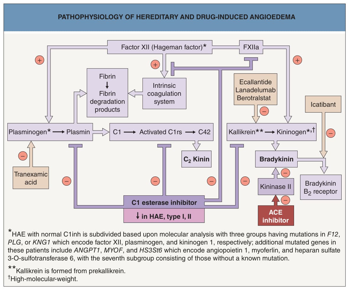
Fig. 18.6 Pathophysiology of hereditary and drug- induced angioedema. Angiotensin-converting enzyme (ACE) inhibitor-induced urticaria is believed to result from the inhibition of endogenous kininase and a subsequent increase in bradykinin. Intravenous C1 inh concentrate (derived from human plasma) or recombinant C1 inh (derived from milk of transgenic rabbits), subcutaneous icatibant (a decapeptide that is a specific bradykinin B receptor antagonist), and subcutaneous ecallantide (a 60-amino acid recombinant kallikrein inhibitor) are options for the emergency treatment of hereditary angioedema (HAE). Subcutaneous or intravenous C1 inh (every 3–4 days), subcutaneous plasma kallikrein inhibitor lanadelumab (every 2 weeks), and oral kallikrein inhibitor berotralstat (150 mg daily) are options for prevention of attacks. ACE, angiotensin-converting enzyme; HAE, hereditary angioedema; inh, inhibitor.
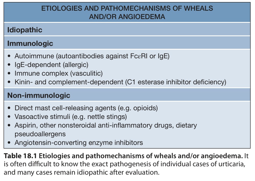
Table 18.1 Etiologies and pathomechanisms of wheals and/or angioedema. It is often difficult to know the exact pathogenesis of individual cases of urticaria, and many cases remain idiopathic after evaluation.