CLINICAL FEATURES
Clinical Diversity
It is important to distinguish urticaria from urticarial dermatoses, such as urticarial drug eruptions, eosinophilic cellulitis, and the urticarial phase of pemphigoid. The individual wheals of urticaria are “here today and gone tomorrow” (i.e. they generally last <24 hours), whereas with urticarial dermatoses, the individual lesions may last for days or longer. Although urticarial vasculitis is often included in classifications of urticaria because the wheals resemble urticaria, it is actually an urticarial dermatosis. Clinically, the lesions last longer than 24 hours (as determined by circling and observing individual wheals), and histologically there is evidence of leukocytoclastic vasculitis (see Ch. 24).
Wheals may be small or large, as well as single or multiple. In the physical urticarias, the distribution pattern and morphology can be helpful in separating the different clinical types (see Inducible Urticarias). Angioedema can merge with wheals and these two can be difficult to separate, especially around the eyelids. Angioedema may also be a feature of anaphylaxis, if the throat is involved. In this sense, wheals, angioedema, and anaphylaxis form parts of a clinical spectrum resulting from mast cell activation.
Classification
Because the etiology is often unknown at the time of the first consultation, the most practical classification of urticaria relies on clinical characteristics rather than etiology when defining groups of patients (Table 18.2). Mostly, spontaneous urticaria can be considered to behave in an ordinary manner (i.e. fluctuating wheals which may be accompanied by angioedema) whatever its etiology or duration, unless it has a predominantly physical trigger, has evidence of vasculitis, or is
caused by exogenous contact. If no cause for spontaneous urticaria can be defined, it is called idiopathic, but the use of this label as an automatic prefix to chronic urticaria is no longer tenable as a significant number of these patients are considered to have an autoimmune etiology as evidenced by functional basophil histamine release and activation assays (Table 18.3) or flow cytometry-based BAT. Elevated thyroid antibodies are also suggestive of autoimmune urticaria. It is worth separating angioedema without wheals from the other patterns of urticaria, because some of these cases will be due to HAE, acquired C1 inh deficiency, or drug reactions (e.g. ACE inhibitors, NSAIDs), and these are managed differently.
Acute Versus Chronic Urticaria
All urticarias are acute initially. Some will become chronic after a period of time that is usually defined as 6 weeks or more. In a study from Korea, only a small proportion (7.8%) of patients with new-onset urticaria developed chronic urticaria during a 10-year follow-up period. The term “chronic urticaria” should only be applied to continuous urticaria
occurring at least twice a week off treatment. Urticaria occurring less frequently than this over a long period is better called episodic (or recurrent), because this presentation may be more likely to have an identifiable environmental cause.
The causes of acute urticaria in adults presenting to emergency dermatology clinics in one survey are shown in Fig. 18.7. Most of these patients probably fall into the spontaneous category, because physical urticarias and urticarial vasculitis tend to persist beyond 6 weeks and contact urticarias would not normally present as an emergency. The high frequency of associated viral infections is notable, as is the lack of food allergy as a cause. The different clinical patterns of chronic urticaria and their causes (where known) are shown in Fig. 18.8. These data reflect the experience of specialist urticaria clinics in dermatology departments and may not represent the experience of other medical practitioners, such as those in general practice, internal medicine, pediatrics, or allergy.
Spontaneous Urticaria
The condition can present at any age. Acute urticaria is common in young atopic children but chronic urticaria peaks in the fourth decade. Multiple pruritic wheals of different sizes erupt anywhere on the body
and then fade within 2–24 hours without bruising; angioedema may last up to 72 hours when severe. In literature reviews, isolated urticaria occurred in ~50% of patients, urticaria plus angioedema in ~40%, and angioedema alone in the remainder. Wheals may occur at any time, but often appear in the evening or are present on waking. Irritation tends to be most intense at night and may disturb or prevent sleep. This, in turn, compounds the distress of the condition. Substantial impairment in quality-of-life measures, including self-image, sexual relationships, and social interactions, has been demonstrated. Women may describe premenstrual exacerbations. Systemic symptoms of fatigue, lassitude, sweats and chills, indigestion, myalgia, or arthralgia may occur with severe attacks, but the occurrence of pyrexia or arthritis should alert the clinician to another explanation, such as urticarial vasculitis or neutrophilic urticarial dermatosis due to Schnitzler syndrome, Still disease, or CAPS.
Associations and comorbidities
Chronic spontaneous urticaria has been associated with autoimmune thyroid disease and other autoimmune conditions, including vitiligo, insulin-dependent diabetes, rheumatoid arthritis, and pernicious anemia. Those chronic urticaria patients with demonstrable histamine-releasing autoantibodies have a very strong association with HLA DRB104 (DR4) and its associated allele DQB10302 (DQ8). A possible association between Helicobacter pylori gastritis and chronic urticaria was suggested by a systematic review of therapeutic studies, which showed a higher frequency of urticaria remission when the infection was eradicated than when it was not. Parasitic infections, in particular protozoa (e.g. Blastocystis hominis, Giardia spp.), are an uncommon cause of urticaria in high-income countries, but may be a significant problem where they are endemic. Acute urticaria due to gastric Anisakis simplex has been reported from Spain. Possible associations between dental infections or gastrointestinal candidiasis and chronic urticaria have not been substantiated by large epidemiologic studies.
Although there have been anecdotal reports linking urticaria to malignancy, no statistically significant association was found in a Swedish survey. However, an increased risk of hematologic malignancies, especially non-Hodgkin lymphoma, was noted in a large retrospective cohort study of a National Insurance database in Taiwan. Atopic diseases, metabolic syndrome, and psychiatric disorders (especially anxiety and depression) are reported comorbidities in chronic urticaria.
Inducible Urticarias (syn. Physical Urticarias)
These urticarias represent a distinct subgroup of urticaria that is induced by an exogenous stimulus rather than occurring spontaneously. This section discusses physical triggers while the next section reviews other chemical and protein stimuli. Because cholinergic urticaria is triggered by factors that lead to sweating (e.g. a rise in core body temperature, stress, spicy foods), rather than an external physical stimulus, it is often classified separately from other inducible urticarias. The inducible urticarias are classified by the predominant stimulus that triggers whealing, angioedema, or anaphylaxis (Table 18.4). Of all the urticarias, they may affect the quality of life most severely, particularly delayed pressure urticaria and cholinergic urticaria. While the lesions of most physical urticarias occur within minutes of provocation and generally resolve within 2 hours, a few physical urticarias (e.g. delayed pressure urticaria, delayed dermographism) develop after a delay of several hours and persist for 24 hours or longer.
The wheals are usually localized to the stimulated area of skin. However, sometimes the physical stimuli need to produce a systemic effect, e.g. a rise or drop in core body temperature, to induce urticaria of a reflex type. Thus, generalized heating of the body can induce cholinergic urticaria (which is common), and generalized cooling, cold
reflex urticaria (which is rare). Here, multiple small wheals occur on widespread areas of the body.
Angioedema may be seen with all the physical urticarias except with symptomatic dermographism. Vibratory angioedema manifests with subcutaneous swellings but not wheals. Also, several forms of physical urticaria can coexist in the same patient. Common combinations include: (1) symptomatic dermographism and cholinergic urticaria; and (2) cold and cholinergic urticaria. In addition, delayed pressure urticaria may coexist with CSU. Some rare forms of physical urticaria, such as familial delayed cold urticaria, are beyond the scope of this chapter.
Urticaria due to mechanical stimuli
Immediate dermographism This is divided into simple or symptomatic. Simple immediate dermographism occurs in at least 5% of the normal population in response to moderate stroking of the skin and may be regarded as an exaggerated physiologic response and is typically not pruritic.
Symptomatic immediate dermographism is the most common type of physical urticaria (Fig. 18.9). It manifests as pruritic linear wheals at sites of scratching or sites of friction, such as collars and cuffs of clothes. Whealing occurs after gentle stroking of the skin in response to a shearing force. Patients, most frequently young adults, often complain of pruritus before the wheals appear and may not associate them with scratching. It is worst in the evening and often occurs in bouts. The lesions usually resolve within an hour. Oral mucosal involvement does not occur, but vulvodynia and swelling during sexual intercourse has been reported.
The general course is unpredictable, but usually there is a gradual tendency towards improvement over several years. Symptomatic dermographism occasionally follows scabies infestation and penicillin allergy reactions. There is no association with systemic disease, atopy, food allergy, or autoimmunity.
Rarer forms of dermographism These include delayed dermographism, which appears at least 30 minutes after a stroking stimulus, red dermographism (in response to rubbing, not stroking, the skin), localized dermographism, cholinergic dermographism, and Darier’s sign (a localized dermographic response after gentle rubbing of cutaneous mastocytosis lesions). The blanching response seen in patients with atopic and other forms of dermatitis, known as white dermographism, is not a form of urticaria nor is black dermographism in which there is dark discoloration of the skin due to surface deposition of metallic particles.
Delayed pressure urticaria (DPU) is important because it can interfere severely with quality of life; it may be underdiagnosed and its treatment is difficult. DPU is characterized by the development of deep
erythematous swellings at sites of sustained pressure to the skin, after a delay of 30 minutes to 12 hours (Fig. 18.10); sometimes it resembles angioedema. The swellings are usually pruritic, painful, or both, and may persist for several days, unlike the spontaneous wheals of ordinary urticaria. Sites frequently involved are the waistline after wearing tight clothing, below the elastic of socks, the feet in tight shoes, the palms after manual work, the soles after walking or climbing ladders, and the genitalia after intercourse.
Rarely, systemic features such as malaise, “flu-like” symptoms, and arthralgia may occur. If the swellings develop over joints, the resulting restriction of motion may be mistaken for arthritis. The prognosis is variable, but the mean duration in different series was 6–9 years. Up to 35% of patients with CSU attending a hospital specialist clinic had associated DPU. Conversely, nearly all patients with DPU have associated spontaneous wheals. Because of this association and the delayed onset, patients may not be aware of any relationship to pressure unless specifically questioned.
This is a very rare form of urticaria, where a vibratory stimulus induces localized swelling and erythema within minutes, lasting 30 minutes. Stimuli include jogging, vigorous rubbing with a towel, and the use of vibrating machinery such as lawnmowers and motorcycles. Avoidance of vibratory stimuli enables patients to live a normal life. Vibratory angioedema may be acquired or familial. The acquired form is often milder, and it may be associated with other physical urticarias such as DPU and symptomatic immediate dermographism. The familial form is dominantly inherited, has its onset during infancy, and intense vibratory stimuli may induce generalized erythema and systemic symptoms such as headaches. Pathogenic variants in ADGRE2, which encodes adhesion G protein-coupled receptor E2, have been detected (see Pathogenesis).
Urticaria due to temperature changes
Heat contact urticaria This is one of the rarest forms of urticaria. Within minutes of contact with heat from any source, itching and whealing occur at the site of contact, lasting up to 1 hour. This must be distinguished from cholinergic urticaria. Patients can present after contact with hot water (e.g. washing dishes), but also after contact with radiant heat or warm sunlight. Systemic symptoms of faintness, headache, nausea, and abdominal pain have occurred when the urticaria is extensive. There is an even rarer delayed form.
Cold urticaria represents a heterogeneous group of conditions in which whealing occurs within minutes of rewarming after cold exposure. There are both typical and atypical forms, with the latter group constituting ~30% of cases and being characterized by negative provocation tests. Typical forms are primary cold contact urticaria and secondary
cold contact urticaria, while atypical forms are systemic atypical acquired cold urticaria, cold-dependent dermographism, cold-induced cholinergic urticaria, delayed cold urticaria, and localized cold reflex urticaria.
Primary cold contact urticaria The most commonly encountered form is primary cold contact urticaria; it accounts for at least 95% of cases (Fig. 18.11). Although cold urticaria may follow respiratory infections, arthropod bites or stings, and perhaps HIV infection, the cause of most cases remains unknown (idiopathic). It may occur at any age, but is most frequently seen in young adults. Itching, burning, and whealing occur in cold-exposed areas minutes after rewarming the skin. Commonly, primary cold contact urticaria occurs in rainy, windy weather and after contact with cold objects or drinking cold liquids. Patients often relate symptoms to changes in temperature as much as to the absolute external temperature. Systemic symptoms of flushing, headache, syncope, and abdominal pain can develop if large areas are affected. Cold baths and swimming should usually be avoided as there is a potential risk of anaphylaxis. The mean duration was 6–9 years in one series, though primary cold contact urticaria may be more transient if it follows a viral infection.
Secondary cold contact urticaria Cases due to cryoproteinemia are rare, and they are associated with other manifestations such as Raynaud phenomenon or purpura. The wheals are usually similar to those of primary cold contact urticaria, but may last longer. The presence of circulating cryoglobulins should be determined and if detected, underlying causes, such as hepatitis B or C viral infections, lymphoproliferative disease, or infectious mononucleosis, should be excluded.
Reflex cold urticaria In this form, generalized cooling of the body induces widespread whealing. Patients with this disorder can experience life-threatening reactions from exposures such as diving into a cold lake. The ice cube test is negative, but placing the patient in a cold room at 4°C can reproduce lesions; however, one should exert caution because anaphylaxis could occur.
Familial cold urticaria Familial cold urticaria, also referred to as familial cold autoinflammatory syndrome (FCAS), belongs to the group of disorders known as cryopyrin-associated periodic syndromes (CAPS) (see Table 45.2). The patients with FCAS1 have mutations in NLRP3, which encodes the inflammasome protein cryopyrin, and they develop cold-induced urticaria wheals. FCAS2 and FCAS3 are due to mutations in NLRP12 and PLCG2, respectively; the latter encodes a phospholipase (see Table 45.7). Lastly, gain-of-function mutations in F12 have been detected in families with either HAE3 or cold-induced urticarial lesions and systemic symptoms.
Urticaria due to sweating or stress
Cholinergic urticaria is characterized by multiple, transient, papular wheals that are 2–3 mm in diameter and surrounded by an obvious flare. They occur within 15 minutes of sweat-inducing stimuli, such as any form of physical exertion, hot baths, or sudden emotional stress (Fig. 18.12). Other eliciting stimuli include moving from a cold to a hot room, drinking alcohol, and eating spicy food. It occurs more frequently in young adults with an atopic diathesis and is unusual in the elderly. It may be associated with acquired generalized hypohidrosis (see Table 39.11).
After the appropriate stimulus, pruritus is followed by the development of small, monomorphic wheals that are symmetrically distributed. They are most prominent on the upper half of the body, but can also affect the lower legs and forearms and even become generalized. Angioedema and systemic manifestations consisting of faintness, headache, palpitations, abdominal pain, and wheezing may occur. Reduced forced expiratory volumes (in the absence of respiratory symptoms) have been recorded by spirometry. In more severely affected individuals, cholinergic urticaria can cause severe personal and occupational disability.
Cold urticaria, symptomatic dermographism, or aquagenic urticaria may be associated with cholinergic urticaria. There is usually a gradual tendency towards improvement, but the condition may last for years. Rarer forms include cholinergic pruritus, cholinergic erythema (in which pruritic, symmetric, small erythematous macules appear to be persistent, but individual lesions actually last for up to 1 hour), and cholinergic dermographism. Severe exercise-induced cholinergic urticaria may sometimes progress to anaphylaxis.
This can occur without the typical wheals of cholinergic urticaria and appears to be a syndrome in its own right. Food- and exercise-induced anaphylaxis (FEIA) is an increasingly recognized syndrome. In FEIA, angioedema and/or anaphylaxis occur within minutes of exercise if it follows either prior ingestion of a specific food (e.g. α-gliadin in wheat), or sometimes within 4 hours of a heavy meal. FEIA may be due to priming of the mast cell by either prior exposure to an allergen or an unknown mechanism. EIA is produced by exercise, but not by an increase in core temperature induced by other means capable of provoking cholinergic urticaria (e.g. passive overheating in a hot bath), and this distinction can be used to differentiate between the two conditions.
Adrenergic urticaria can be distinguished from cholinergic urticaria by the presence of blanched vasoconstricted skin surrounding small pink wheals induced by sudden stress, as opposed to pale wheals surrounded by a pink halo. The lesions can be reproduced by intradermal injections of norepinephrine (noradrenaline).
Urticaria due to other exposures
Itching and whealing occur within minutes of exposure to ultraviolet or visible wavelengths of solar radiation specific to the patient (see Ch. 87).
In aquagenic urticaria, contact with water of any temperature induces an urticarial eruption resembling a sparse form of cholinergic urticaria, with large flares around papular wheals. Lesions occur most frequently on the upper part of the body and last for less than an hour. Other physical urticarias must be excluded and the condition must be differentiated from aquagenic pruritus (see Ch. 6).
Contact Urticaria (see Ch. 16)
Contact urticaria is defined by the development of urticaria at the site(s) of contact with skin or mucosa but contact erythema and even burning and stinging without erythema are sometimes embraced in the definition. Percutaneous or mucosal penetration of the urticant may have distant effects, including acute urticaria or even anaphylaxis. Contact urticaria may be more common than reported, as patients often do not present to the hospital or tertiary care clinics, because the diagnosis is usually self-evident.
Immunologic and non-immunologic forms are recognized, depending on whether the contact urticaria is due to allergen interaction with specific IgE or is IgE-independent. Immunologic (i.e. allergic) contact urticaria can be seen in children with atopic dermatitis who become sensitized to environmental allergens, such as grass, animals and foods, or in glove-wearers who are allergic to latex. In those individuals repeatedly exposed to latex via mucosal surfaces (e.g. urinary catheterization in spina bifida patients), allergic contact urticaria can be complicated by anaphylaxis. Non-immunologic (i.e. non-allergic) contact urticaria is due to direct effects of urticants on blood vessels, such as from exposure to either sorbic acid and benzoic acid in eye solutions and foods or cinnamic aldehyde in cosmetics; it may be mediated by PGD and can be inhibited by NSAIDs. Percutaneous microinjection of histamine, acetylcholine, and/or serotonin by nettle stings as well as contact with histamine liberators that degranulate mast cells (e.g. dimethylsulfoxide, cobalt chloride) can result in contact urticaria.
Food contact hypersensitivity syndrome
The oral allergy syndrome, also referred to as pollen–food allergy syndrome, presents with itching and mild swelling of the mouth, tongue, and soft palate within minutes of eating fresh fruits such as apples, pears, peaches, and cherries, but not cooked fruit. It usually occurs in hay fever sufferers and represents a cross-reaction between fruit proteins and panallergens in grass (profilin) or birch pollen allergens, in particular Bet v 1, a member of the PR-10 family. Angioedema and anaphylaxis are fortunately very rare. The term “food contact hypersensitivity syndrome” was proposed to include all forms of immediate mucosal food contact reactions, including oral allergy syndrome.
Angioedema Without Wheals
Recurrent angioedema without wheals is in most instances idiopathic, but it is important to exclude a drug reaction and to consider the possibility of C1 inhibitor (inh) deficiency. The latter can be inherited or acquired (e.g. B cell lymphoproliferative disorders, plasma cell dyscrasias, autoantibodies against C1 inh).
Drug reactions
A number of drugs can cause angioedema without wheals. The most common are NSAIDs and ACE inhibitors (see Fig. 18.6). Aspirin intolerance may present with angioedema alone, or with urticaria or anaphylaxis; cross-reactions with other NSAIDs can occur. A causative link to ACE inhibitors may be overlooked because angioedema can present over a year after starting the medication and occasionally even years later. An increased risk of ACE inhibitor-induced angioedema has been observed when the latter is coadministered with dipeptidyl peptidase-4 inhibitors (“gliptins”) and mTOR inhibitors. ACE inhibitors are contraindicated in patients with HAE and acquired C1 inh deficiency, and for practical purposes, ACE inhibitors should be discontinued in favor of other antihypertensive medications if angioedema occurs without wheals.
Angiotensin receptor blockers (ARBs) such as valsartan and losartan are also associated with development of angioedema. Depending upon the study, the risk of ARB-induced angioedema in patients with ACE inhibitor-induced angioedema varies from 0–15%. As a result, ARBs are avoided in such patients. As with other drug eruptions, it is important to consider exposure via combination drugs, in this case sacubitril–valsartan, which is increasingly being used to treat congestive heart failure. Of note, an increased incidence has been reported in Black patients.
Hereditary angioedema (HAE), types I and II (inherited C1
The likelihood of inherited C1 inh deficiency as a cause of recurrent angioedema is increased if there is a family history or previous laryngeal edema or colicky abdominal pain (sometimes presenting as a surgical emergency). Trauma (emotional or physical) and estrogens can trigger attacks. The usual duration is 48–72 hours, and episodes are often followed by a refractory period. Some families with HAE describe a transient, prodromal erythematous eruption, but urticaria whealing does not occur and so a history of recurrent wheals effectively excludes the disorder.
Hereditary angioedema (HAE) with normal C1 inh activity (nC1-INH-HAE; previously type III)
This form of HAE currently has seven subgroups, six of which are associated with mutations in the genes that encode factor XII, plasminogen, angiopoietin-1, kininogen 1, myoferlin, and heparan sulfate 3-O-sulfotransferase 6 (see above). The seventh group includes those whose genetic basis is unknown. Compared to patients with an inherited deficiency of C1 inh, the age of onset is later (usually in or after the second decade) and there is a higher frequency of facial angioedema.
Distinctive Syndromes with Urticarial Lesions
Hereditary periodic fever syndromes
The allelic disorders Muckle–Wells syndrome, familial cold autoinflammatory syndrome (familial cold urticaria), and neonatal-onset multi-system inflammatory disease (NOMID/CINCA) are all now known to be due to mutations in NLRP3, which encodes the protein cryopyrin (see Table 45.2). These patients can develop wheals from an early age that are resistant to antihistamines.
In hyperimmunoglobulinemia D with periodic fever syndrome (HIDS) due to partial mevalonate kinase deficiency as well as TNF receptorassociated periodic syndrome (TRAPS), erythematous and edematous urticarial plaques can occur in addition to periorbital edema (in TRAPS). The cutaneous lesions in familial Mediterranean fever are more erysipelas-like in appearance (see Table 45.2).
Acquired autoinflammatory syndromes
Schnitzler and colleagues in 1974 described an unusual variant of chronic urticaria characterized by recurrent, non-pruritic wheals plus intermittent fevers, bone pain, arthralgia or arthritis, an increased ESR, and an IgM monoclonal gammopathy. Occasionally, there is an IgG paraprotein. The Strasbourg validated diagnostic criteria for Schnitzler syndrome consist of two major criteria – urticarial rash and IgM or IgG monoclonal gammopathy – and four minor criteria – recurrent fever >38°C without any other cause, changes in bone structure visible in imaging ± bone remodelling, neutrophilic dermal infiltrate, and leukocytosis and/or elevated CRP. Confirming the diagnosis requires both major and at least two minor criteria (if IgM) or at least three minor criteria (if IgG). If the two major criteria are accompanied by only one minor criterion (if IgM) or two minor criteria (if IgG), the diagnosis is considered probable. Some of the patients with Schnitzler syndrome will have an MYD88 L265P variant.
The clinical presentation overlaps with that of the CAPS group of inherited autoinflammatory disorders. While skin biopsy specimens usually show a neutrophil-predominant dermal infiltrate without edema, fully developed small vessel vasculitis is not seen. Some clinicians believe that these patients are best considered as having a variant of neutrophilic urticaria while others prefer the term neutrophilic urticarial dermatosis (see Table 45.6). Schnitzler syndrome responds very well to IL-1 blockade, e.g. anakinra, canakinumab, rilonacept.
This is a rare acquired autoinflammatory disorder in which patients present with fever, a sore throat, an often evanescent urticarial eruption, arthritis, lymphadenopathy, and hyperferritinemia (see Ch. 45).
Episodic angioedema with eosinophilia (Gleich syndrome)
A syndrome of episodic angioedema with hypereosinophilia, weight gain, and fever has been described. The cause is uncertain, but increased serum levels of IL-5 have been noted.
Systemic capillary leak syndrome
Also known as Clarkson syndrome, this rare acquired disorder is characterized by episodic massive plasma exudation from blood vessels, leading to potentially life-threatening hypotension, analogous to anaphylaxis. Angioedema may be a feature. It is associated with an IgG paraproteinemia. Medications, in particular IL-2, can also produce a systemic capillary leak syndrome.
Urticarial vasculitis (see Ch. 24)
Urticarial vasculitis is a clinicopathologic entity in which cutaneous lesions resemble urticaria clinically, but last longer than 24 hours and may resolve with postinflammatory hyperpigmentation or purpura (Fig. 18.13). Histologically, there is evidence of leukocytoclastic vasculitis. Extracutaneous manifestations, especially in the setting of hypocomplementemia, may include arthralgias, abdominal pain, obstructive pulmonary disease, glomerulonephritis, and uveitis or episcleritis.
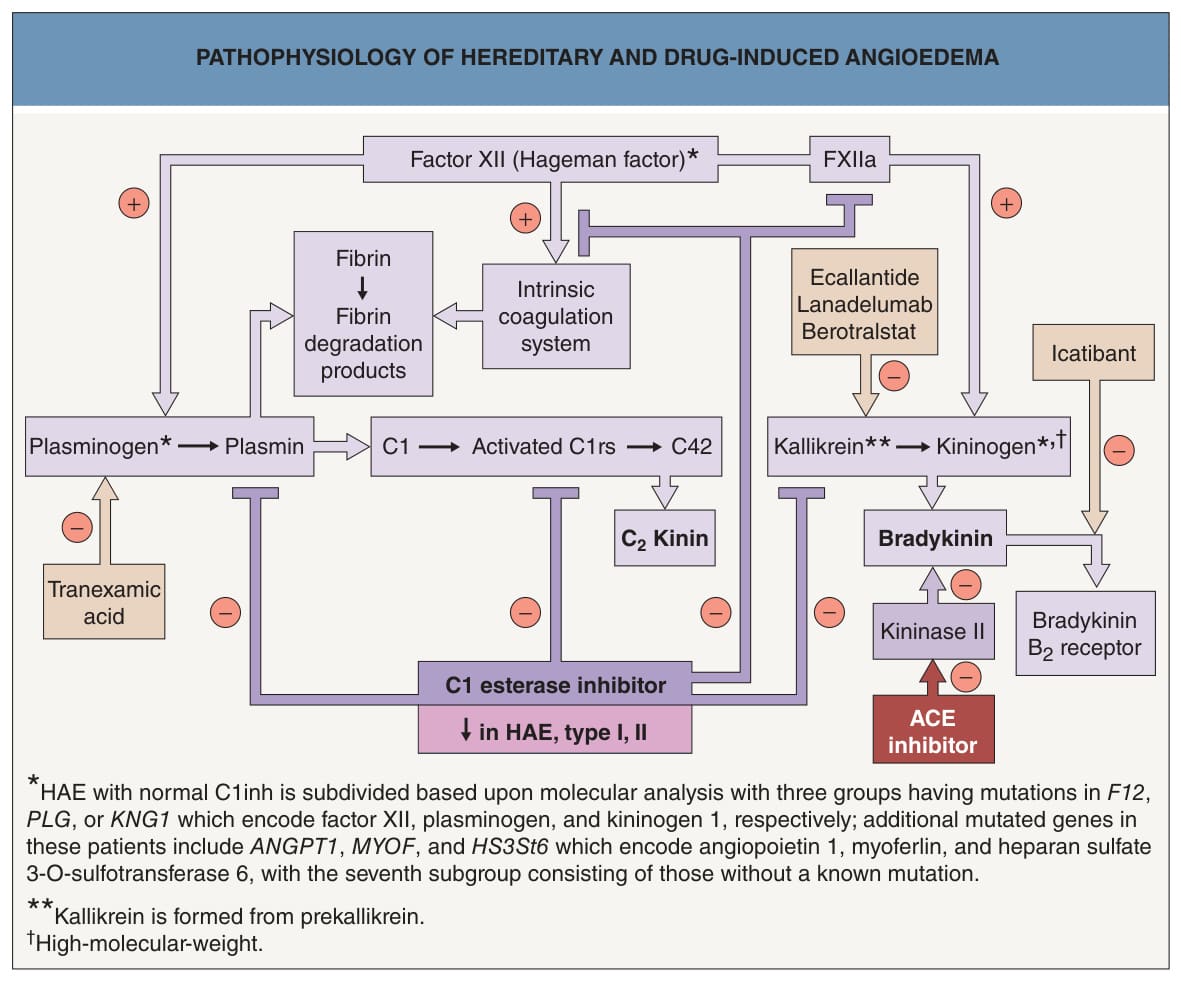
Fig. 18.6 Pathophysiology of hereditary and drug- induced angioedema. Angiotensin-converting enzyme (ACE) inhibitor-induced urticaria is believed to result from the inhibition of endogenous kininase and a subsequent increase in bradykinin. Intravenous C1 inh concentrate (derived from human plasma) or recombinant C1 inh (derived from milk of transgenic rabbits), subcutaneous icatibant (a decapeptide that is a specific bradykinin B receptor antagonist), and subcutaneous ecallantide (a 60-amino acid recombinant kallikrein inhibitor) are options for the emergency treatment of hereditary angioedema (HAE). Subcutaneous or intravenous C1 inh (every 3–4 days), subcutaneous plasma kallikrein inhibitor lanadelumab (every 2 weeks), and oral kallikrein inhibitor berotralstat (150 mg daily) are options for prevention of attacks. ACE, angiotensin-converting enzyme; HAE, hereditary angioedema; inh, inhibitor.
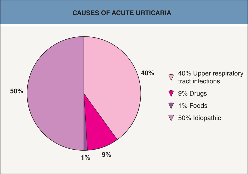
Fig. 18.7 Causes of acute urticaria.Data from Zuberbier T, Iffländer J, Semmler C, Henz BM. Acute urticaria: clinical aspects and therapeutic responsiveness. Acta Derm Venereol 1996;76:295–7.
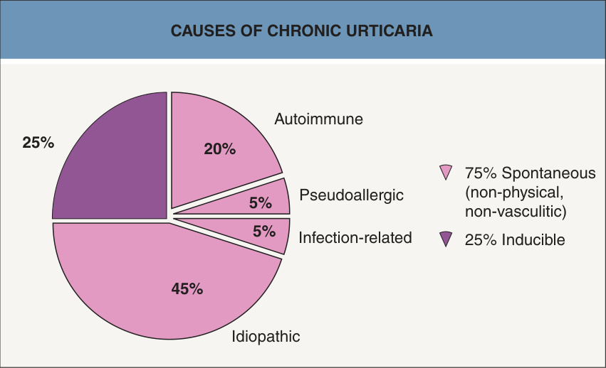
Fig. 18.8 Causes of chronic urticaria. Autoimmune represents those patients with functional autoantibodies against FcεRI or the Fc portion of IgE.
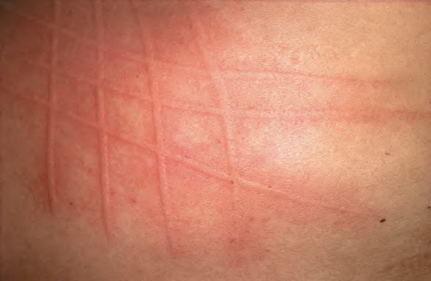
Fig. 18.9 Symptomatic immediate dermographism. Itchy wheals appear very soon after scratching. Courtesy Jean L. Bolognia, MD.
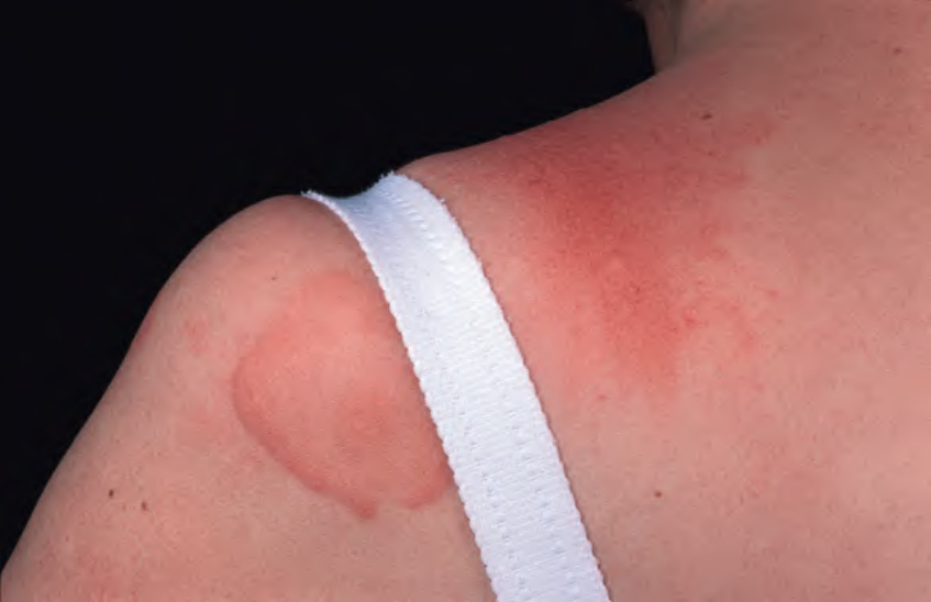
Fig. 18.10 Delayed pressure urticaria.
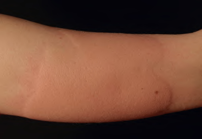
Fig. 18.11 Cold urticaria. Wheals developed on the forearm after placement of an ice cube for 10 minutes, followed by rewarming. Courtesy Thomas Schwarz, MD.
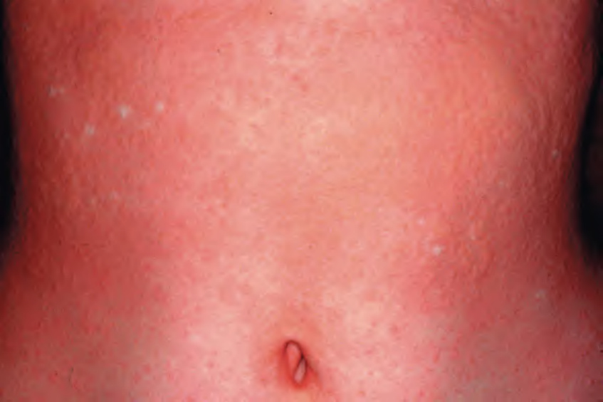
Fig. 18.12 Cholinergic urticaria on the trunk after hot bath provocation.
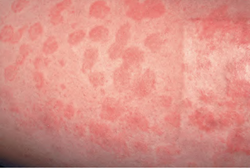
Fig. 18.13 Urticarial vasculitis. Lesions look like spontaneous urticaria but last longer and may bruise. An incidental finding is a skin graft donor site.
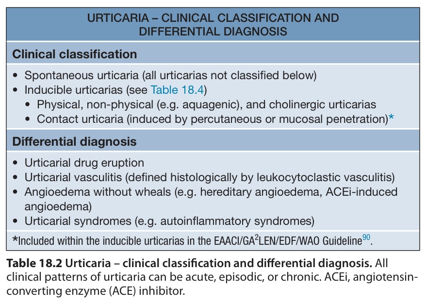
Table 18.2 Urticaria – clinical classification and differential diagnosis. All clinical patterns of urticaria can be acute, episodic, or chronic. ACEi, angiotensinconverting enzyme (ACE) inhibitor.
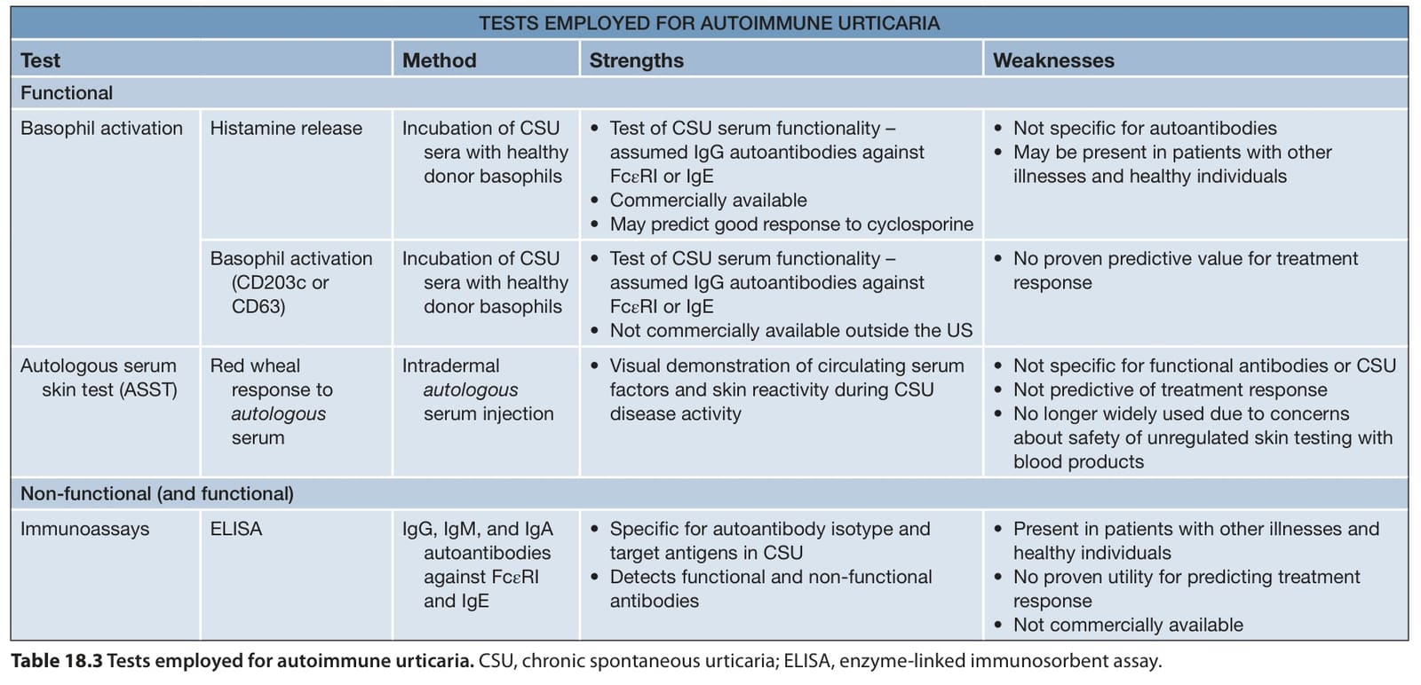
Table 18.3 Tests employed for autoimmune urticaria. CSU, chronic spontaneous urticaria; ELISA, enzyme-linked immunosorbent assay.
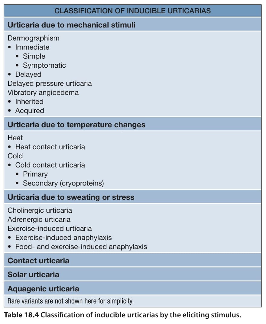
Table 18.4 Classification of inducible urticarias by the eliciting stimulus.