PATHOLOGY
The histopathology of urticaria is characterized by upper dermal edema with a mild mixed perivascular infiltrate of lymphocytes, eosinophils, basophils, and some neutrophils (Fig. 18.14). A predominantly neutrophilic pattern has been described in a minority of patients (see below). Previous reviews of biopsy specimens from large series of patients with chronic urticaria have emphasized a spectrum of changes, ranging from mild lymphocytic infiltrates with rare neutrophils to occasionally showing fully developed leukocytoclastic vasculitis, pointing to some histopathologic overlap between chronic urticaria and urticarial vasculitis. Features of the latter include endothelial cell damage, a perivenular cellular infiltrate rich in neutrophils, swelling of vessel walls, and leukocytoclasis. Extravasation of erythrocytes and fibrinoid deposits in and around blood vessels may also be seen.
The term neutrophilic urticaria is sometimes used when biopsy specimens of urticaria contain predominantly neutrophilic dermal infiltrates but lack features of vasculitis. Although this pattern did not appear to have clinical or etiologic significance, neutrophilic urticaria overlaps with neutrophilic urticarial dermatosis. This term is used to describe an aseptic, interstitial, perivascular, and peri-appendageal neutrophilic infiltrate with nuclear dust and neutrophils within dilated dermal blood vessels, but without vasculitis. The latter occurs
in the setting of inherited acquired and inherited autoinflammatory syndromes (e.g. CAPS, Schnitzler syndrome, Still disease; see Table 45.6) as well as occasionally in systemic lupus erythematosus.
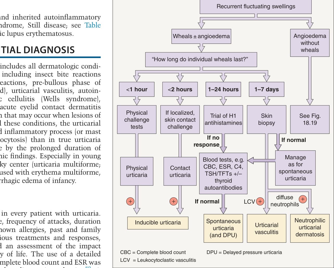
Fig. 18.14 Spontaneous urticaria – histologic findings. A sparse, mainly perivascular inflammatory infiltrate and scant edema within the superficial and mid dermis. Note several neutrophils and a few eosinophils (inset). Courtesy Lorenzo Cerroni, MD.