DIAGNOSIS AND DIFFERENTIAL DIAGNOSIS
The differential diagnosis of urticaria includes all dermatologic conditions with an urticarial component, including insect bite reactions (papular urticaria), urticarial drug reactions, pre-bullous phase of pemphigoid (i.e. urticarial pemphigoid), urticarial vasculitis, autoinflammatory syndromes, eosinophilic cellulitis (Wells syndrome), polymorphic eruption of pregnancy, acute eyelid contact dermatitis (versus angioedema), and the urtication that may occur when lesions of urticaria pigmentosa are rubbed. In all these conditions, the urticarial component is part of a more prolonged inflammatory process (or mast cell proliferation in the case of mastocytosis) than in true urticaria and should be readily distinguishable by the prolonged duration of individual lesions or associated systemic findings. Especially in young children, urticaria may develop a dusky center (urticaria multiforme; Fig. 18.15; see Table 20.3) and be confused with erythema multiforme, serum sickness-like reaction, or hemorrhagic edema of infancy.
Diagnosis
A comprehensive history is essential in every patient with urticaria. It should cover the duration of disease, frequency of attacks, duration of individual lesions, medications, known allergies, past and family history including comorbidities, previous treatments and responses, occupation and leisure activities, and an assessment of the impact of the disease on the patient’s quality of life. The use of a detailed questionnaire in conjunction with a complete blood count and ESR was found to be almost as good as a complete diagnostic evaluation. A full physical examination looking for the morphology and duration (by circling individual lesions) of wheals, bruising, livedo reticularis, and signs of systemic disease should always be undertaken, but it is often normal. The patient’s own photographs can often be useful because it is common for urticaria to have cleared by the time of consultation. Investigation should be targeted at specific features identified in the clinical assessment, as outlined below, and may include blood tests, skin biopsy (obtained in all patients with lesions consistently lasting
more than 24 hours), challenges for physical urticarias, food additives, and drugs, and, infrequently as an extended investigation, skin tests for allergy and measurement of histamine-releasing autoantibodies. Most patients with antihistamine-responsive urticaria do not need an extensive investigation. A diagnostic algorithm is given in Fig. 18.16.
Acute urticaria
No routine laboratory tests are recommended for acute urticaria but when a type 1 allergic reaction to environmental allergens is suspected as the cause of acute urticaria or contact urticaria, skin prick testing and measurement of specific IgE antibodies (e.g. ImmunoCAP®) can be performed.
Chronic urticaria
No investigations are routinely required for the majority of patients with mild disease that responds to antihistamines. A useful screening profile for non-responders with more severe disease could include a complete blood count with differential (for instance, to detect the eosinophilia of intestinal parasitosis) and ESR (usually normal in CSU). The ESR may be elevated in urticarial vasculitis, and it is consistently high in symptomatic acquired and genetic autoinflammatory syndromes in which patients also have an elevated C-reactive protein level (usually >30 mg/l). Measurement of thyroid autoantibodies and thyroid function tests should be performed if indicated clinically. The prevalence of thyroid autoantibodies is higher in patients with chronic urticaria than in controls and may be an indicator of autoimmune urticaria. Tests for helminths may be considered in the appropriate clinical setting. Although evidence is equivocal that Helicobacter pylori infection can be associated with CSU, patients with hyperacidity should be tested since eradication therapies may lead to urticaria remission.
Although the term “autoimmune” urticaria is increasingly being used for patients with evidence of functional serum autoantibodies (see Fig. 18.3 & Table 18.3), there is still a lack of agreement with regard to definitively establishing such a diagnosis. Demonstration of autoreactivity in the form of a localized wheal and flare response to intradermal injection of autologous serum (autologous serum skin test [ASST]) provides useful evidence of functional factors in the blood but is not specifically a test for functional autoantibodies (Fig. 18.17). A negative test, however, has very good negative predictive value for the absence of functional autoantibodies. However, stricter regulatory guidelines regarding the handling of blood products will increasingly limit performance of this assay.
Immunoassays for anti-FcεRI and anti-IgE antibodies do not necessarily correlate with basophil and mast cell histamine-releasing assays because non-functional as well as functional autoantibodies have been detected by Western blot and ELISA (see Table 18.3). Fig. 18.18 illustrates the overlap between: (1) autoreactivity by ASST; (2) functional autoantibodies as detected via basophil or mast cell activation assays; and (3) immunoassays (“immunoreactivity”). The most convincing evidence for an autoimmune etiology of urticaria is provided by demonstrating all three features, but some clinicians would accept a positive functional assay as providing sufficient proof of autoimmunity to guide therapies and prognosis. Because CSU patients can have increased rates of IgE sensitization to autoantigens such as thyroid peroxidase and interleukin-24 and some have a very rapid response to anti-IgE antibody (e.g. omalizumab), the concept of autoallergic urticaria has been proposed.
Physical urticarias
International standards for the diagnosis of physical urticarias and definitions of challenge testing have been proposed. Symptomatic dermographism can be tested for by stroking the skin of the back lightly with the rounded edge of a wooden spatula or a plastic device with round pegs that have different lengths (FricTest®). A calibrated instrument called a dermographometer, which is a spring-loaded stylus, the pressure of which can be adjusted to a predetermined setting, can be used to confirm the diagnosis of symptomatic dermographism (if ≤36 g/mm). Delayed pressure urticaria is confirmed if applying either a 2.5-kg weight to the thigh or back for 15 minutes (using a rod with a diameter of 1.5 cm) or a dermographometer set at 100 g/mm for 70 seconds results in a palpable wheal at 30 minutes to 8 hours (usually at 6 hours).
Provocation tests for cholinergic urticaria include exercise to the point of sweating in an overheated environment, preferably on a treadmill or exercise bicycle, or partial immersion in a hot bath at 42°C for
At 30 minutes, a red wheal response is present at the site of an intradermal injection of autologous serum, but not at that of a normal saline control. For the test to be positive, the wheal response at the serum injection site must be at least 1.5 mm greater in diameter than the control site. This criterion is a reasonably sensitive and specific screening test for functional autoantibodies as detected by the basophil release assay. Spontaneous wheals are also present at the venipuncture site and above the serum skin test.
10 minutes. A positive hot bath test excludes exercise-induced anaphylaxis. Cold contact urticaria is confirmed either by the development of whealing at the site of application of a melting ice cube in a thin polyethylene bag or glove for 5 minutes (the most sensitive individuals may react after 30 seconds, with the least sensitive requiring up to 20 minutes of ice application; see Fig. 18.11) or by assessment of the critical temperature threshold by an instrument such as TempTest®.
Phototesting confirms the diagnosis of solar urticaria, with the development of wheals within minutes of exposure to natural or artificial radiation. The action spectra of the inducing wavelengths can be determined by narrowband monochromators (see Ch. 87). The diagnosis of aquagenic urticaria is made by inducing a limited number of small pruritic wheals with a large flare after a bath or shower at body temperature, or under wet gauze applied at body temperature for 20 minutes. Heat contact urticaria is confirmed by applying a heated cylinder (temperature increments up to 44°C) to the skin for 30 seconds to 5 minutes or assessment of the critical temperature threshold by an instrument such as TempTest®.
Urticarial vasculitis
Lesional skin biopsy is essential and should be undertaken if circled lesions consistently last >24 hours, in order to confirm the presence or absence of leukocytoclastic vasculitis histologically. The sampled lesion should have been present for 12–24 hours. Direct immunofluorescence microscopy for C3 and immunoglobulin deposits in blood vessels is generally uninformative and does not need to be performed routinely. Patients with urticarial vasculitis need a full vasculitis screen (see Ch. 24).
Angioedema without wheals
Detection of a low serum level of C4 is a very sensitive, but nonspecific, screening test for types I and II HAE and acquired C1 inh deficiency (the level is normal in nC1-INH-HAE). Measurement of the amount of C1 inh by immunochemical assay for type I HAE and C1 inh function to detect type II HAE should be performed if indicated by the history and supported by a low serum C4 level. Serum C1q levels are also reduced in acquired C1 inh deficiency (Fig. 18.19).
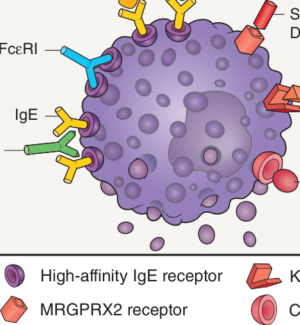
Fig. 18.3 Mast cell degranulating stimuli. Both immunologic and non-immunologic stimuli can lead to release of mediators. Substance P also binds to neurokinin 1 receptor; stem cell factor is also known as KIT ligand. MRGPRX2, MAS-related G-protein coupled receptor X2.
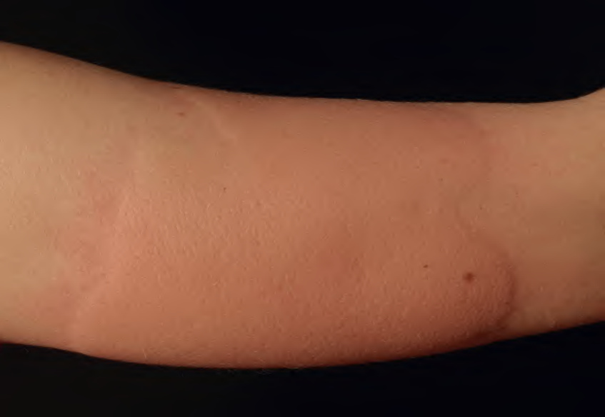
Fig. 18.11 Cold urticaria. Wheals developed on the forearm after placement of an ice cube for 10 minutes, followed by rewarming. Courtesy Thomas Schwarz, MD.
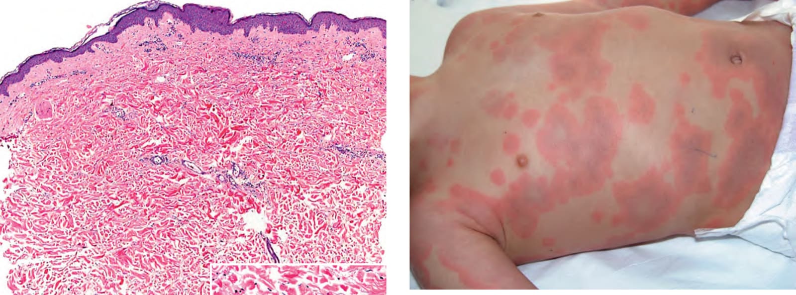
Fig. 18.15 Urticaria in a young child. This clinical presentation of urticaria is sometimes misdiagnosed as erythema multiforme because of the dusky centers, with some clinicians referring to it as urticaria multiforme. As lesions expand, central clearing can occur, which is not seen in erythema multiforme. Courtesy Luis Requena, MD.
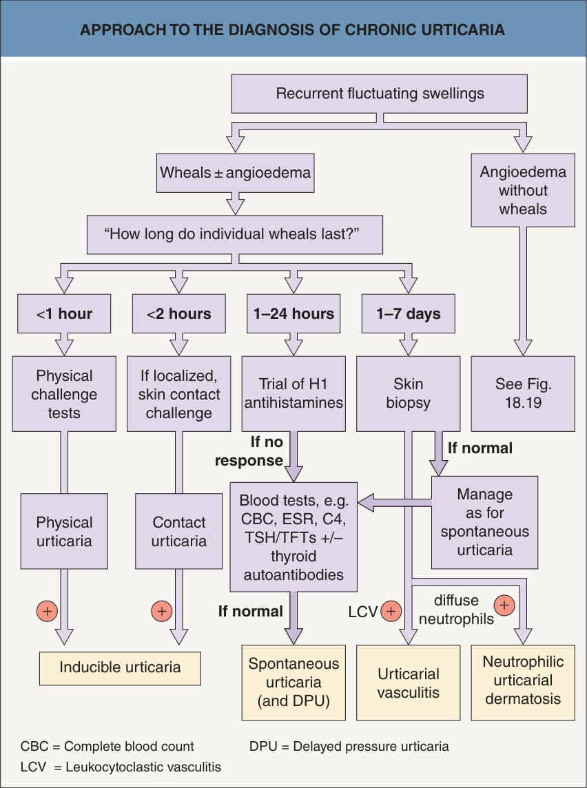
Fig. 18.16 Approach to the diagnosis of chronic urticaria.
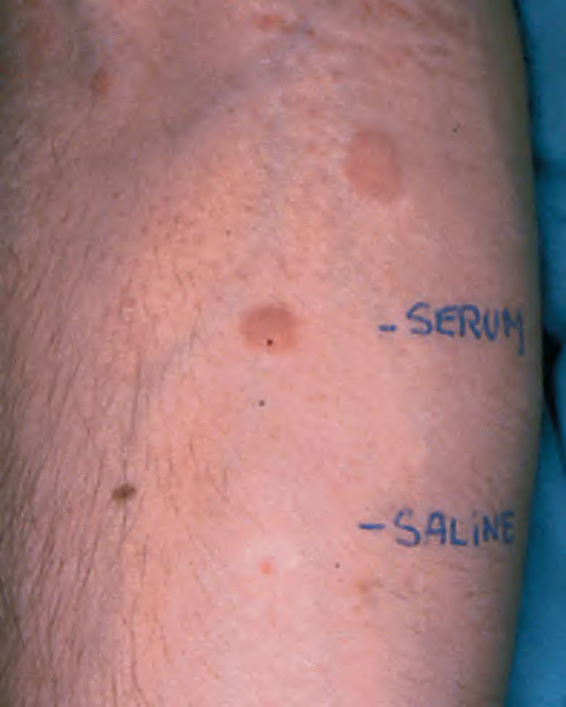
Fig. 18.17 Autologous serum skin test (ASST).
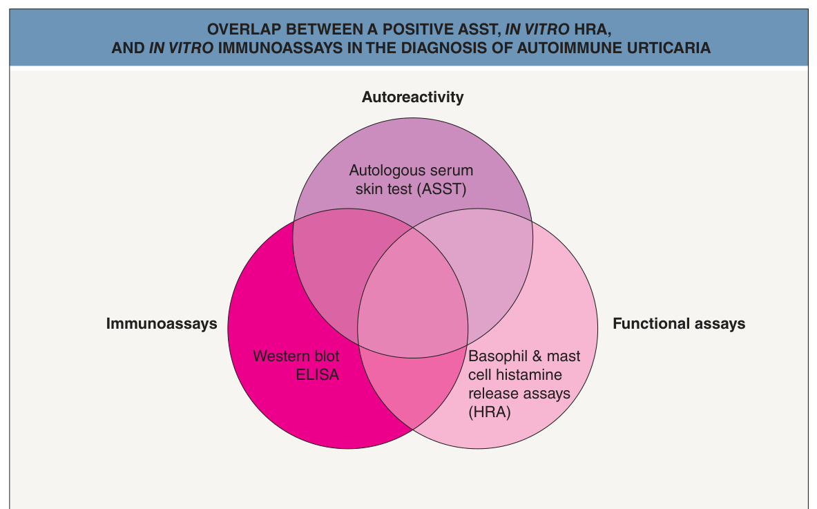
Fig. 18.18 Overlap between a positive autologous serum skin test (ASST), in vitro histamine release assays (HRA), and in vitro immunoassays in the diagnosis of autoimmune urticaria. The ASST will become increasingly difficult to perform in the future as regulations regarding blood products become more restrictive.
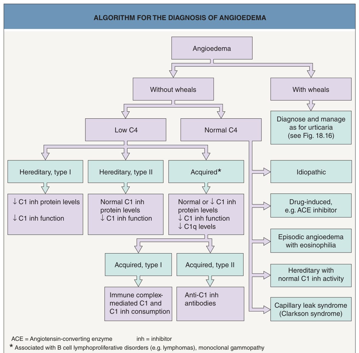
Fig. 18.19 Algorithm for the diagnosis of angio- edema. Episodic angioedema with hypereosinophilia, along with weight gain and fever, is known as Gleich syndrome. There are currently 7 subgroups of HAE with normal C1 inh activity (nC1-INH-HAE, previously referred to as type III HAE); the six subgroups that have a known underlying genetic mutation are termed HAE3 to HAE8 (see Fig. 18.6), while the seventh subgroup includes those whose genetic basis is unknown.
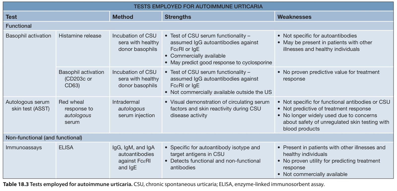
Table 18.3 Tests employed for autoimmune urticaria. CSU, chronic spontaneous urticaria; ELISA, enzyme-linked immunosorbent assay.