ERYTHEMA ANNULARE CENTRIFUGUM
Synonyms: Superficial or deep gyrate erythema Erythema perstans Palpable migrating (arciform) erythema
Key features
Erythematous annular lesions that migrate centrifugally
Superficial lesions can have the classic “trailing” white scale, while the deep gyrate erythemas have a more infiltrated border
The disorder occurs more commonly in adults, and superficial lesions favor the thighs and hips
Individual lesions usually last for several days to a few months
Although often idiopathic in nature, it can be associated with infections (e.g. tinea pedis) and anecdotally with other disorders or exposures
Introduction
The term “erythema annulare centrifugum” (EAC) has come to encompass more than just annular erythematous plaques with trailing scale. One explanation for this is that once the other three major figurate erythemas (with specific etiologies; see below) and the disorders listed in Table 19.1 are excluded, EAC often becomes a “default” diagnosis. Unfortunately, this has led to some confusion, but until a specific “trigger” can be identified for each individual patient, this situation will persist. As a consequence, some authors have come to the conclusion that EAC, especially the deeper form, represents a clinical reaction pattern rather than a specific clinicopathologic entity. Adding to the confusion is the school of thought in which the term EAC is reserved for only the superficial form.
History
In 1881, Colcott-Fox described persistent, ring-shaped lesions with pruritus, to which he gave the name “erythema gyratum perstans”. The term “EAC” was introduced by Darier in 1916 while the name “erythema perstans” has been used by some authors to describe similar annular erythemas. It is now thought that all of these terms refer to clinical or pathologic variants of the entity now referred to as EAC. However, Ackerman preferred to call the two histologically different forms (superficial and deep) of EAC “superficial gyrate erythema” and “deep gyrate erythema”, while Weyers et al. believed that the superficial and deep forms of EAC were unrelated to one another and should not be referred to by the same name. These latter authors recommended that the term EAC be reserved for the superficial type, which in their opinion represented a specific clinicopathologic entity.
Epidemiology
Although EAC can appear in any age group, its peak incidence is in the fifth decade of life. There is no known sex difference. A rare autosomal dominantly inherited form of EAC, referred to as “familial annular erythema”, has also been described.
Pathogenesis
It has been suggested that EAC, especially the superficial form with epidermal spongiosis histologically, represents a reaction pattern or “hypersensitivity” to one of many antigens. EAC has been associated with infectious agents, particularly dermatophytes, but also with other fungi (e.g. Candida, Penicillium in blue cheese), viruses (e.g. poxvirus, EBV, varicella–zoster virus, HIV), bacteria (e.g. Pseudomonas, post-tonsillitis), parasites, and ectoparasites (e.g. Phthirus pubis). Less commonly, EAC has been linked to drugs (e.g. diuretics, nonsteroidal anti-inflammatory drugs, antimalarials, finasteride, amitriptyline, nivolumab, sorafenib, rituximab, pegylated interferon-α-2a plus ribavirin, ustekinumab [often as single case reports]), Crohn disease, pregnancy, autoimmune endocrinopathies, hypereosinophilic syndrome and, occasionally, neoplasms (e.g. lymphomas, leukemias, solid organ malignancies). The latter has been referred to as paraneoplastic EAC eruption (PEACE). However, most of these associations are anecdotal, and, in the last group, Curth’s postulates have not been consistently fulfilled (see Ch. 53). There are patients with EAC who noted resolution of their lesions after the diseases that presumably triggered the EAC were successfully treated. In one series, an associated disease was identified in only one-third of patients.
The peripheral migration of EAC lesions is thought to reflect localized production of proinflammatory cytokines and vasoactive peptides, but the precise mechanism is unknown.
Clinical Features
The initial lesions of EAC begin as firm pink papules that expand centrifugally and then develop central clearing. An individual lesion can enlarge to greater than 6 cm in diameter over a period of 1 to 2 weeks. If expansion of the annular plaque is not uniform, incomplete arcs appear, as do polycyclic lesions, or simply festooned bands. In the superficial form, the lesions are minimally elevated, and there is desquamation at the inner margin, i.e. trailing scale (Fig. 19.1A,B). The scale may not be present in all the lesions in a particular patient (Fig. 19.2A). There may be associated pruritus, especially in lesions that histologically show spongiosis.
Occasionally, vesicles develop within the peripheral margin. In deep gyrate erythema, the advancing edges are obviously elevated (Fig. 19.1C) and there is usually no associated scale or pruritus.
As lesions resolve, there is no residual scarring, but postinflammatory hyperpigmentation can be seen and, on occasion, purpura. Although the lesions of EAC may be localized or generalized, they rarely, if ever, involve the palms, soles, scalp or mucous membranes. Individual lesions can persist for weeks to months, but associated systemic manifestations are uncommon. While new lesions can appear just as older ones resolve, there may be prolonged intervals of time between flares. Compared to the deep variant, the superficial form tends to recur more frequently but the individual lesions are often shorter-lived. The total duration of the disorder ranges from days to decades, and an unusual form of EAC has been described that recurs annually.
When EAC is due to an underlying disorder, flares of the former may correlate with recurrences of the latter. However, most patients with EAC do not have an identifiable underlying disorder, nor can the onset, fluctuations, or duration be related to a specific antigen.
Pathology
In superficial lesions of EAC, the findings are nonspecific, with mild spongiosis and microvesiculation plus associated focal parakeratosis, as well as a mild superficial perivascular lymphohistiocytic infiltrate. These histologic features correspond to the scale on the inner margin of the advancing erythematous arc. Characteristically, the inflammatory cells form a fairly tight aggregate around vessels, the so-called “coat sleeve” appearance (Fig. 19.1D). Rarely, eosinophils are found in the perivascular infiltrate. The advancing edge, which is slightly raised, may also show edema in the papillary dermis. The central area of clearing may contain dermal melanophages.
In deep lesions of EAC, the epidermis is usually unremarkable, and a mononuclear cell infiltrate with a sharply demarcated perivascular arrangement is present primarily within the mid and lower dermis. For this reason, the lesions are generally elevated and are more indurated than in the superficial form of EAC, and they do not have trailing scale.
Differential Diagnosis
EAC must be differentiated from other annular erythematous lesions (see Table 19.1), particularly tinea corporis and annular psoriasis, when there is associated scale. Additional entities to consider include annular urticaria (Fig. 19.2B), allergic urticarial eruption, symmetric acral annular erythema, cutaneous lymphoid hyperplasia (pseudolymphoma), and lymphoma cutis. Patients with autoimmune disorders, including linear IgA bullous dermatosis, bullous pemphigoid, Sjögren
syndrome, and lupus erythematosus (LE tumidus, subacute cutaneous LE, neonatal LE), can also have erythematous annular, arciform, and polycyclic lesions.
Treatment
If EAC is due to an underlying disorder, the skin lesions will usually resolve once the latter has been successfully treated. Topical corticosteroids applied to the advancing border of the lesions may be of benefit. Topical antipruritics and sedating antihistamines can be prescribed if there is associated pruritus. Even in the absence of an identifiable cause, some authors have advocated the empiric use of antibiotics (e.g. macro lides) or antifungal agents. Although systemic corticosteroids can induce a clinical remission, recurrences are common when the medication is discontinued. Based solely on case reports, the following have been reported as beneficial: topical tacrolimus, topical calcipotriene (calcipotriol), NB-UVB, oral metronidazole, apremilast, subcutaneous etanercept, and subcutaneous interferon-α. However, systemic therapies are seldom necessary.
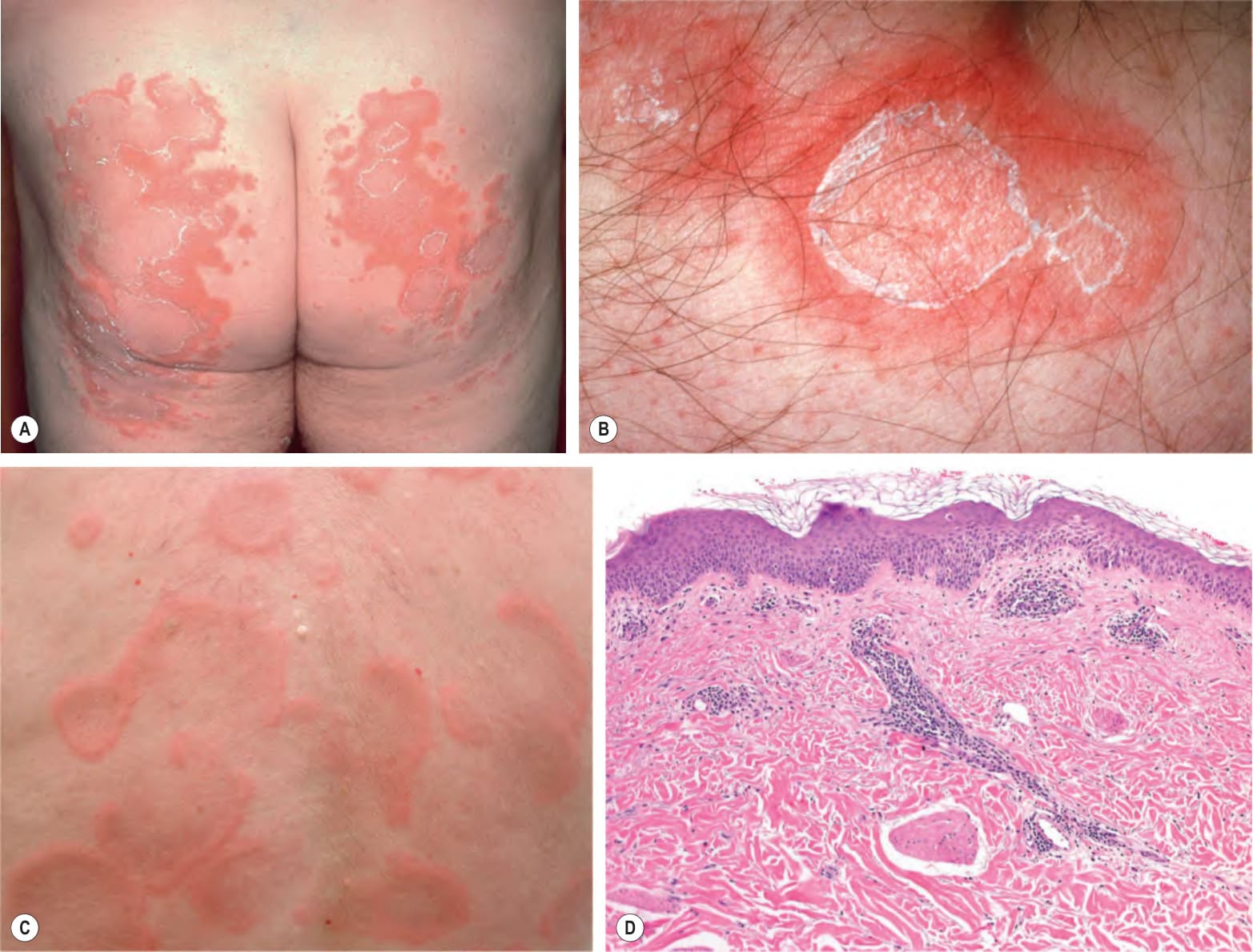
Fig. 19.1 Erythema annulare centrifugum.A, B Superficial form, with polycyclic plaque and delicate scale on the inner margin of the advancing edge (trailing scale). The scale is detached centrally. C Deep form, with obvious elevation of advancing edges and without trailing scale. D Photomicrograph of the superficial form with dermal perivascular lymphohistiocytic infiltrates; note the tight “coat sleeve”-like appearance of the infiltrate. D, Courtesy Lorenzo Cerroni, MD.
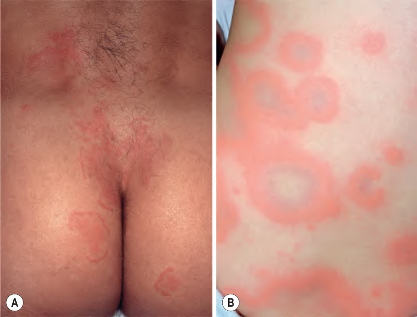
Fig. 19.2 A comparison of erythema annulare centrifugum and urticaria. The lesions in A have no scaling and may be confused with annular urticaria (B). However, individual lesions of urticaria are evanescent. Placement of an ink circle around a lesion, followed by longitudinal observation, allows this distinction.
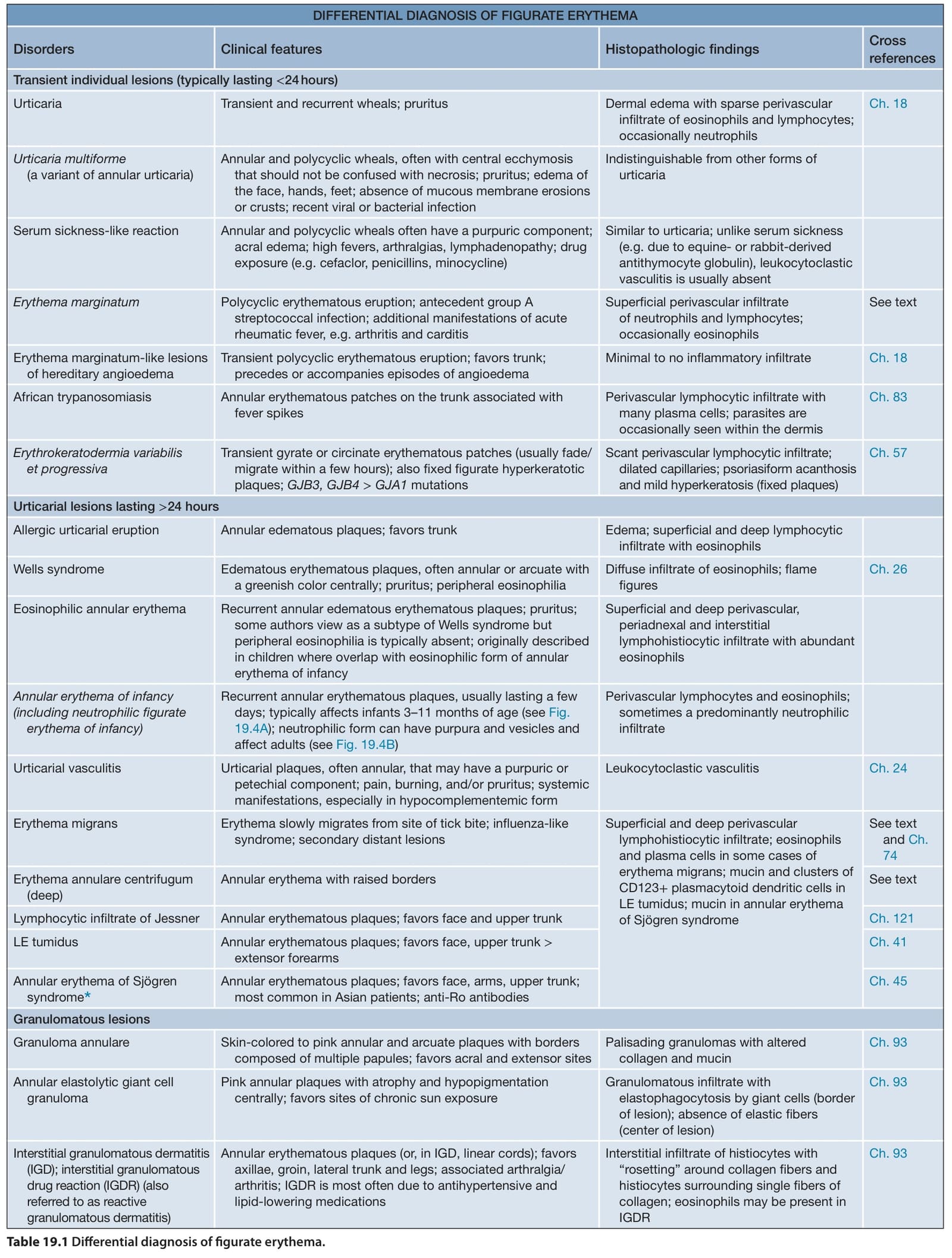
Table 19.1 Differential diagnosis of figurate erythema.