ERYTHEMA MARGINATUM
Synonyms: Erythema marginatum rheumaticum Erythema annulare rheumaticum
Key features
Migratory, annular and polycyclic, erythematous eruption
Cutaneous manifestation of acute rheumatic fever
Associated findings include carditis, migratory polyarthritis,
Sydenham chorea, and subcutaneous nodules
Seen more commonly in children than in adults
Introduction
Rheumatic fever (RF) is characterized by an abnormal immunologic response to a preceding infection with group A β-hemolytic streptococci and by the triad of fever, arthritis, and carditis. Cutaneous manifestations include erythema marginatum and subcutaneous nodules, but they are seen in a minority of patients. The diagnostic criteria of RF were recently revised in 2015 by the American Heart Association: major criteria in low-risk populations are carditis or valvulitis (by echocardiography), polyarthritis, Sydenham chorea, erythema marginatum, and subcutaneous nodules. Minor criteria include fever, polyarthralgia, elevated acute phase reactants (ESR ≥60 mm, C-reactive protein ≥3 mg/dL), and prolonged PR interval on ECG. To establish the diagnosis of acute RF, two major or one major and two minor criteria must be accompanied by evidence of an antecedent group A streptococcal infection, e.g. a positive culture, an elevated or rising antistreptolysin O or anti-DNase B titer. In moderate- to high-risk populations, monoarticular arthritis is also considered a major criterion, with monoarthralgia and an ESR ≥30 mm representing additional minor criteria.
History
In 1831, Bright noted the presence of annular erythematous lesions in patients with RF. In 1889, Cheadle proposed the name “erythema marginatum rheumaticum”. Carol and van Krieken first described the histopathologic findings in 1935. Two years later, Perry reported that rapid migration of the lesions was a characteristic finding. In 1944, Jones established criteria for diagnosing RF.
Epidemiology
Attacks of acute RF are associated with antecedent group A β-hemolytic streptococcal infections of the pharynx. Approximately 3% of patients with untreated infections can develop acute RF. It is estimated that in high-income countries, i.e. low-risk populations, the incidence is 5 per 100 000, whereas in low-income countries, it ranges from 100 to 1000 per 100 000. Erythema marginatum is seen in less than 10% of patients with acute RF. The higher incidence of erythema marginatum in children (as compared to adults) reflects the predominance of RF during childhood; the peak age-related incidence is between 5 and 15 years.
Pathogenesis
The mechanisms that underlie erythema marginatum are unknown. Presumably, there is an abnormal humoral and cellular immune response to one or more antigens associated with group A β-hemolytic streptococci (e.g. M protein). Antigenic mimicry may play a role in that epitopes that cross-react with group A streptococcal antigens have been identified in human myosin, actin, tropomyosin, keratin, laminin, N-acetylglucosamine, and vimentin. Specific characteristics of certain strains of group A β-hemolytic streptococci, such as higher M protein content and mucoid colony formation, may facilitate the development of acute RF. Enhanced expression of genes that encode cysteine proteases (e.g. Streptococcus pyogenes exotoxin B [SpeB], immunoglobulin G degrading enzyme of Str. pyogenes [IdeS]) is thought to contribute to cardiotoxicity and virulence. The role of host genetic factors is currently under investigation via genome-wide association studies.
Clinical Features
Following the initial episode of streptococcal pharyngitis, there is usually a latency period of 2–5 weeks before the development of the acute attack of RF. The lesions of erythema marginatum begin as
erythematous macules that spread peripherally and become patches or plaques with no scale. Erythema marginatum can also have a polycyclic arrangement (Fig. 19.3). The lesions are usually asymptomatic, and, over a period of 12 hours, they can migrate from 2 to 12 mm; in areas of previous involvement, the skin may appear pale or lightly pigmented. As with other erythemas, exposure to heat can accentuate lesions.
The most common locations for erythema marginatum are the trunk, axillae and proximal extremities, with sparing of the face. New lesions usually last from a few hours to a few days and are often most noticeable during the afternoon. Recurrent crops can occur over a number of weeks. Erythema marginatum is associated primarily with the active phase of RF, and, in general, it is seen in conjunction with carditis. Of note, children less than 5 years of age appear to have a higher incidence of erythema marginatum as well as carditis and arthritis.
Subcutaneous nodules appear primarily over bony prominences, especially the wrists, elbows, knees and ankles, in patients with long-standing disease. The nodules are usually painless and are rare during the initial attack of RF. Similar nodules can be seen in patients with polyarticular juvenile idiopathic arthritis (see Ch. 45). In addition to the major and minor criteria (see above), patients may also have malaise, chest or abdominal pain, epistaxis, tachycardia and anemia, as well as a nonspecific urticarial eruption.
Pathology
An interstitial and perivascular infiltrate composed predominantly of neutrophils without vasculitis is observed. Occasionally, eosinophils are seen and in later stages, there can be extravasation of erythrocytes. Direct immunofluorescence microscopy for immunoglobulin and complement deposits is negative. These histologic findings are not unique to erythema marginatum, and overlapping features with the neutrophilic variant of annular erythema of infancy (neutrophilic figurate erythema of infancy) and the neutrophilic urticarial dermatosis seen in several autoinflammatory disorders may be observed.
Differential Diagnosis
The clinical differential diagnosis includes primarily annular urticaria (including urticaria multiforme) and annular erythema of infancy (Fig. 19.4A), as well as its variant neutrophilic figurate erythema of infancy. Occasionally, neutrophilic figurate erythema is seen in adults and can be accompanied by vesicles and purpura (Fig. 19.4B). Less often, Still disease, hereditary periodic fever syndromes (particularly TNF receptor-associated periodic syndrome [TRAPS]; see Table 45.2), EAC, Kawasaki disease, and other entities listed in Table 19.1 need to be considered. Annular erythema with an appearance similar to erythema marginatum may precede or accompany episodes of hereditary angioedema and is seen only occasionally in patients with cat scratch disease or psittacosis. The angioedemaassociated figurate erythema may be related to deposits of bradykinin within the dermis while the latter may reflect an overlap between erythema marginatum and EAC. In a patient receiving sorafenib, figurate lesions with associated scale-crust and hemorrhage developed and the name “erythema marginatum hemorrhagicum” was proposed, but clinically these lesions can easily be distinguished from erythema marginatum.
Treatment
There is no specific therapy for this dermatosis. In general, its clinical course is unaltered by treatment of the underlying acute RF; however, symptoms are usually mild and lesions resolve spontaneously.
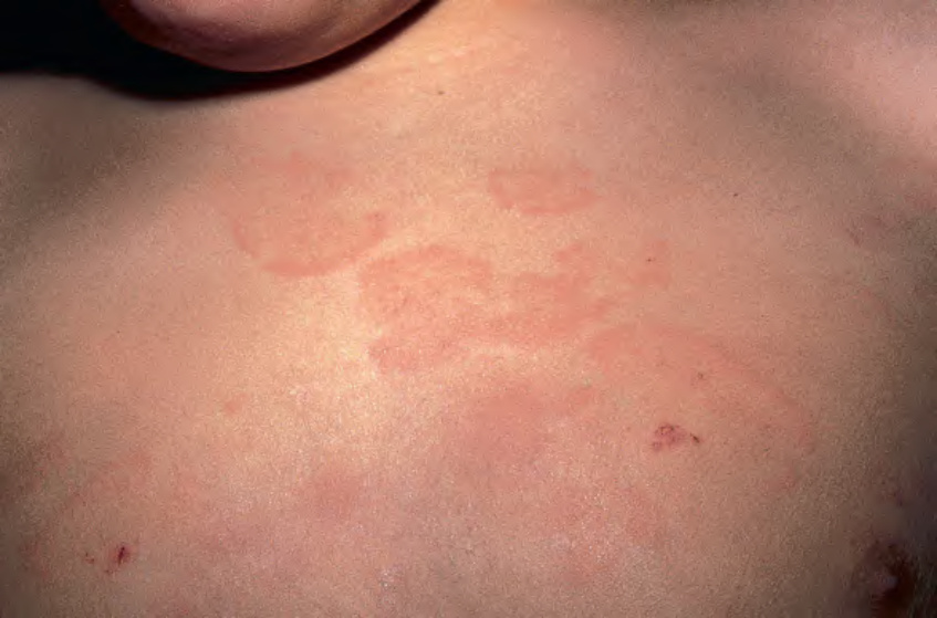
Fig. 19.3 Erythema marginatum. Polycyclic and evanescent annular lesions are seen on the trunk of this young patient.
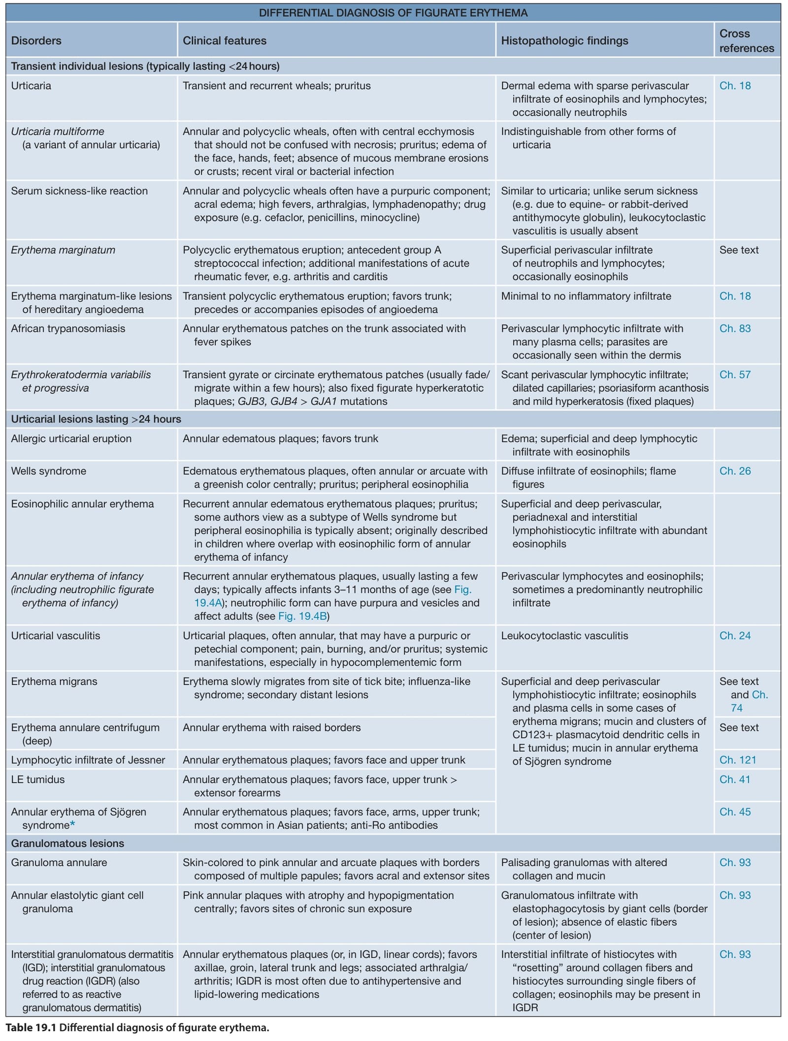
Table 19.1 Differential diagnosis of figurate erythema.