ERYTHEMA MIGRANS
Synonyms: Erythema chronicum migrans Lyme borreliosis (early stage) Lyme disease (early stage)
Key features
Annular erythema develops at the site of the bite of a Borrelia- infected tick
Several species of Ixodes ticks are infected with different genospecies of Borrelia burgdorferi
Erythema migrans represents the initial cutaneous manifestation of
Lyme disease and is seen in 60%–80% of patients diagnosed with the disease
Multiple secondary lesions that may be smaller in size can occur as a result of spirochetemia or lymphatic spread
Introduction
Lyme disease (LD) is an infection due to Borrelia burgdorferi spirochetes that are transmitted by bites from several species of Ixodes ticks (e.g. I. scapularis, I. pacificus, I. ricinus; see Chs. 76 & 85). Erythema migrans represents the initial cutaneous manifestation.
History
The first report of an annular erythema developing after a tick bite was by Afzelius in 1909 and he coined the term “erythema migrans”. Lipschütz then referred to these cutaneous lesions as “erythema
chronicum migrans”. In the US, Steere described an epidemic form of childhood arthritis preceded by erythema migrans in several communities in the vicinity of Lyme, Connecticut. In the 1980s, Burgdorfer isolated a new spirochete in lesions of erythema migrans and the organism was subsequently named B. burgdorferi.
Epidemiology
Although LD has a worldwide distribution, it is seen most commonly in the US (Northeast, mid-Atlantic, and Great Lakes regions) and in northern and eastern Europe. The competent hosts that serve as a reservoir for Borrelia include mice, chipmunks and birds, with deer playing a role in sustaining the life cycle. In temperate regions, the peak incidence for LD is during the spring and the summer, and it occurs more commonly in those who spend time outdoors in or near fields and woods. Approximately 30 000 cases of Lyme disease are currently reported to the CDC each year, with 95% of confirmed cases coming from 14 states (www.cdc.gov/lyme/). Based upon recent US insurance claim data, an estimated 475 000 people were treated yearly for Lyme disease, but this number obviously includes noninfected individuals. In Europe, ~320 000 cases of LD are diagnosed each year, but the incidence varies depending upon the country. Not all patients who have positive serologic tests for B. burgdorferi develop LD.
Pathogenesis
In mice, B. burgdorferi has been shown to usurp a tick salivary protein, Salp15, as a means of enhancing transmission. By binding to the outer surface protein C (OspC) of B. burgdorferi, Salp15 provides protection against antibody-mediated killing. In addition, Salp15 inhibits adaptive immune responses against both Borrelia and tick antigens. Of note, one of the proposed strategies for preventing LD is the development of anti-tick vaccines that utilize tick salivary proteins.
Once the Borrelia organisms have entered the body, spirochetal lipoproteins trigger an innate immune response, with cytokines being produced by macrophages. In addition, a Th1 response is triggered and B cell responses facilitate the synthesis of antibodies against different antigens of B. burgdorferi. Spirochetemia is present in ~45% of patients at the time of their presentation with erythema migrans, and organisms can disseminate widely due to their ability to resist elimination by macrophages, evade complement, adhere to brain and epithelial cells, penetrate into the cytoplasm via intracellular junctions, and induce TNF production.
Even though the lesion expands centrifugally, B. burgdorferi organisms have been found both in the center and at the periphery of lesions of erythema migrans. The persistence of the spirochete in the skin after the tick bite may be due to an absence of the production of interferon-γ and an ineffective immune response. Also, different immune responses to the various genospecies of the B. burgdorferi sensu lato complex (B. burgdorferi sensu stricto, B. afzelii, B. garinii) may lead to different clinical presentations (e.g. borrelial lymphocytoma in Europe but not the US; see Ch. 74). More recently, an additional genospecies was identified in the upper midwestern US, B. mayonii, which may lead to a more severe clinical presentation due to an unusually high spirochetemia.
Clinical Features
LD is divided into three clinical stages: (1) early localized disease; (2) early disseminated disease; and (3) chronic disease (Table 19.2). Erythema migrans is an important clinical feature of early disease. Typically, 7–15 days (range, 3–30 days) after tick detachment, an erythematous, expanding, circular or annular plaque appears that may have a lighter-colored central area or a bull’s-eye appearance (Fig. 19.5). The eventual diameter is usually at least 5 cm, and the center may become darker red to violaceous in color, crusted, or even vesicular. Lesions of primary erythema migrans favor the trunk, axilla, groin, and popliteal fossa. Untreated, the lesions usually last less than 6 weeks (median, 4 weeks). In 20%–25% of patients, multiple lesions can appear due to multiple tick bites or as a result of disseminated disease secondary to spirochetemia. Disseminated lesions tend to be smaller in size and less pronounced and usually appear days to weeks after the appearance of the primary erythema migrans (Fig. 19.6).
Erythema migrans is seen in 60%–80% of patients diagnosed with LD. In the US, the systemic manifestations of this initial phase tend to be acute, with influenza-like symptoms, including fatigue, headache, arthralgia, myalgia and fever, as well as lymphadenopathy, conjunctivitis or hepatitis (see Table 19.2). In Europe, this initial clinical phase is usually less severe, and the lesions of erythema migrans tend to last longer. If untreated, ~60% of patients will go on to have monoarticular or oligoarticular arthritis (usually the knee) within weeks to months after the initial infection; ~10%, neurologic manifestations (most commonly facial nerve palsy); and ~5%, cardiac complications (usually varying degrees of atrioventricular block). For a discussion of borrelial lymphocytoma and acrodermatitis chronica atrophicans, see Chapter 74.
Depending upon the geographic region, Ixodes ticks can be co-infected with the following microbes (in addition to B. burgdorferi): Babesia microti (babesiosis), Anaplasma phagocytophilum (anaplasmosis), Borrelia miyamotoi, or Powassan virus. Co-infections should be considered when patients with LD have prolonged or severe manifestations of infection, cytopenias, or an unusually high or persistent fever.
Pathology
The histologic features of erythema migrans are similar to those of a deep gyrate erythema (Fig. 19.7). While routine histology is often not specific, many specimens will show superficial and deep lymphoid infiltrates admixed with a few eosinophils and plasma cells. Immunohistochemical studies have shown a reduction in the number of Langerhans cells; in the dermis, the inflammatory infiltrate contains macrophages, CD4+ T helper cells and CD45RO+ memory T cells. PCR analysis of tissue specimens can be performed to confirm the presence of Borrelia spirochetes within a lesion.
Differential Diagnosis
The 2022 revised CDC case definition for LD now includes three categories: “confirmed”, “probable”, and “suspected” (Table 19.3). These categories are based upon laboratory evidence and whether a person resides in a low-incidence versus high-incidence jurisdiction (state). Confirmatory laboratory evidence consists of one of the following: (1) isolation of B. burgdorferi sensu stricto or B. mayonii in culture; (2) detection of B. burgdorferi sensu stricto or B. mayonii in a clinical specimen by a B. burgdorferi group-specific nucleic acid amplification test (NAAT) assay; (3) detection of B. burgdorferi group-specific anti-gens by immunohistochemical assay on biopsy or autopsy tissues; or (4) positive serologic tests in a two-tier or equivalent format. The latter includes either: (1) the standard two-tier test (STTT) - a positive or equivocal first-tier screening assay, often an enzyme immunoassay (EIA) or immunofluorescence assay (IFA) for IgM, IgG or combination, followed by a concordant positive IgM or IgG immunoblot; or (2) modified two-tier test (MTTT) - positive or equivocal first-tier screen, followed by a different sequential positive or equivocal EIA in lieu of an immunoblot. In 2019, the MTTT methodology was introduced in which an FDA-cleared second EIA (e.g. for VlsE1-IgG and pepC10-IgM) could be used in place of the Western immunoblot. Presumptive laboratory evidence consists of a positive IgG immunoblot, interpreted by established criteria, without positive or equivocal first-tier screening.
Of note, the peak specific IgM response (usually directed against the 41 kDa flagellar antigen) occurs 3–6 weeks into the infection. As a result, serologic testing has a low sensitivity, e.g. only 25%– 40% of patients with erythema migrans during the acute phase (but without evidence of dissemination) have positive test results, with a slightly higher percentage in those with dissemination (~50%). During convalescence following antibiotic therapy, positive test results are still only observed in approximately half of those who did not have dissemination, presumably reflecting eradication of the spirochetes.
Although culture of tissue from the perimeter of erythema migrans lesions is essentially 100% specific and can distinguish live from dead organisms, it requires special agar (modified Barbour–Stoenner–Kelly medium) and prolonged observation, making it impractical in the
standard clinical setting. As noted previously, detection of B. burgdorferi in skin biopsy specimens of erythema migrans via PCR is possible.
Erythema migrans must be distinguished from exaggerated local arthropod reactions (including to the tick bite), erysipelas, cellulitis, allergic contact dermatitis and non-pigmented fixed drug eruption, and less often, other entities outlined in Table 19.1. An erythema migranslike skin lesion can be seen in southern tick-associated rash illness (STARI or Masters disease) which follows the bite of the lone star tick, Amblyomma americanum (see Ch. 85).
Treatment
Recommended antibiotic regimens are outlined in Table 19.4. It is estimated that even in highly endemic areas, the risk of disease transmission from a recognized tick bite is low (1%–3%). This may be due to the observation that in general transmission from infected nymph
CDC, Centers for Disease Control and Prevention. https://ndc.services.cdc.gov/ case-definitions/lyme-disease-2022/ ticks requires 36–48 hours of attachment and for adult ticks, ≥48 hours of attachment. Therefore, routine treatment of tick bites with 2- to 3-week courses of antibiotics is not recommended. In patients from endemic areas (>20% of ticks infected) in whom the tick, identified as a nymphal or adult I. scapularis, has been attached for >36 hours and prophylaxis can be started within 72 hours of tick removal, a single 200 mg dose of doxycycline may decrease the risk of developing LD (from 3.2% in the placebo group to 0.4% in the treatment group in one study).
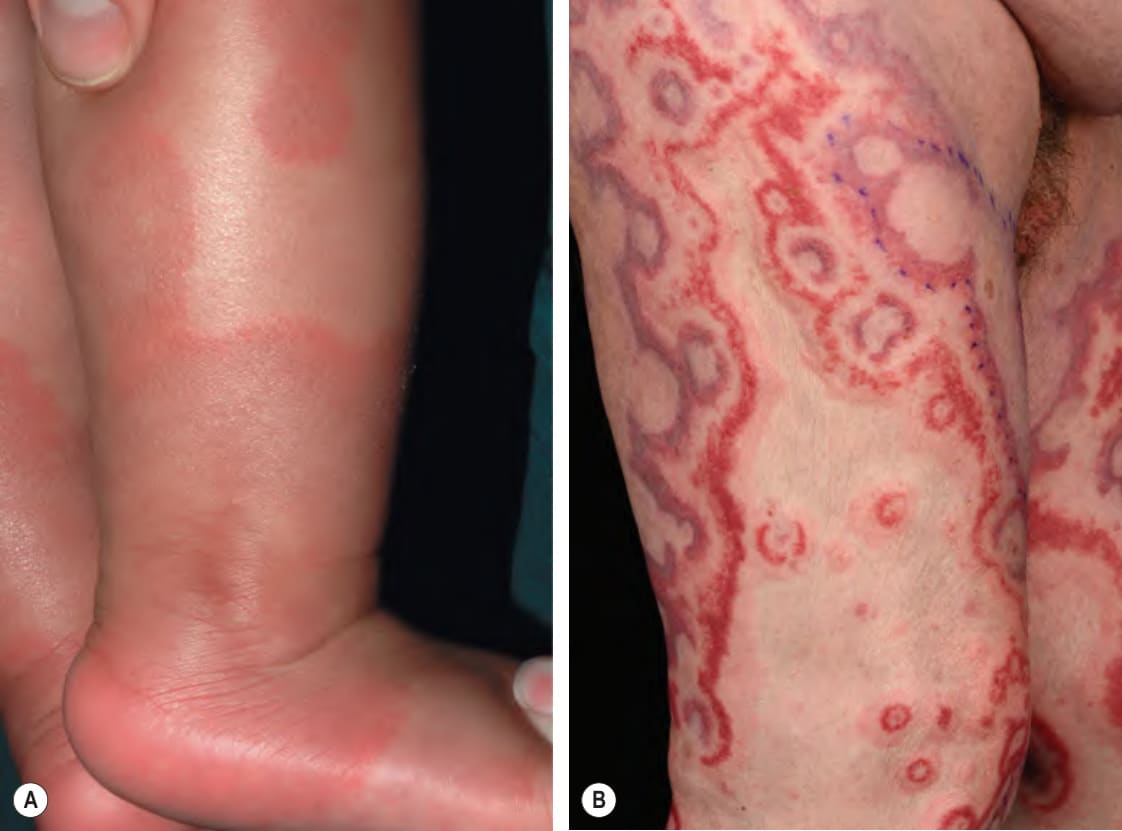
Fig. 19.4 Annular erythema of infancy and neutrophilic figurate erythema.A Expanding annular urticarial plaques in a 6-month-old boy. Lesions are typically asymptomatic, expand slowly, and then resolve over a few days. New plaques may continue to appear for several months. B A neutrophilic variant occurs in both infants and adults and in the latter, there can be striking purpura within the lesions. A, Courtesy Julie V. Schaffer, MD; B, Courtesy Lorenzo Cerroni, MD.
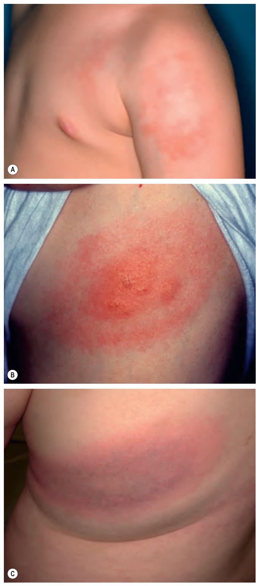
Fig. 19.5 Erythema migrans.A, B Annular erythema that spreads centrifugally; the peripheral erythematous border may or may not be sharply demarcated and is usually 1–2 cm in width. Concentric rings can form, leading to a bull’s-eye appearance, but some lesions of EM have a more uniform color. Vesiculation centrally can also be seen (B). C The center may become violaceous in color. Patients may not report a history of a tick bite. A, Courtesy Department of Dermatology, Medical University of Graz; C, Courtesy Dennis Cooper, MD.
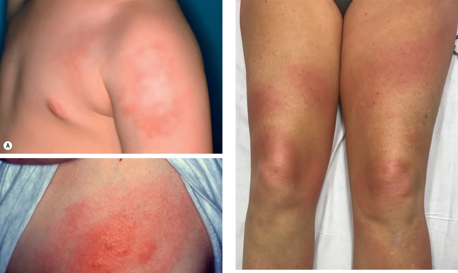
Fig. 19.6 Disseminated erythema migrans. Multiple pink circular and ovalshaped thin plaques with coalescence. Patients often have influenza-like symptoms. Courtesy Ian W. Tattersall, MD, PhD.
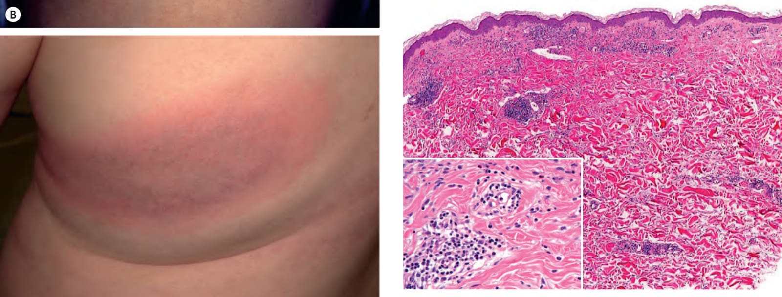
Fig. 19.7 Erythema migrans. A superficial and deep perivascular infiltrate is present within the dermis as well as a scattered interstitial pattern. The infiltrate is composed of lymphocytes and plasma cells (inset). Courtesy Lorenzo Cerroni, MD.
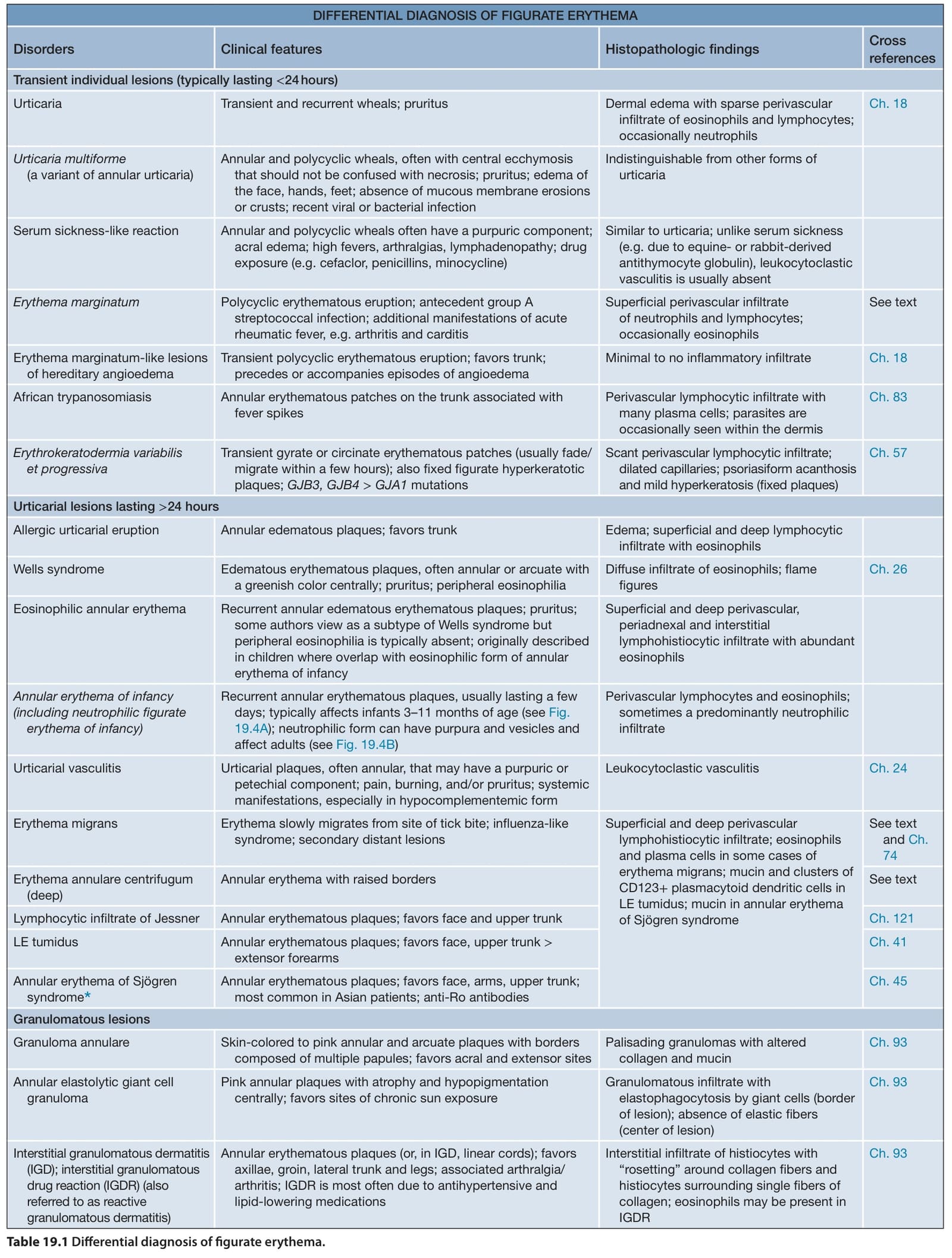
Table 19.1 Differential diagnosis of figurate erythema.
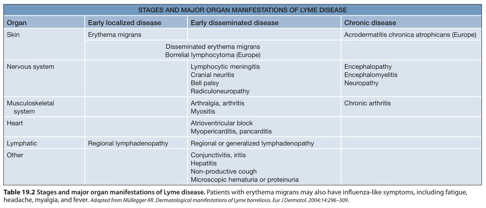
Table 19.2 Stages and major organ manifestations of Lyme disease. Patients with erythema migrans may also have influenza-like symptoms, including fatigue, headache, myalgia, and fever. Adapted from Müllegger RR. Dermatological manifestations of Lyme borreliosis. Eur J Dermatol. 2004;14:296–309.
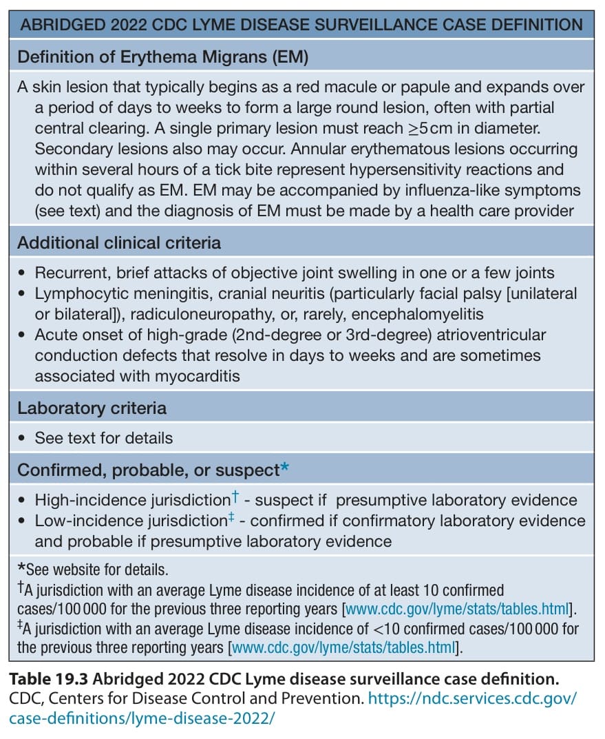
Table 19.3 Abridged 2022 CDC Lyme disease surveillance case definition.
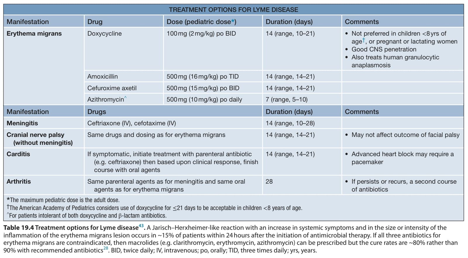
Table 19.4 Treatment options for Lyme disease. A Jarisch–Herxheimer-like reaction with an increase in systemic symptoms and in the size or intensity of the inflammation of the erythema migrans lesion occurs in ~15% of patients within 24 hours after the initiation of antimicrobial therapy. If all three antibiotics for erythema migrans are contraindicated, then macrolides (e.g. clarithromycin, erythromycin, azithromycin) can be prescribed but the cure rates are ~80% rather than 90% with recommended antibiotics. BID, twice daily; IV, intravenous; po, orally; TID, three times daily; yrs, years.