ERYTHEMA GYRATUM REPENS
Synonym: Gammel disease
Key features
A figurate erythema that is migratory and composed of concentric rings with a wood-grain appearance
It represents a paraneoplastic phenomenon and the most common underlying neoplasm is carcinoma of the lung
The lesions may have associated pruritus and scale, and they characteristically exhibit rapid migration (up to 1 cm per day)
The cutaneous lesions resolve when the neoplasm is successfully treated
Introduction
In the vast majority of patients, erythema gyratum repens (EGR) represents a paraneoplastic figurate erythema. In theory, the cutaneous lesions arise as a result of an immune reaction against tumor-associated antigens with the subsequent recognition of similar antigens in the skin.
History
EGR was first reported by Gammel in 1952, and his original description clearly identified its key clinical features.
Epidemiology
EGR is a very rare disorder seen primarily in adults. Risk factors parallel those for the underlying neoplasms.
Pathogenesis
A leading hypothesis is that EGR represents an immune reaction in which there is a cross-reaction between tumor and cutaneous antigens. In a few patients, direct immunofluorescence (DIF) has demonstrated deposits of IgG and C3 in the basement membrane zone (BMZ) of the skin, and, in isolated cases, similar deposits were seen in the associated neoplasm. However, this could simply represent a nonspecific finding. Caux et al. suggested that the tumor produces a modification in its basement membrane and this subsequently induces an immune response. Recognition of similar antigens in the BMZ of the skin then leads to the cutaneous eruption. The responsible antigen(s) is not known, but a curious finding is an accumulation of active Langerhans cells in the upper layers of the epidermis. Recently, a link between glutamine metabolism within the skin and the distinct pattern of EGR lesions was proposed.
Clinical Features
Patients usually have multiple, annular or polycyclic, erythematous lesions that develop scale at their edges and advance at a rapid rate (up to 1 cm per day). This rate of peripheral spread is significantly faster than that of EAC. Lesions have a wood-grain or zebra-like pattern, due to the development of “rings within rings” (Fig. 19.8). In some patients, the eruption is also pruritic. Additional findings in patients with EGR include acquired ichthyosis, palmoplantar keratoderma, and peripheral eosinophilia.
In at least 70% of patients, EGR is associated with an underlying neoplasm, most commonly of the lung then breast, esophagus or stomach. The cutaneous lesions usually develop between 1 year prior to 1 year after the diagnosis of the neoplasm. EGR may coincide with under-lying pulmonary tuberculosis, and patients with other disorders (see below) can also develop EGR-like lesions. Occasionally, no underlying disorder or malignancy can be identified.
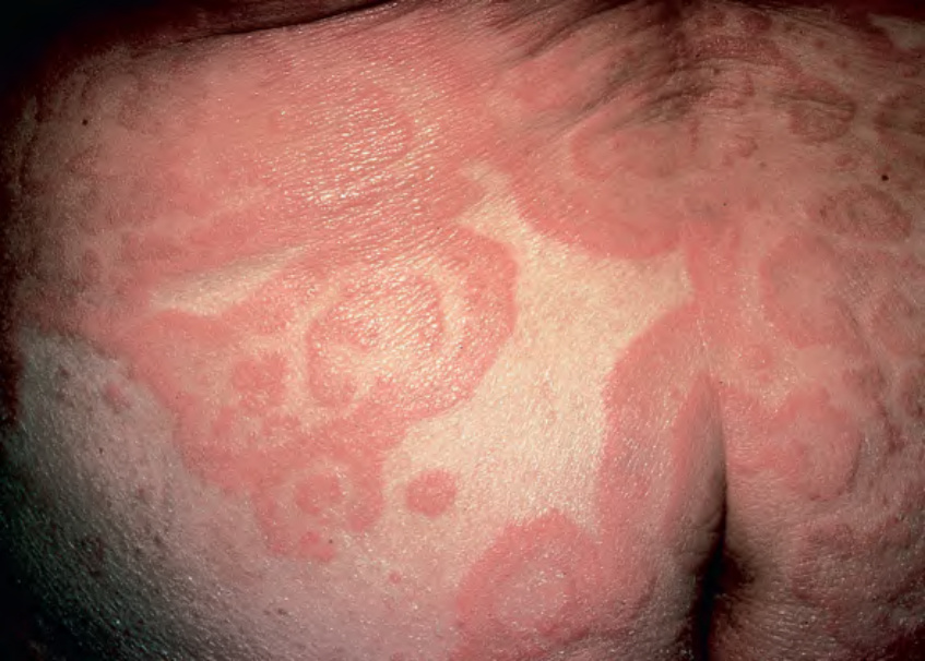
Fig. 19.8 Erythema gyratum repens. Multiple gyrate erythematous plaques, showing a wood-grain pattern. Courtesy Agustin Alomar, MD.
Table 19.4 Treatment options for Lyme disease. A Jarisch–Herxheimer-like reaction with an increase in systemic symptoms and in the size or intensity of the inflammation of the erythema migrans lesion occurs in ~15% of patients within 24 hours after the initiation of antimicrobial therapy. If all three antibiotics for erythema migrans are contraindicated, then macrolides (e.g. clarithromycin, erythromycin, azithromycin) can be prescribed but the cure rates are ~80% rather than 90% with recommended antibiotics. BID, twice daily; IV, intravenous; po, orally; TID, three times daily; yrs, years.
The histopathologic findings are nonspecific and include hyperkeratosis, focal parakeratosis, moderate patchy spongiosis and a mild, perivascular lymphohistiocytic infiltrate. In some cases, variable numbers of eosinophils are seen in the dermis.
In addition to excluding the other types of figurate erythema, EGR-like lesions may be seen in patients with erythrokeratodermia variabilis et progressiva and resolving pityriasis rubra pilaris (Fig. 19.9) as well as autoimmune bullous diseases, in particular bullous pemphigoid (both paraneoplastic and classic) and epidermolysis bullosa acquisita. Similar appearing lesions can occasionally be seen in mycosis fungoides, resolving psoriasis, hypersensitivity reactions to azathioprine, urticarial vasculitis, and autoimmune connective tissue diseases, primarily lupus erythematosus (subacute variant [also referred to as LE gyratus repens] or an associated neutrophilic dermatosis or small vessel vasculitis), and Sjögren syndrome. In certain regions of the world (e.g. maritime and mainland Southeast Asia), tinea imbricata would also be included in the differential diagnosis.
EGR resolves when the associated neoplasm is successfully treated. There may be a return of cutaneous lesions in association with the development of metastases or local recurrences of the malignancy.
Disease. Philadelphia: Lea-Febiger; 1978:174–175, 231–3, 283–4.
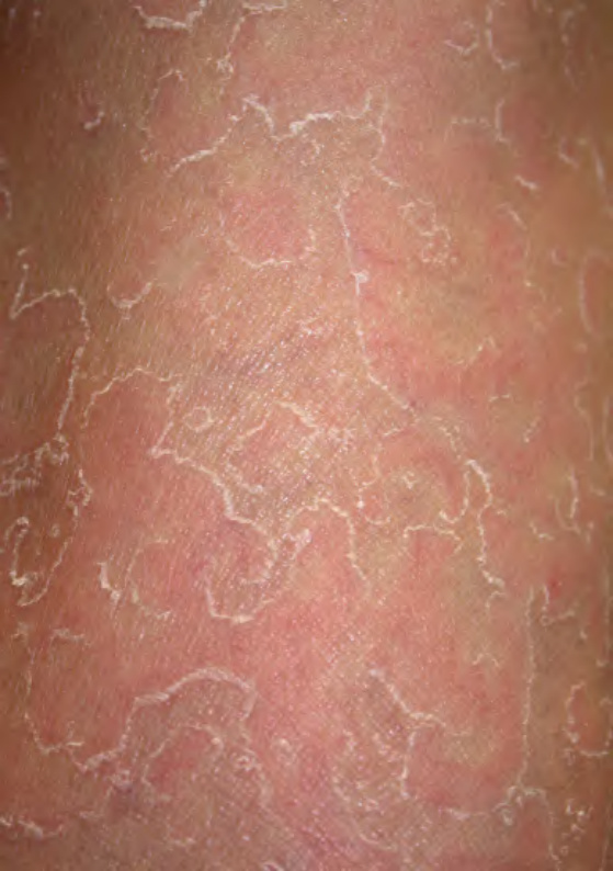
Fig. 19.9 Resolving pityriasis rubra pilaris. The lesions can resemble erythema gyratum repens. Courtesy Irwin Braverman, MD.