DIAGNOSTIC FEATURES
Drug eruptions, both suspected and unsuspected, frequently lead to a dermatologic consultation, and it is often (although not always) possible to categorize a drug as having a high, medium, or low probability of being the culprit. A rigorous approach begins with an accurate description of the skin lesions and their distribution, in addition to associated signs and symptoms (Table 21.5). Data regarding all the drugs taken by the patient, including prescription, non-prescription/over-the-counter, and complementary or alternative treatments, as well as the dates of administration and doses need to be collected on a timeline. The chronology of drug administration is of paramount importance (Table 21.6). The time between initiation of the drug and the onset of the skin eruption is a key element in identifying the culprit drug, as most immunologically mediated reactions occur 7 to 21 days after initiation of a new medication.
Evolution after drug withdrawal may be helpful, as common cutaneous eruptions (e.g. exanthematous) often clear when the suspected drug is discontinued. However, this assessment may prove difficult in the case of drugs with a long half-life or “persistent” drug reactions such as lichenoid and photoallergic eruptions, drug-induced pemphigus foliaceus and subacute cutaneous lupus erythematosus, or even SCARs, once the disease process has been definitively triggered.
The culprit drug should be withdrawn as soon as possible. The usual practice is to discontinue all other drugs that are non-essential. However, in some instances, it is necessary to weigh the risks versus the benefits of each drug and to determine if a similar-acting, but non-cross-reactive, drug is available as a substitute.
In the process of identifying the responsible drug, access to drug databases or drug agency websites is very helpful. However, new or unusual drug reactions may not be identified. Moreover, the drug most frequently associated with adverse reactions may be innocent in a particular patient, and the physician dealing with a suspected drug reaction must remain open-minded.
OTC, over-the-counter.
With the exception of assays for IgE antibodies, diagnostic or confirmatory assays to establish the responsible drug are not readily available. A number of in vitro tests have been designed, including the histamine release test, migration inhibition factor test, lymphocyte toxicity assay, lymphocyte transformation test, and basophil degranulation test. However, their sensitivity and specificity have not been assessed in a reliable way with relevant controls. As a result, they are of limited value in the clinical setting.
Results of patch testing, in which drugs (usually with petrolatum or alcohol as the vehicle) are applied to the upper back for 48 hours, vary depending upon the responsible drug and the type of eruption (Table 21.7). Prick and intradermal tests can be performed in patients with urticaria and angioedema, but they are contraindicated in SJS/ TEN due to the risk of relapse. Of note, delayed reading of prick, intradermal, and patch tests is particularly important in the case of amoxicillin-induced morbilliform eruptions. Unfortunately, investigations involving series of patients with several different types of drug reactions have shown heterogeneous results. In general, these tests, when positive, may be helpful in preventing re-administration of the culprit drug, but with the exception of prick and intradermal tests in patients with urticaria and perhaps patch tests in a few disorders (see Table 21.7), their specificity and/or sensitivity are still controversial. Of note, delayed intradermal testing is increasingly being performed in patients with DRESS when there is uncertainty regarding the responsible drug and pretest probability is high.
Rechallenge carries the risk of inducing a more severe reaction, thus it is contraindicated after SCARs. In critical situations, rechallenge may be useful, e.g. a patient with active tuberculosis who develops a rash while receiving a cocktail of antituberculous drugs; a step-by-step reintroduction may be performed in specialized centers. However, the recurrence rate is not 100% with rechallenge and a negative result may give an erroneous sense of security.
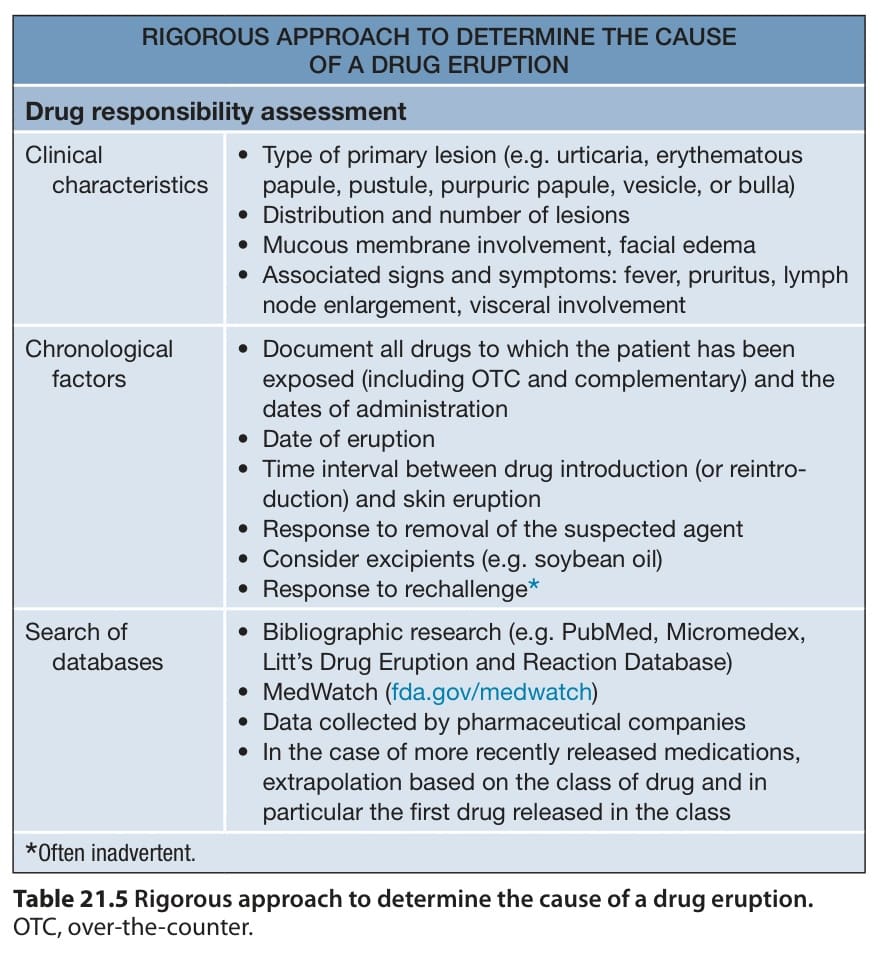
Table 21.5 Rigorous approach to determine the cause of a drug eruption.
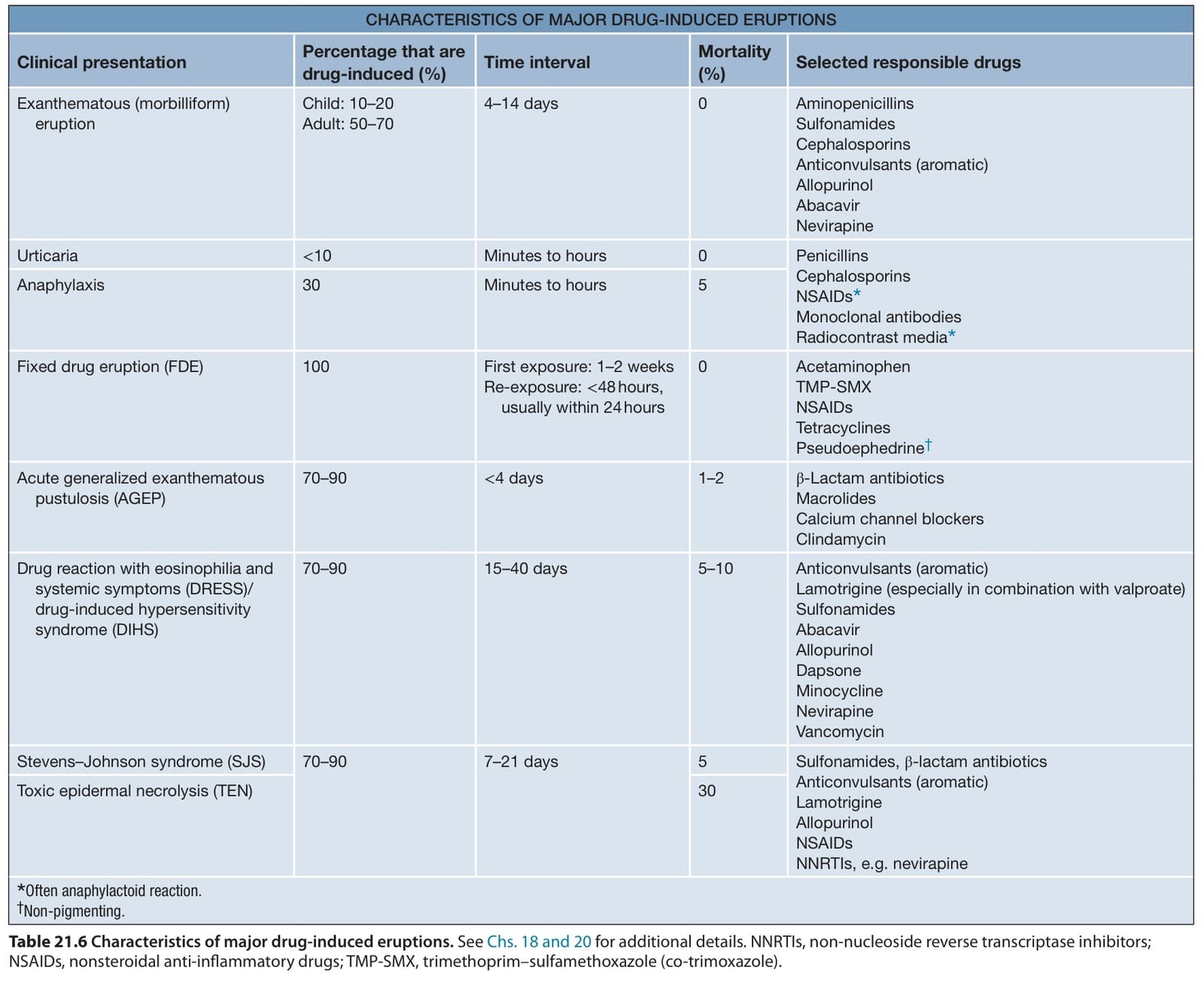
Table 21.6 Characteristics of major drug-induced eruptions. See Chs. 18 and 20 for additional details. NNRTIs, non-nucleoside reverse transcriptase inhibitors; NSAIDs, nonsteroidal anti-inflammatory drugs; TMP-SMX, trimethoprim–sulfamethoxazole (co-trimoxazole).
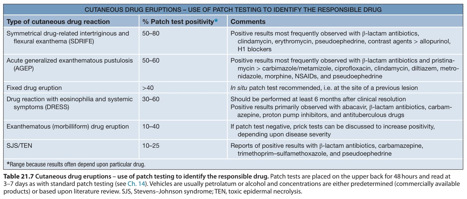
Table 21.7 Cutaneous drug eruptions – use of patch testing to identify the responsible drug. Patch tests are placed on the upper back for 48 hours and read at 3–7 days as with standard patch testing (see Ch. 14). Vehicles are usually petrolatum or alcohol and concentrations are either predetermined (commercially available products) or based upon literature review. SJS, Stevens–Johnson syndrome; TEN, toxic epidermal necrolysis.