CLINICAL FEATURES
Urticaria (see Ch. 18 for details)
Urticaria presents as transient, pruritic, erythematous, and edematous papules and plaques that may appear anywhere on the body. Lesions can vary significantly in size and number and may assume a figurate configuration (Fig. 21.2).
Although drugs are thought to be responsible for <10% of all cases of urticaria, they are more often associated with acute rather than chronic urticaria. Drug-induced urticaria may either occur immediately, i.e. less than one hour after drug intake (see below), or have a delayed appearance (late urticaria). Of note, patients with chronic urticaria should avoid acetylsalicylic acid (ASA; aspirin), as well as other NSAIDs, as they can lead to an exacerbation.
In IgE-mediated urticaria, lesions typically appear within minutes to less than an hour after drug administration, especially when there has been prior sensitization. Both immunologic assays, such as radio allergosorbent tests (RAST) that detect specific IgE antibodies, and skin tests (prick tests) are useful in confirming the diagnosis. Unfortunately, US commercial RAST panels only test for a limited number of drugs. Prick tests should be performed under appropriate medical supervision due to the risk of an anaphylactic reaction. Of note, in some series, only 10%–20% of patients who reported a history of penicillin allergy were truly allergic when assessed by skin testing.
Drugs that most frequently produce immunologically based urticaria are antibiotics, especially penicillins and cephalosporins, and less often, sulfonamides and minocycline. As the use of monoclonal antibodies for neoplastic and inflammatory diseases increases, so will cases of urticaria (and vasculitis; see below) due to these exogenous proteins (see Ch. 128).
In anaphylactoid reactions, which are not IgE-mediated but resemble anaphylaxis clinically, vasodilation results from the liberation of large amounts of histamine, bradykinin, and/or leukotrienes. Acetylsalicylic acid is the classic example of a drug that induces an anaphylactoid reaction and it does so via cyclooxygenase inhibition and subsequent accumulation of leukotrienes. The majority of urticarial reactions to radiocontrast media are also non-immunologic (Table 21.8), as are many, but not all, reactions to NSAIDs (e.g. ibuprofen, naproxen). Infusion of chemotherapy (e.g. taxanes) can also result in anaphylactoid reactions.
The most important step in the treatment of drug-induced urticaria is withdrawal of the causative agent. Treatment consists primarily of H1 antihistamines. Additional therapeutic options are discussed in Chapter 18 as is the differential diagnosis which includes the immune complex-mediated disorders urticarial vasculitis and serum sickness.
Angioedema (see Ch. 18 for details)
Angioedema is a consequence of transient edema of the deep dermal, subcutaneous, and submucosal tissues. It is associated with urticaria in 50% of cases and may be complicated by life-threatening anaphylaxis. Angioedema occurs in 1 to 2 per 1000 new users of angiotensinconverting enzyme (ACE) inhibitors and is due to an accumulation of bradykinin (see Fig. 18.6). The most severe cases of angioedema may start within a few minutes after drug administration. However, in the case of ACE inhibitor-induced angioedema, lesions may appear from 1 day to several years after starting the drug; most appear within the first year. Blacks and women are at increased risk for developing ACE inhibitor-induced angioedema.
The most common clinical presentation is an acute, asymmetric, pale or pink, subcutaneous swelling involving the face. Involvement of the oropharynx, larynx, and epiglottis can lead to impaired swallowing and stridor. Occasionally, there is edema of the intestinal wall with abdominal pain, nausea, vomiting, and diarrhea.
The major drugs implicated in angioedema, besides penicillins and ACE inhibitors, are NSAIDs, radiographic contrast media, and more recently, monoclonal antibodies (see Ch. 128). Although angiotensin
II receptor antagonists do not increase levels of bradykinin, they are also associated with angioedema, albeit less frequently. It is important to note that drug-induced angioedema may actually represent an unmasking of another cause for angioedema, e.g. acquired C1 inhibitor deficiency due to autoimmune or lymphoproliferative disorders.
Anaphylaxis
Anaphylaxis consists of an acute life-threatening reaction that occurs within minutes of drug administration, usually parenteral. It occurs in about 1 per 5000 exposures to penicillin and combines skin signs (urticaria and/or angioedema) with systemic manifestations such as hypotension and tachycardia. Occasionally, there is hypotension in the absence of cutaneous lesions. In severe cases, the patient becomes unconscious as a result of cardiovascular shock. Prompt discontinuation of the culprit drug is mandatory, as is strict avoidance of the drug in the future. Subcutaneous epinephrine (adrenaline) is the primary treatment for life-threatening angioedema and anaphylaxis, along with careful monitoring. Systemic corticosteroids and H1 antihistamines are also beneficial. Of note, patients taking β-blockers may have a limited response to epinephrine.
The most frequently incriminated drugs are antibiotics, in particular the penicillins/aminopenicillins but also cephalosporins and quinolones, muscle relaxants, NSAIDs, and gadolinium-based contrast media. Anaphylaxis can also be seen following exposure to latex (see Ch. 16) while anaphylactoid reactions are usually seen with NSAIDs and radiocontrast media (see Table 21.8).
Exanthematous Drug Eruptions
Synonyms: Morbilliform drug eruption Maculopapular drug eruption Urticarial drug eruption
Exanthematous or morbilliform eruptions are the most common adverse drug reactions affecting the skin. They are often referred to as maculopapular drug eruptions or, in the case of non-dermatologists, “drug rashes”.
The primary underlying pathomechanisms are most likely immunologic, complex, and cell-mediated. Several mechanisms have been proposed (see above) in which the drug or drug-peptide hapten presented by dendritic cells to T lymphocytes can either bind covalently or non-covalently to MHC molecules. CD4+ and CD8+ T cells that strongly express perforin and granzyme are then recruited and their cytotoxic activity leads to death of keratinocytes.
Overall, most drug classes can induce an exanthematous eruption in ~1% of treated patients. Higher risk medications (>3% of treated patients) include aminopenicillins, allopurinol, sulfonamides, cephalosporins, and anticonvulsants (aromatic and lamotrigine). Certain viral infections are also known to increase the incidence of drug reactions. Depending upon the series, the frequency of aminopenicillin-induced exanthematous eruptions in patients with infectious mononucleosis ranges from 33% to 100% (see Ch. 80). One theory is that reactive drug metabolites disturb the balance between cytotoxic and regulatory immune responses, leading to a cytotoxic reaction that targets virally infected keratinocytes.
A morbilliform eruption classically begins 4 to 14 days after the initial drug administration, but appears earlier (less than 2 days) in the case of rechallenge. Symmetrically distributed erythematous macules, papules, and/or urticarial lesions initially appear on the trunk and upper extremities; over time they can become confluent (Fig. 21.3A). Sometimes, the lesions on the distal lower extremities become petechial or purpuric (Fig. 21.3B). Mucous membranes are usually spared but pruritus and a low-grade fever are often present. There may also be annular plaques (Fig. 21.3C) or atypical “target” lesions, leading to a misdiagnosis of erythema multiforme. Once the culprit drug is discontinued, the eruption gradually resolves over one to two weeks, without complications and/or sequelae. However, for 1 to 3 days immediately following discontinuation of the responsible medication, an increase in extent and intensity may be observed.
Signs and symptoms that point to the possibility of a more severe drug-induced eruption (see Table 21.1) include skin pain, edema of the face, pustules, vesicles, purpuric macules, skin fragility (Nikolsky sign), mucous membrane involvement, and/or marked peripheral blood eosinophilia. Histologically, nonspecific findings are typically seen in morbilliform drug eruptions, i.e. a mild superficial perivascular and interstitial
lymphocytic infiltrate that may contain eosinophils (up to 70% of cases) in addition to interface changes.
The major entity in the differential diagnosis of a morbilliform drug eruption is a viral exanthem (e.g. Epstein–Barr virus [EBV], enterovirus, adenovirus, early HIV infection, human herpesvirus type 6 [HHV-6]; see Fig. 81.2). Peripheral blood eosinophilia and a polymorphous appearance point to a drug eruption, and in the absence of definitive evidence, drug eruptions are favored in adults whereas viral exanthems are favored in children. Occasionally, there is a viral infection that might enhance the risk of developing a drug eruption (see above). However, “idiopathic”
exanthematous eruptions still remain a frequent occurence. Toxic shock syndrome, scarlet fever, acute GVHD, Kawasaki disease, and SCARs should be excluded on the basis of associated clinical features (Fig. 21.4).
Treatment is supportive. Topical steroids may help to alleviate pruritus and reduce erythema. Discontinuing the culprit agent is the first therapeutic intervention. “Treating through”, i.e. continuing the drug despite the cutaneous eruption, can be considered when there are no diagnostic criteria for a SCAR, the suspected drug is of paramount importance for the patient, and there is no satisfactory substitute drug. Desensitization may be considered in HIV-infected patients who require sulfonamides. Unfortunately, in vitro assays (see above) and patch testing (see Table 21.7) are of limited clinical utility.
Drug Reaction With Eosinophilia and Systemic Symptoms (DRESS)
Synonyms: Drug-induced hypersensitivity syndrome (DIHS) Drug-induced delayed multi-organ hypersensitivity syndrome (DIDMOHS)
DRESS is an unusual, potentially life-threatening, multi-organ adverse drug reaction. Its incidence has been estimated to be between 1 in 1000 and 1 in 10 000 exposures to drugs such as aromatic anticonvulsants (e.g. phenytoin, carbamazepine, phenobarbital) and sulfonamides. Additional names for this syndrome have arisen in part because not all patients have peripheral blood eosinophilia. To aid in the clinical diagnosis of this disorder, two accepted scoring systems have been proposed as outlined in Tables 21.9 and 21.10.
Although the precise pathogenesis of DRESS is still not fully under-stood, several underlying mechanisms have been proposed, including a specific alteration in the metabolism of particular drugs. For example, genetic polymorphisms that affect detoxification of anticonvulsants and sulfonamides have been identified in patients recovering from DRESS. For aromatic anticonvulsants, the inability to detoxify toxic arene oxide metabolites is probably a key factor and would provide an explanation for the cross-reactivity between phenytoin, carbamazepine, and phenobarbital which has been well documented, both in vivo and in vitro.
Distinct HLA alleles have been associated with a significantly increased risk of developing drug-specific DRESS in certain populations (see Table 21.3). In addition, IL-5 plays a role in the generation of eosinophilia and drug-specific T cells that are activated in the skin and internal organs serve to mediate the disorder.
Also implicated in the pathogenesis of DRESS is reactivation of human herpes viruses, primarily HHV-6 and HHV-7, but also CMV and EBV. In patients with DRESS, transmission of HHV-6 to skininfiltrating CD4+ T cells (and subsequent replication) was found to require recruitment of HHV-6+ peripheral monomyeloid cells to damaged skin. A study of 40 patients with DRESS noted reactivation of EBV, HHV-6, or HHV-7 in 76% of patients; activated circulating CD8+ T cells with cutaneous homing markers secreted large amounts of TNF and IFN-γ, especially in those with the most severe visceral involvement. In patients’ EBV-transformed B cells, culprit drugs triggered the production of EBV.
Clinically, DRESS develops 2 to 8 weeks after drug initiation, i.e. later than most other immunologically mediated skin reactions. With re-exposure and perhaps depending upon the particular drug, there can be a shorter time to onset. Fever and a cutaneous eruption are the most
common symptoms, seen in 85% and 75% of patients, respectively. Cutaneous involvement usually begins as a morbilliform eruption (Fig. 21.5A), which later becomes edematous and oftentimes more extensive and sometimes erythrodermic. Less common manifestations include vesicles (Fig. 21.5B), follicular or non-follicular pustules (~20% of patients, overlapping with AGEP), and erythema multiforme-like or purpuric lesions. The face, upper trunk, and extremities are usually the initial sites of involvement. Edema of the face is a frequent finding and is a hallmark of DRESS whereas mucosal involvement, if present, is mild.
The major internal manifestations include lymphadenopathy and hepatitis (~80% of patients); rarely, the latter may become lifethreatening (see Tables 21.9 and 21.10). Patients may also develop renal involvement (e.g. interstitial nephritis), myocarditis, interstitial pneumonitis, myositis, thyroiditis, and even infiltration of the brain by eosinophils. Of note, DRESS has overlap with and/or may progress to hemophagocytic lymphohistiocytosis. The cutaneous and visceral involvement may persist for several weeks or months after drug with drawal, and additional sites of involvement (e.g. cardiac, thyroid) may
due to carbamazepine.A Exanthematous eruption with confluence on the thighs. B Edema and vesiculation on the forearm. Courtesy Alicia Little, MD.
develop weeks or months later, including following a taper of cortico steroids. Overall mortality due to DRESS ranges from 2% to 10%. Possible predictors of more severe disease include a higher fever (>38.5°C), purpura, erythema multiforme-like lesions, and histologic features of severe dyskeratosis, interface dermatitis, and an intense lymphocytic infiltrate with atypical lymphocytes.
(drug reaction with eosinophilia and systemic symptoms). BM, bone marrow; BUN, blood urea nitrogen; CBC, complete blood count; CK, creatine kinase; CRP, C-reactive protein; LDH, lactic dehydrogenase; LFTs, liver function tests; PFT, pulmonary function test; PT, prothrombin time; PTT, partial thromboplastin time; TSH, thyroid stimulating hormone.
Histologically, various inflammatory patterns can be seen, including eczematous, interface dermatitis, AGEP-like, and erythema multi-forme-like. Prominent peripheral blood eosinophilia is common and is a very characteristic feature. It is often accompanied, and sometimes replaced, by mononucleosis-like atypical lymphocytosis. Elevation of hepatic enzymes can be a worrisome finding and requires serial evaluation. Thyroid and cardiac dysfunction (as detected by an ECG and echocardiogram) may develop as delayed complications and patients should therefore undergo longitudinal evaluation (Table 21.11).
The major entities in the differential diagnosis include other cutaneous drug eruptions, viral infections, hypereosinophilic syndrome, lymphoma (especially angioimmunoblastic T cell lymphoma), and pseudolymphoma (e.g. HIV-related CD8+ cutaneous pseudolymphoma). Involvement of multiple internal organs differentiates DRESS from the more common morbilliform eruptions. In addition to the most common etiologies – the aromatic anticonvulsants (phenobarbital, carbamazepine, and phenytoin), lamotrigine (especially when coadministered with valproic acid), and sulfonamides – vancomycin, minocycline, allopurinol, and dapsone may also induce this syndrome, as well as drugs used to treat HIV infection (e.g. abacavir) and targeted therapies for cancer (e.g. vemurafenib; Table 21.12).
Early withdrawal of the culprit drug is mandatory, but this may not result in a rapid complete recovery. Although guidelines for the treatment of DRESS are still lacking, systemic corticosteroids (oral or intravenous) typically represent first-line therapy, at least in moderate-to-severe DRESS. Because relapse can occur when the dosage is reduced, a slow taper of corticosteroids over a period of 3 to 6 months is required. In milder cases of DRESS, even in the setting of mild hepatitis, topical high-potency corticosteroids may be helpful and may lead to less viral reactivation. In a recent trial of IVIg (without corticosteroids), there was an increased risk of complications, and little therapeutic benefit. There are also reports of successful treatment with cyclosporine or JAK inhibitors30a,30b. To date, antiviral strategies have only been proposed in case reports.
Serum Sickness-Like Eruption
This syndrome is more commonly observed in children and typically includes fever, arthralgias, arthritis, rash (urticarial, morbilliform), and lymphadenopathy. It occurs 1 to 3 weeks after drug exposure. Unlike in “true” serum sickness due to non-human proteins (e.g. antithymocyte globulin, tositumomab, infliximab; Fig. 21.6), hypocomplementemia, circulating immune complexes, vasculitis, and renal disease are absent. This reaction occurs in approximately 1 in 2000 children given cefaclor. Other drugs associated with serum sickness-like reactions are penicillins, NSAIDs, bupropion, phenytoin, sulfonamides, minocycline, and propranolol.
Vasculitis (see Ch. 24)
Drug-induced cutaneous small vessel vasculitis (CSVV) accounts for ~10% of all cases of CSVV and typically involves small vessels and in some patients medium-sized vessels. Drug-induced CSVV may be either a type II (cytotoxic) or a type III (immune complex) drug reaction.
Clinically, drug-induced CSVV usually presents as purpuric papules, primarily on the lower extremities. Urticaria-like lesions, hemorrhagic blisters, pustules, digital necrosis, and ulcers may also be seen. Systemic involvement is unusual, but suggestive symptoms are fever, myalgias, arthralgias, and/or headache. Internal manifestations include arthritis, nephritis, peripheral neuropathy, and gastrointestinal bleeding. Histopathologic examination demonstrates leukocytoclastic vasculitis.
Vasculitis usually develops 7 to 21 days after drug administration and within 3 days if a rechallenge. Systemic corticosteroids may benefit patients with systemic involvement; otherwise, discontinuing the culprit drug is usually sufficient. Additional management options are discussed in Chapter 24.
The primary medications associated with drug-induced CSVV include penicillins, NSAIDs (both oral and topical), sulfonamides, and cephalosporins; additional drugs are propylthiouracil, thiazide diuretics, furosemide, allopurinol, quinolones, levamisole, bortezomib, and systemic immunomodulators (e.g. granulocyte and granulocyte– macrophage colony-stimulating factors [G-CSF, GM-CSF], interferons, TNF inhibitors). ANCA-positive vasculitis with anti-myeloperoxidase antibodies has been associated with several drugs, including propylthiouracil, hydralazine, levamisole, and minocycline, and polyarteritis nodosa has been observed following hepatitis B vaccination and in patients receiving minocycline.
Neutrophilic Drug Eruptions
Acute generalized exanthematous pustulosis
Synonyms: Pustular drug eruption Toxic pustuloderma
Acute generalized exanthematous pustulosis (AGEP) is an acute febrile drug eruption characterized by numerous small, primarily non-follicular, sterile pustules arising within large areas of edematous erythema. More than 90% of cases of AGEP are drug-induced. Occasionally, AGEP is due to other causes, e.g. enteroviral infection, exposure to mercury.
HLA-B5, -DR11, and -DQ3 have been found more frequently in patients with AGEP, and mutations in IL36RN may be a risk factor as well. Prior sensitization (including contact sensitization) would explain the short interval (<4 days) between drug administration and the onset of the eruption, as this suggests an immunologic recall phenomenon. The percentage of positive patch tests to an incriminated drug is relatively high (50%–60%) (Fig. 21.7A). Blood neutrophilia and the accumulation of neutrophils within the lesions suggest the release of neutrophil-activating cytokines by drug-specific T lymphocytes (e.g. IL-3, IL-8, IL-17, G-CSF), but the precise underlying mechanisms of AGEP are still unknown.
Clinically, AGEP is characterized by a high fever, with skin lesions beginning on the face or in the major intertriginous zones (i.e. axillae and groin), followed by dissemination over a few hours. Numerous small (<5 mm), primarily non-follicular, sterile pustules arise within large areas of edematous erythema (Fig. 21.7B,C). There may be associated burning and/or pruritus. Lesions typically last from 1 to 2 weeks and are followed by superficial desquamation. In a series of 58 patients, systemic involvement was detected in a minority (17%) of patients, with the primary sites being the liver and kidneys followed by the lungs (acute respiratory distress). A validation score developed
A Positive patch test result 4 days following the application of 0.75% metronidazole in a patient with a previous pustular drug eruption to that medication. B Diffuse erythema of the buttock with multiple small sterile pustules due to a cephalosporin. C Numerous sterile pustules with superficial epidermal detachment in areas where the pustules have become confluent in a patient receiving amoxicillin. D Subcorneal pustule composed of neutrophils as well as scattered neutrophils within the epidermis (inset). A, Courtesy Kalman Watsky, MD; C, Courtesy Laurence Valeyrie-Allanore, MD; D, Courtesy Lorenzo Cerroni, MD.
by the EuroSCAR study group assists in categorizing cases as definite, probable, possible, or no AGEP.
Rarely, a localized variant of AGEP, referred to as acute localized exanthematous pustulosis (ALEP), occurs. There is involvement of a single site, most commonly the face, neck, or trunk, with ALEP most often due to b-lactam antibiotics. Fever and peripheral neutrophilia are associated findings.
Histologically, spongiform pustules are seen within the superficial layers of the epidermis (Fig. 21.7D). Edema of the papillary dermis and a perivascular mixed infiltrate with neutrophils and some eosinophils are usually present. A complete blood count typically reveals a marked leukocytosis with an elevated neutrophil count.
AGEP must be differentiated from acute pustular psoriasis of the von Zumbusch type; no personal history of psoriasis, rapid appearance, and recent drug exposure all favor AGEP. In severe forms of AGEP, the confluence of the pustules and subsequent superficial epidermal detachment may lead to confusion with TEN. However, the presence of subcorneal pustules in biopsy specimens allows one to distinguish between the two entities. In addition to facial edema, pustules may also be observed in DRESS, but the prolonged evolution, atypical lymphocytosis, marked hypereosinophilia, and common visceral involvement usually permit differentiation. That said, a true overlap drug reaction with features of both AGEP and DRESS may be observed.
Medications that most frequently induce AGEP include: (1) antibiotics, e.g. aminopenicillins, cephalosporins, clindamycin, and outside of the US, pristinamycin; and (2) calcium channel blockers, e.g. diltiazem. Additional associated drugs are listed in Table 21.13. Withdrawal of the responsible drug is the major therapeutic intervention, in conjunction with topical corticosteroids and antipyretics.
Sweet syndrome (acute febrile neutrophilic dermatosis) (see Ch. 25)
Drug-induced Sweet syndrome is increasingly being recognized, but still remains a relatively unusual condition. Sweet syndrome is characterized by fever, peripheral blood neutrophilia, and painful erythematous plaques that favor the face and upper extremities and contain dense neutrophilic dermal infiltrates. Neutrophilic infiltrates can be seen elsewhere, including the muscles and lung. In drug-induced Sweet syndrome, the lesions usually develop about a week after initial drug administration and neutrophilia is often absent. The latter finding most likely reflects the fact that drug-induced Sweet syndrome is frequently due to granulocyte growth factors used to reverse chemotherapy-induced neutropenia.
The list of drugs associated with drug-induced Sweet syndrome continues to expand. It includes granulocyte growth factors (G-CSF, GM-CSF, pegfilgrastim), all-trans-retinoic acid, other antineoplastic medications (e.g. bortezomib, FLT3 inhibitors, immune checkpoint inhibitors, imatinib, decitabine, azacitidine), antibiotics (e.g. trimethoprim–sulfamethoxazole,
Edematous erythematous vesiculopustules on the buttocks with central crusts.
minocycline, quinolones), abacavir, azathioprine, furosemide, hydralazine, interferon-α, and NSAIDs. Following withdrawal of the responsible drug, the fever abates in 1 to 3 days and the lesions disappear within 3 to 30 days. Systemic corticosteroids may be required in severe cases. Of note, when these medications are used to treat Sweet syndrome-associated disorders or treatment-associated neutropenia, timing of the eruption and degree of control of the underlying disorder need to be considered when determining underlying etiology.
Halogenoderma
Bromoderma, fluoroderma, and iododerma are rare dermatoses that develop following exposure to bromides, fluorides, iodides, and iodinecontaining compounds. The latter two groups are used as radiation protectants, in combination therapy for hyperthyroidism, and as expectorants (saturated solution of potassium iodide; SSKI). Systemic exposure to iodine-containing contrast media, irrigation of wounds with povidoneiodine, ingestion of iodide-containing supplements, and the use of amiodarone are additional causes of iododerma. One important risk factor for the development of these reactions is acute or chronic renal failure.
Halogens classically induce acneiform eruptions and pustules, but granulomatous or vegetative plaques, ulcers, and even bullae can be seen (Fig. 21.8). Although cutaneous lesions often appear after long-term exposure, they can appear in as quickly as a few days. Histologically, accumulation of neutrophils within the dermis is seen and exocytosis of neutrophils into the epidermis can lead to intraepidermal abscesses. In longer-standing lesions, papillomatosis develops. Histologic evaluation and measurement of blood levels of iodine and bromide aid in establishing the diagnosis. Halogenodermas may persist for weeks after drug withdrawal because of the slow elimination rate of iodides and bromides. Topical and systemic corticosteroids, in addition to diuretics, may hasten resolution, and, in severe cases, cyclosporine may be administered.
Other Neutrophilic Eruptions
Neutrophilic eccrine hidradenitis (NEH) is characterized by erythematous, often painful papules and plaques due to neutrophilic infiltrates around and within eccrine glands (Fig. 21.9). Common sites of involvement include the face, upper trunk, and dorsal aspect of the hands. NEH is most frequently seen 7–14 days after initiating chemotherapy, in particular cytarabine plus an anthracycline (e.g. idarubicin, mitoxantrone) for acute myelogenous leukemia; additional culprit drugs include bleomycin, imatinib, and G-CSF (see Ch. 39). Facial pustular neutrophilic eruption can be seen following SARS-CoV-2 vaccine administration.
Symmetrical Drug-Related Intertriginous and Flexural Exanthema (SDRIFE)
In SDRIFE, sharply demarcated, symmetrical areas of erythema develop in the anogenital region following exposure to a systemically administered drug, most commonly amoxicillin or another β-lactam antibiotic. Often based upon case reports, it has also been associated with other antimicrobials, radiocontrast media, and H2 blockers as well as the
Erythematous plaques on the leg, which may be confused with Sweet syndrome. Courtesy Laurence Valeyrie-Allanore, MD and Jean Revuz, MD.
medications listed in Table 21.14. Typically, there is involvement of at least one other flexural site. Confusion arises because some patients with systemic contact dermatitis may have erythema of the buttocks and upper inner thighs, with both the latter clinical presentation – as well as SDRIFE – sometimes referred to as baboon syndrome. The differential diagnosis also includes fixed drug eruption and toxic erythema of chemotherapy, but the history of chemotherapy administration 2–4 weeks earlier permits distinction.
Bullous Eruptions
Fixed drug eruption
In fixed drug eruption (FDE), lesions develop from a few days to two weeks after the initial exposure. With subsequent exposures, they usually appear within 24 hours. However, because the first episode is often undiagnosed or underdiagnosed, a brief time interval is most commonly observed. Clinically, one or a few round to oval, sharply demarcated, erythematous to dusky violet plaques are seen (Fig. 21.10A,B). Associated edema varies and vesiculobullae may develop centrally (bullous FDE), followed by erosions due to epidermal detachment (Fig. 21.10B–E).
Lesions can appear anywhere on the body, as well as the mucous membranes (Fig. 21.10F), with common sites being the lips, face, hands, feet, and genitalia. Over several days, the lesions fade and often, but not always, leave residual postinflammatory brown pigmentation (Fig. 21.10G,H). Upon readministration of the causative drug, lesions recur in exactly the same sites. With each subsequent recurrence, additional sites of involvement may appear or the number of lesions may remain constant. The presence of numerous lesions, involving more than two or three anatomical sites, is referred to as generalized bullous FDE (GBFDE; see Fig. 21.10D,E), and it may be difficult to distinguish from TEN (especially when the oral mucosa is also involved), but an asymmetric distribution pattern and large skip areas point to GBFDE. GBFDE occurs more commonly in older patients, and prognosis correlates with the extent of skin detachment.
A non-pigmenting variant of FDE, characterized by large erythematous edematous plaques, has also been described. It occurs primarily after administration of pseudoephedrine, but has been observed with other drugs, e.g. NSAIDs, acetaminophen, tetrahydrozoline (eye drops). Linear FDE is a rare variant that may be confused with linear lichen planus. The proposed pathogenic events in FDE are depicted in Fig. 11.3.
Histopathology reveals a lichenoid infiltrate composed of lymphocytes admixed with eosinophils, and sometimes neutrophils. There are usually scattered necrotic keratinocytes in the epidermis (Fig. 21.11A) or rarely extensive epidermal necrosis that is indistinguishable from
(SDRIFE) – clinical criteria and reported culprit drugs. This entity is also referred to as drug-induced intertrigo, flexural drug eruption, and baboon syndrome. The latter term is also used to describe a form of systemic contact dermatitis. More common culprit drugs are in bold. Adapted from Häusermann P, Harr TH, Bircher AJ. Baboon syndrome resulting from systemic drugs: is there strife between SDRIFE and allergic contact dermatitis syndrome? Contact Derm 2004;51:297–310.
SJS/TEN (Fig. 21.11B); deeper extension of the infiltrate and more prominent melanin incontinence point to FDE. Dermal melanophages are often the only histologic finding in non-inflammatory lesions.
The drugs most frequently associated with FDE include antibiotics (sulfonamides, tetracyclines >β-lactams, fluoroquinolones, macrolides), NSAIDs, acetaminophen, aspirin, barbiturates, dapsone, proton pump inhibitors, and azole antifungal drugs. In some patients, the responsible drug can be readministered without inducing an exacerbation, and there may be a refractory period after the occurrence of a fixed eruption. Provocation via patch testing in a previously involved site may be useful in determining the responsible drug (as long as not performed during the refractory period; see Table 21.7).
If there is a single lesion, the differential diagnosis includes a spider or arthropod bite reaction whereas when there are multiple lesions, it includes erythema multiforme and TEN. Genital and periungual lesions may be misdiagnosed as a recurrent HSV infection or paronychia, respectively.
Linear IgA bullous dermatosis
Linear IgA bullous dermatosis (LABD) is an autoantibody-mediated subepidermal blistering disease that may be drug-induced. Tense vesicles and bullae appear 24 hours to 15 days after the culprit medication is begun. The blisters often have an annular configuration, as in the idiopathic form of the disease (see Ch. 31), but there can also be a TEN-like clinical presentation, especially when due to vancomycin. Histologic examination shows subepidermal bullae with neutrophils in the dermis. Direct immunofluorescence reveals linear deposition of IgA (+/− IgG, C3) within the basement membrane zone. Most patients lack circulating IgA autoantibodies. Resolution occurs within 2 to 5 weeks of discontinuation of the responsible medication. Vancomycin is the most common cause of drug-induced LABD; less often β-lactam antibiotics, captopril, NSAIDs, phenytoin, sulfonamide antibiotics, amiodarone, furosemide, lithium, rifampin, and G-CSF are the inciting drugs (see Table 31.5). The origin of the drug exposure may initially be obscure as in vancomycin-loaded bone cement spacers.
Drug-induced bullous pemphigoid
Drug-induced bullous pemphigoid (BP) may occur up to months after initial administration of a new medication. The clinical presentation is similar to the spontaneous form of this autoimmune disorder. Three major classes of inducers have been identified – diuretics (e.g. furosemide, spironolactone), neuroleptics (e.g. phenothiazine), and more recently, dipeptidyl peptidase 4 inhibitors (“gliptins”) that function as hypoglycemic agents. Anti-PD-1/PD-L1 antibodies have also been recognized as triggers of BP (see Ch. 30). The histologic and direct immunofluorescence findings are similar to those observed in BP, and the possibility of drug-induced disease should always be considered.
Drug-induced pemphigus vulgaris and pemphigus foliaceus are discussed in Chapter 29.
Stevens–Johnson syndrome and toxic epidermal necrolysis (see Ch. 20)
Stevens–Johnson syndrome (SJS) and toxic epidermal necrolysis (TEN) are considered rare, life-threatening, drug-induced skin reactions within a clinical spectrum and early detection is essential. During the acute phase, the mean mortality rate is ~15% and depends upon body surface area involved as well as the level of supportive care.
These diseases are discussed in detail in Chapter 20.
Photosensitivity (see Ch. 87)
Cutaneous photosensitivity may be idiopathic, due to endogenous photosensitizers (e.g. porphyrins), or result from exogenous photosensitizers (e.g. medications). The combination of light (ultraviolet or visible) plus drug can lead to a range of cutaneous findings, but classically photosensitivity drug reactions are divided into two major types – phototoxic and photoallergic. The former is more common than the latter.
Phototoxicity
Phototoxic drug reactions are usually predictable. They occur immediately after exposure to ultraviolet radiation (UVR) and are often dependent on the dose of the drug and/or UVR. There is a direct inter-action of UVR with the drug (or its metabolite) within the skin such that an unstable singlet or triplet state is created, leading to the generation of reactive oxygen species. The latter are responsible for cellular damage.
Clinically, a phototoxic drug reaction usually resembles an exaggerated sunburn limited to sun-exposed sites (Fig. 21.12) and is often followed by postinflammatory hyperpigmentation. Histologically, necrotic keratinocytes are present as well as variable degrees of edema and vasodilation and a sparse lymphocytic infiltrate within the dermis.
Photo-onycholysis and pseudoporphyria are less common clinical presentations. In the former, the nail plate separates from the nail bed of several digits, with minimal nail bed changes. Fragility, erosions, and vesicles of the dorsal hands and face are observed in pseudoporphyria, but plasma porphyrins are normal (see Ch. 49).
The drugs most commonly responsible for phototoxic reactions contain ringed chemical structures and include doxycycline, quinolones, NSAIDs, thiazide diuretics, amiodarone, voriconazole, chlorpromazine, and vemurafenib (see Table 87.5). Pseudoporphyria is most frequently linked to naproxen (see Table 49.5). For drugs with a short half-life, administration of the drug in the evening may decrease the risk below the clinical threshold for phototoxicity.
Photoallergy
Photoallergic drug reactions occur as a result of cell-mediated hypersensitivity to an allergen activated or produced by the effect of light on a drug (or its metabolite). UVR is required to convert the drug into an immunologically active compound (photoallergen) that induces the immune response. These reactions are idiosyncratic and can be maintained in the absence of excessive sun exposure.
Clinically, the lesions are pruritic and resemble eczema or lichen planus (Fig. 21.13); they are primarily in sun-exposed sites but may extend to non-sun-exposed areas. Chronic exposure to photoallergic drugs can, in an occasional patient, lead to extreme photosensitivity that persists for months to even years after the responsible drug is eliminated. These patients then fall within the spectrum of chronic actinic dermatitis (see Ch. 87). Photopatch testing can be useful in the clinical evaluation.
Classes of drugs commonly associated with photoallergic reactions include thiazide diuretics, sulfonamide antibiotics, sulfonylureas, and phenothiazines, all of which contain a sulfur moiety (Fig. 21.14). Additional medications that have been reported as photoallergens include quinine, quinidine, tricyclic antidepressants, griseofulvin, and both oral and topical NSAIDs. For example, topical ketoprofen is a frequent cause of photoallergic contact dermatitis.
In photoallergic reactions, drug withdrawal is recommended, given the risk of exacerbation even with relatively low-dose exposures to UVR and the risk of chronicity. Topical corticosteroids are a mainstay of therapy, along with physical barriers, reduced sun exposure, and broadspectrum sunscreens.
Reactions to Anti-Neoplastic Agents – Chemotherapy, Targeted Therapy, and Immunotherapy
This group of medications can induce cutaneous reactions that range from benign to life-threatening, and their occurrence continues to rise as new antineoplastic agents are introduced.
Chemotherapy
One of the most common adverse reactions to chemotherapy is alopecia due to anagen effluvium in which there is an abrupt cessation of mitotic activity (Table 21.15). The alopecia involves primarily the scalp hairs, but other sites may be affected, such as the eyebrows and axillary and pubic regions. The hair loss is typically reversible, and the severity of alopecia depends primarily upon the specific drugs administered. However, drugs such as taxanes and busulfan can lead to irreversible diffuse thinning of scalp hair. Occasionally, patients with straight hair will develop curly hair when it regrows.
Approximately 10% of the patients with a history of a drug reaction to antimicrobial sulfonamides will also react to the non-arylamine sulfonamides. However, this is thought to be a reflection of a predisposition to allergic reactions rather than cross-reactivity. *Often combined with other antihypertensives, e.g. triamterene, angiotensin-converting enzyme inhibitors, β-blockers, angiotensin II receptor blockers. **Eyedrops.
Scalp cooling during chemotherapy administration may diminish alopecia. Possible mechanisms include local vasoconstriction of blood vessels, decreased follicle cell metabolic rate, reduced delivery of chemotherapy, and reduced cellular drug uptake. Cooling is begun 30 minutes prior to administration of chemotherapy and continued for at least 90 minutes following conclusion of the chemotherapy infusion. While scalp cooling is generally well tolerated, there are possible side effects including patient discomfort (feeling cold) and headache. In addition, it is to be avoided in patients with head and neck, CNS, or hematologic malignancies.
Mucositis of the oral and gastrointestinal mucosae is a major, often dose-limiting, toxic effect of chemotherapy and it can be particularly severe when chemotherapy is combined with radiotherapy. Stomatitis occurs in over 40% of patients receiving chemotherapy and can be explained by:
●Direct drug-induced cytotoxic effects on the oral epithelial cells, which have a high mitotic index (Fig. 21.15).
●Indirect effects on the oral mucosa due to superimposed infections (e.g. Candida, herpes simplex virus) and/or hemorrhage, reflections of bone marrow suppression. These effects are typically observed at the therapeutic nadir of the white blood cell count. However, the prophylactic use of antivirals (e.g. acyclovir) and antifungals (e.g. fluconazole) as well as colony-stimulating factors (which reduce the duration of neutropenia) have decreased the impact of this second mechanism. Dental evaluation and treatment prior to administering chemotherapy can reduce the risk of oral infections. Preventive measures include maintaining oral hygiene via the use of soft toothbrushes and rinsing with water and sodium bicarbonate. In addition to specific antimicrobial agents, topical anesthetics and anti-inflammatory agents may be helpful; in some patients, systemic analgesics are required. Occasionally, palifermin is prescribed, but its use can lead to white coating of the tongue and transient keratoses.
Chemotherapeutic agents are also responsible for other mucocutaneous reactions such as ulcerations at sites of extravasation of intravenous
medications; skin, oral, and nail hyperpigmentation (Fig. 21.16A); and acral erythema (Fig. 21.16C,D), also known as erythrodysesthesia. The frequency of extravasations has been markedly reduced by the use of “permanent” central venous catheters, whereas the incidence of acral erythema has increased in the setting of prolonged infusions of drugs and the chronic use of oral multi-kinase inhibitors (see below).
A variety of terms have been used to describe the cutaneous toxic effects of chemotherapy, e.g. “palmoplantar erythrodysesthesia”, “hand– foot syndrome”, “eccrine squamous syringometaplasia”, “intertrigo eruption of chemotherapy”, “malignant intertrigo”, and “epidermal dysmaturation”, creating some confusion. There is considerable overlap in the appearance of the symmetric erythematous to dusky patches which can develop edema, desquamation, and/or purpura (depending on the patient’s platelet count), whether they favor acral sites, inter-triginous zones, the scrotum, or the elbows and knees (Fig. 21.17). “Toxic erythema of chemotherapy” has been suggested as an encompassing term that allows simplification (Fig. 21.18). Treatment options include reduction of the chemotherapy dose, analgesia, topical medications (e.g. corticosteroids), high-dose vitamin D, low-dose acitretin for hyperkeratosis, and perhaps regional cooling as a preventive measure.
Additional reactions to chemotherapy include radiation recall or enhancement; photosensitivity (see Fig. 21.12); inflammation of pre-existing actinic or seborrheic keratoses (Fig. 21.16B); necrosis of plaques of psoriasis (see Fig. 21.1B) or mycosis fungoides; Raynaud phenomenon with digital necrosis (Fig. 21.16E); flagellate erythema or hyperpigmentation (Fig. 21.16F); and neutrophilic eccrine hidradenitis (see Fig. 21.9). Long-term administration of hydroxyurea is associated with the development of painful ulcerations of the malleolar region of the lower extremities as well as the development of cutaneous malignancies (see Fig. 105.20F). The ulcers may be difficult to heal unless the hydroxyurea is discontinued.
Targeted therapy
The use of targeted therapies that act on cell-surface receptors and intra-cellular signaling pathways is rapidly increasing. Examples include specific inhibitors of VEGFR and EGFR and multi-kinase inhibitors that target the KIT receptor and mitogen-activated protein (MAP) kinase pathway (see Ch. 113). The side effects of targeted therapies and newer antineoplastic agents are outlined in Tables 21.16 and 21.17, respectively. Hand–foot skin reaction (HFSR), presenting with acral hyperkeratosis, erythema and occasionally blisters, is particularly prevalent with
certain multi-kinase inhibitors (e.g. sorafenib, regorafenib). It can be treated with topical corticosteroids and keratolytics, avoidance of heat, trauma and irritants, and a drug holiday followed by dose reduction in severe cases.
Immunotherapy
As with targeted agents, the use of immunotherapies is expanding exponentially. ICIs are being used for common malignancies such as non-small-cell lung cancer as well as metastatic melanoma. The side effects of anti-CTLA-4 (e.g. ipilimumab), anti-PD-1 (e.g. nivolumab), and anti-PD-L1 (e.g. atezolizumab) antibodies are outlined in Table 21.18. The skin is the most common site for immune-related adverse events (irAEs; Fig. 21.19) and often is the initial site, with an onset within six weeks. However, appearance of cutaneous side effects can be delayed and may even appear after discontinuation of the immunotherapy. Along with pruritus, morbilliform, psoriasiform, eczematous, and lichenoid dermatoses are most frequently observed and they are usually managed with topical therapies and continuation of immunotherapy (see Ch. 11). Less often, patients develop bullous pemphigoid or SCARs (e.g. SJS/TEN, DRESS). The appearance of irAEs, in particular vitiligo-like depigmentation in patients with melanoma, has been associated with improved tumor response rate and survival.
Drug-Induced Adverse Reactions Involving Hair and Mucosae
Hair
A considerable number of medications induce hair loss. They affect the hair follicles via two major mechanisms: anagen effluvium (abrupt inter-ruption of the active growth phase) and telogen effluvium (an increased number of hairs in the resting phase). In the former, hair loss occurs within 2 to 3 weeks of drug administration as with cytotoxic chemotherapy, whereas in telogen effluvium, there is usually a delay of 2 to 4 months. Clinically, both are diffuse non-scarring alopecias (see Ch. 69). The hair loss is usually reversible after discontinuation of the responsible agent. Diagnosis of drug-induced telogen effluvium can be difficult and requires observation of improvement after the suspected drug has been withdrawn. Table 21.19 lists a number of the drugs associated with alopecia, including β-blockers, lithium, retinoids, and heparin.
With regard to drug-induced hair growth, it is important to distinguish between hypertrichosis and hirsutism. The associated medications for both entities are discussed in Chapter 70.
Mucosal ulcerations
Stomatitis with erosions and ulcerations may occur either as part of a drug-induced mucocutaneous syndrome (e.g. FDE, SJS/TEN) or as a side effect of chemotherapy (see above & Table 21.15). In addition to cytotoxic drugs (e.g. methotrexate), several drug classes can cause oral ulcerations, including calcineurin inhibitors, anticholinergic bronchodilators, NSAIDs including acetylsalicylic acid, antihypertensives (e.g. β-blockers), antiretrovirals (e.g. protease inhibitors), bisphosphonates, platelet aggregation inhibitors, and vasodilators (e.g. nicorandil). Medications more commonly prescribed in the past such as metamizole, phenylbutazone, D-penicillamine, and gold salts can also produce oral ulcers. Additionally, allergic reactions to dental materials and exposure to metals such as mercuric chloride or copper sulfate may cause stomatitis. Irritation secondary to drugs excreted in the urine, such as foscarnet, can lead to penile ulceration.
Other Drug-Induced Cutaneous Reactions
Acneiform eruptions (including folliculitis)
Acneiform eruptions represent ~1% of drug-induced skin eruptions. Clinically, papules and/or pustules are seen primarily on the face and upper trunk, the same sites favored by acne; comedones are usually absent unless androgens are the cause. The interval between drug
exposure and the acneiform eruption depends on the responsible agent. Major drugs implicated in acneiform eruptions include corticosteroids, androgens, hydantoins, lithium, halogenides, and oral contraceptives (more often those that contain progestins with androgen-like effects) (see Table 36.1). Less commonly, JAK inhibitors, azathioprine, quinidine, and adrenocorticotropic hormone are the culprits.
With the introduction of EGFR and MEK inhibitors (Fig. 21.21A), there has been an increase in drug-induced follicular eruptions, inasmuch as papulopustular eruptions can occur in up to 80% of patients receiving these medications, in particular 1st and 2nd generation EGFR inhibitors (see Table 21.16). Lesions often appear within a few weeks of administration but may not develop until several months later. Areas rich in sebaceous glands, in particular the central face, scalp and trunk, are most commonly affected (Fig. 21.21B,C). Symptoms include stinging, pruritus, and pain. Although the initial pustules are sterile, secondary infection by Staphylococcus aureus, herpes simplex virus, and dermatophytes may occur as well as an increased density of Demodex folliculorum. Late-onset papulopustular acneiform eruptions that favor the lower extremities may appear months into therapy as can non-follicular purpuric papulopustular eruptions.
It has been suggested that papulopustular eruptions are a surrogate marker for efficacy of EGFR inhibitors as several studies have reported an association with increased overall response rate or survival. Of note, at the initiation of EGFR or MEK inhibitor therapy, some clinicians prescribe a prophylactic regimen consisting of oral doxycycline or minocycline plus low- to mid-potency topical corticosteroids. Severe or recalcitrant papulopustular disease can be treated with higher potency topical corticosteroids, oral corticosteroids, or low-dose oral isotretinoin.
Anticoagulant-induced skin necrosis
Anticoagulant-induced skin necrosis is a rare, potentially life-threatening reaction induced by warfarin or heparin. Warfarin-induced necrosis typically begins 2 to 5 days after therapy is initiated and coincides with
the expected early drop in protein C function (see Ch. 22). One in every 10 000 individuals who receives warfarin will develop this side effect, with middle-aged obese women and those with a hereditary deficiency of protein C being at highest risk.
Clinically, erythematous painful plaques evolve into hemorrhagic blisters and necrotic ulcers as a consequence of ischemic infarcts. The latter are due to occlusive thrombi within blood vessels of the skin and subcutaneous tissue. The most common sites of involvement are areas with greater subcutaneous fat such as the breasts, thighs, abdomen, and buttocks. Therapy includes discontinuing warfarin and administering vitamin K, heparin (as the anticoagulant), and intravenous infusions of protein C concentrate. Warfarin necrosis should not be confused with anticoagulant-induced cholesterol emboli, which, despite the name “warfarin blue toe syndrome”, is not restricted to this medication.
Heparin-induced cutaneous necrosis is due to antibodies that bind to complexes of heparin and platelet factor 4 and induce platelet aggregation and consumption (see Ch. 23). Platelet counts are usually depressed, but, unless the baseline platelet count is known, this may not be appreciated. In addition to relative thrombocytopenia, heparin can
A EGFR signal transduction and sites of action of inhibitors. In addition to the tyrosine kinase inhibitors listed, there are drugs that block EGFR plus other targets such as ALK or VEGFR (e.g. vandetanib). B Numerous pustules on an erythematous base with coalescence on the central upper back. C Multiple follicular papulopustules, hyperpigmented scars, and a large multiloculated inflamed cyst on the cheek. gen, generation; P, phosphorylation; Tyr, tyrosine. B, Courtesy Lauren Levy, MD, and Jonathan Leventhal, MD; C, Courtesy Kalman Watsky, MD.
induce thrombosis and cutaneous necrosis, both at the site of injection and at distant sites (Fig. 21.22) as well as in internal organs (e.g. CNS). This is sometimes referred to as heparin-induced thrombocytopenia (HIT) syndrome. Discontinuation of the heparin and administration of anticoagulants, such as argatroban or bivalirudin, is recommended.
Granulomatous reactions
Granulomatous drug reactions can mimic interstitial granulomatous dermatitis and granuloma annulare and are discussed in Chapter 93. In addition, sarcoidal reactions have been seen in patients with hepatitis C viral infections who received interferon-α and ribavirin as well as immune checkpoint inhibitors.
Drug reactions in HIV infection
Patients with HIV infection are at increased risk for drug eruptions, especially when the CD4+ count is between 100 and 400/mm (see Ch. 78). As an example, the incidence of SJS/TEN in HIV-infected patients is 1/1000 per year (versus 1/1 000 000 per year in the general population). One possible explanation for this increased risk is a loss of skin-protective
A Ischemia and necrosis of the foot. B Petechiae due to thrombocytopenia and an irregular area of cutaneous necrosis due to thrombosis. A, Courtesy Kalman Watsky, MD; B, Courtesy Jean L. Bolognia, MD.
CD4+/CD25+ T regulatory cells in that HIV-infected patients were found to have an increased ratio of CD8+ : CD4+ T cells within TEN lesions as well as a decreased number of skin-directed CD4+ T cells.
The major culprit drugs include TMP-SMX, aminopenicillins, dapsone, abacavir, nevirapine, antituberculous drugs, and anticonvulsants. For example, up to 40% of HIV-infected patients may develop a “rash” during treatment with TMP-SMX. These adverse cutaneous reactions, especially when severe, necessitate discontinuation of the drug. In the case of mild reactions, “treating through”, i.e. continuing the drug despite the cutaneous eruption, is possible and has been followed by the disappearance of the rash. Corticosteroids may lower the rate of reactions to sulfonamides in AIDS patients. Desensitization has been performed in these patients with varying results.
Drug-induced lupus erythematosus (LE)
A range of medications can induce systemic LE (SLE) and cutaneous LE, most commonly subacute cutaneous LE (SCLE) and occasionally discoid lupus erythematosus (DLE). In drug-induced SLE, patients may develop fever, malaise, polyarthritis, and serositis from drugs such as hydralazine, minocycline, and procainamide, but associated acute cutaneous LE is rare. Although symptoms typically appear at least one month after drug initiation and resolve days to weeks after discontinuation of the responsible medication, the skin lesions of SCLE are sometimes more persistent. Tables 41.2 and 41.10 review the drugs that have been reported to induce SLE and SCLE.
The pathogenesis of drug-induced SLE is not well understood, but one possibility is that reactive drug metabolites, interacting with nuclear histones, could act as haptens and activate the complement cascade. For example, procainamide-induced SLE occurs more frequently in patients who are slow acetylators as compared to rapid acetylators.
While drug-induced SLE is characterized by the presence of antihistone antibodies in up to 95% of cases, these antibodies are not specific and may be seen in patients with idiopathic SLE. Antibodies against double-stranded DNA are typically absent, with the exception of TNF inhibitor-induced SLE. The seroconversion from negativity to positivity for anti-nuclear antibodies alone is not sufficient to discontinue a particular medication, but if symptoms develop, the culprit drug should be withdrawn. However, the anti-nuclear antibodies may persist for 6 to 12 months.
In drug-induced SCLE, anti-SSA/Ro and anti-SSB/La antibodies are often present and the cutaneous and histologic findings are indistinguishable from those seen in the idiopathic form of the disease. It therefore behooves the clinician to carefully review all medications in patients with the diagnosis of SCLE, in particular terbinafine, thiazide diuretics, proton pump inhibitors, calcium channel blockers, and taxanes (see Table 41.2). Of note, patients receiving TNF inhibitors may develop cutaneous lesions of chronic (discoid), subacute, or acute cutaneous LE as well as anti-nuclear and anti-DNA antibodies.
Lymphomatoid drug reaction (see Ch. 121)
Synonyms: Pseudolymphomatous drug reaction Drug-induced cutaneous lymphoid hyperplasia
A lymphomatoid drug reaction has a benign biological behavior and does not satisfy the criteria for non-Hodgkin lymphoma. Although this disorder was originally thought to represent a hypersensitivity reaction, current evidence indicates that the responsible drugs may depress immunologic functions and impair immunosurveillance, leading to an abnormal clonal or non-clonal proliferation of B cells or T cells.
Lymphomatoid drug reactions develop insidiously over a period of months or even years after initial administration of the culprit drug. Cutaneous lesions may be solitary or multiple, localized or generalized, and consist of erythematous to violet papules, plaques, or nodules. Numerous widespread tumors are rare as is an erythroderma simulating Sézary syndrome. There is often associated lymphadenopathy, but it may be the sole finding.
Histologically, a dense lymphocytic infiltrate is seen within the dermis that can mimic a T or B cell lymphoma. In some patients, the lymphocytic infiltrate is band-like, resembling mycosis fungoides, while in others there is a B cell pattern with reactive germinal centers.
Lesions resolve within weeks to months following withdrawal of the responsible medication. The majority of lymphomatoid drug reactions have been reported with anticonvulsants (phenytoin, phenobarbital, carbamazepine, lamotrigine), antipsychotics (e.g. chlorpromazine), and imatinib (see Table 121.1).
Pigmentary changes (see Ch. 67)
Drug-induced cutaneous hyperpigmentation may result from a variety of mechanisms, including: (1) enhanced melanin production; (2) deposition of drugs or their metabolites – sometimes complexed with melanin or iron and in some instances better described as discoloration; and (3) simply postinflammatory changes. The hyperpigmentation may be more pronounced in sun-exposed areas. The drugs most commonly implicated in cutaneous hyperpigmentation (or discoloration) include minocycline, antimalarials, amiodarone (Fig. 21.23), oral contraceptives, imipramine, chemotherapeutic agents, clofazimine, zidovudine, and less often fluoroquinolones. Exposure to heavy metals such as silver and gold as well as arsenic may also induce darkening of the skin, and bleomycin can lead to linear “flagellate” hyperpigmentation.
Hypopigmentation can occur with the chronic use of several topical medications, including retinoic acid and corticosteroids; depigmentation is associated primarily with the application of monobenzyl ether of hydroquinone and imiquimod or exposure to catechols, phenols, and quinones (i.e. contact or occupational leukoderma; see Ch. 66). Cutaneous hypopigmentation can result from oral tyrosine kinase inhibitors, in particular imatinib and cabozantinib.
A few drugs are capable of changing the color of hair. For example, chloroquine, imatinib, dasatinib, and sunitinib can lead to lightening or even depigmentation (see Table 21.16). Imatinib can also lead to darkening of the hair.
Drug-induced psoriasis
Drugs may be associated with a precipitation or exacerbation of psoriasis. The clinical manifestations of drug-induced psoriasis span the spectrum of psoriasis, from limited or generalized plaques to erythroderma and pustulosis of the palms and soles. Nail changes and scalp
involvement may also be seen as well as plaques at the sites of injections (e.g. interferon). While a wide range of drugs have been implicated, those with the strongest causal relationship include lithium, β-blockers, NSAIDs (oral and topical), TNF inhibitors (especially infliximab), and interferons. Its association with additional medications (e.g. antimalarials, GM-CSF, potassium iodide) has been debated.
Lesions of drug-induced psoriasis usually regress within weeks to a few months of discontinuing the inciting drug. However, psoriasiform eruptions, including palmoplantar pustulosis, may be more persistent in patients receiving TNF inhibitors (see Ch. 128). The latter occur not just in individuals with psoriasis or rheumatoid arthritis, but also in patients being treated for other conditions, e.g. GVHD or inflammatory bowel disease, who have no personal or family history of psoriasis (Fig. 21.24). These eruptions may or may not follow a reduction in immunosuppression.
Histologic findings are similar to those in conventional psoriasis or pustular psoriasis. However, medications may induce more than one type of skin lesion, e.g. eczematous as well as psoriasiform in patients receiving TNF inhibitors, and this is reflected in the histologic findings.
The mainstay of treatment is recognition and discontinuation of the culprit drug when possible. If the psoriatic lesions fail to improve following discontinuation of the suspected drug, routine therapies for psoriasis are initiated. For mild TNF inhibitor-induced disease, a “treat through” approach can be considered, along with the addition of other anti-psoriatic treatments (e.g. methotrexate). In the case of moderate to severe TNF inhibitor-induced paradoxical psoriasis, switching to a different TNF inhibitor (moderate disease) or completely different class of immunomodulators (“biologics”) is recommended.
Additional uncommon drug reactions
Examples of such drug reactions are outlined in Table 21.20.
Cutaneous Side Effects of Vaccines and Injected Medications
Vaccine-induced reactions
With the discontinuation of vaccinations for smallpox in the general population, the incidence of significant cutaneous side effects due to vaccines had markedly decreased. However, the introduction of lipid nanoparticle-encapsulated mRNA-based SARS-CoV-2 vaccines has led to a resurgence. Local inflammatory reactions consisting of erythema, swelling, and tenderness can be seen, as well as urticaria, angioedema, and anaphylaxis; in the past, the latter occurred primarily with live measles vaccines. Excessive limb swelling, mimicking cellulitis, has also been described, particularly with vaccines containing a pertussis component (Table 21.21). Lichenoid eruptions, erythema multiforme, and occasionally autoimmune reactions, e.g. polyarteritis nodosa, bullous pemphigoid, have been observed, with a wide range of reactions reported after the SARS-CoV-2 vaccine, from petechial exanthems to inflammatory reactions to hyaluronic acid soft tissue fillers. In addition, the commonly administered influenza vaccine has been associated with a serum sickness-like reaction, acute febrile neutrophilic dermatosis, and linear IgA bullous dermatosis.
See Table 21.22 and Table 89.4 for reactions to insulin and illicit drugs, respectively. Local injection site reactions consisting of an erythematous plaque and induration can be seen with systemic immunomodulators including ustekinumab, secukinumab, ixekizumab, risankizumab, and dupilumab. CSF, colony-stimulating factor; G, granulocyte; GM, granulocyte–macrophage; SCIg, subcutaneous immunoglobulin.
Vaccination with BCG can lead to a benign, self-limited local reaction consisting of a plaque, pustule, or even ulcer (see Ch. 75). Occasionally, local abscess formation may follow vaccination of strongly reactive individuals, administration of a large amount of vaccine, or a deep injection. Delayed type hypersensitivity to a vaccine’s aluminum adjuvant can appear weeks to months following vaccination, presenting as persistent erythema, a sterile abscess, or subcutaneous nodules.
Localized reactions to injected medications
In addition to the cutaneous reactions to vaccines discussed previously, Table 21.21 outlines cutaneous reactions that are localized to the sites of injections of medications and Table 21.22 reviews the side effects of insulin injections.
Treatment
A limited number of drug eruptions are life-threatening or lead to disabling sequelae. Management begins with the withdrawal of the suspect drug as soon as possible. Frequently, however, several drugs may be incriminated. As there is no clinical or laboratory test which permits identification of the culprit agent with certainty, the decision is usually made to discontinue all drugs that are non-essential as well as the “high-probability” drugs. Medications in the same chemical family are avoided as substitutions. Additional evaluation can be done, e.g. patch tests, prick, and intradermal tests, but results often return after decisions regarding drugs to discontinue have been made.
For mild drug eruptions, topical corticosteroids and oral antihistamines may be helpful. In the case of “acute skin failure” as in TEN, management must be undertaken in specialized intensive care units. Supportive interventions include warming of the environment, correction of electrolyte disturbances, high caloric supplementation, and prevention of sepsis.
The presumed immunologic etiology for drug eruptions has led to the use of systemic corticosteroids, immunosuppressive, and anticytokine therapies. There is minimal evidence for the usefulness of systemic corticosteroids, either as a preventive or as a therapeutic agent, in the most common forms of drug reactions (e.g. exanthematous) or SJS/TEN. However, topical or systemic corticosteroids represent the cornerstone of treatment for DRESS. In patients with mild exanthematous drug eruptions that occur during the treatment of a severe disease, it is sometimes possible to “treat through” the rash and continue the incriminated drug. That said, there is no agreement regarding this clinical approach and, in the case of severe reactions, resumption of the culprit drug, including chemically similar ones, is contraindicated. An allergy card and a detailed list of drugs to avoid ad infinitum should be given to the patient, with modifications made based upon additional testing (see above).
Public health and drug policies should focus on the avoidance and prevention of SCARs at the population level. These measures include: (1) pharmacogenetic tests to identify patients at risk for SCARs in specific subpopulations (see Table 21.3); and (2) pharmacovigilance, which requires systematic reporting of culprit drugs to health authorities, pharmaceutical companies, and independent registries. Guidelines should be followed when prescribing drugs, especially allopurinol and trimethoprim–sulfamethoxazole, and both pharmaceutical companies and government agencies should avoid allowing look-alike and soundalike drugs that could result in dispensing errors.
The authors wish to thank Drs. Laurence Valeyrie-Allanore, Grace Obeid, and Jean Revuz for their valuable contributions to this chapter in the previous editions.
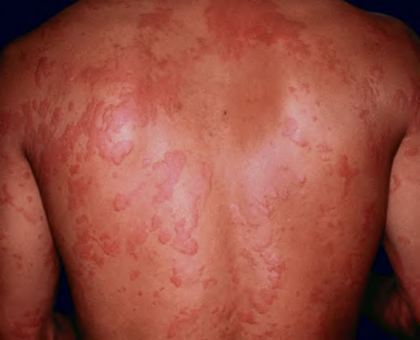
Fig. 21.2 Urticaria secondary to penicillin. Several of the lesions have a figurate appearance.
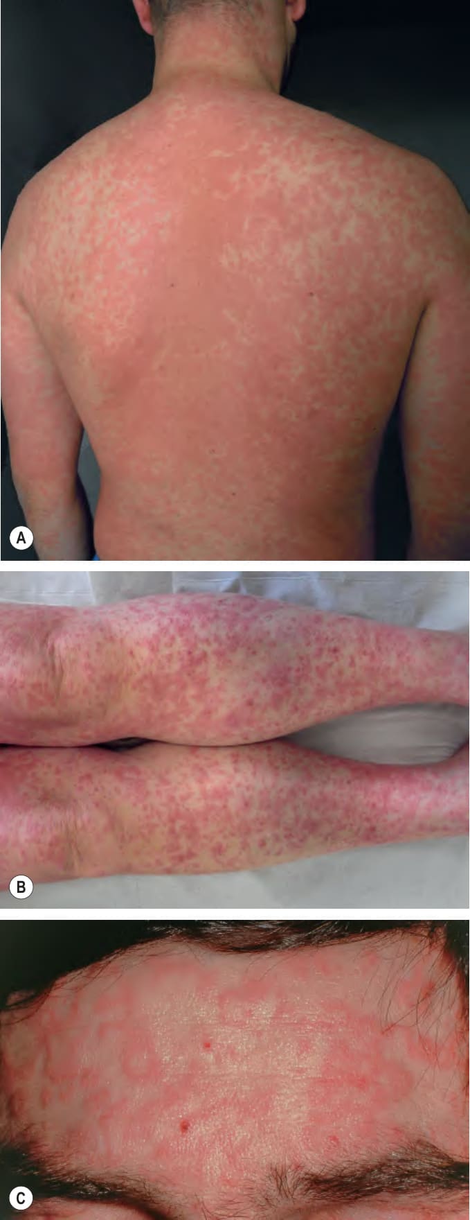
Fig. 21.3 Morbilliform (exanthematous) drug eruptions.A Erythematous papules and urticarial lesions with confluence on the midback induced by amoxicillin. B Due to dependency, lesions on the distal lower extremities can become petechial or purpuric. C Pink papules and annular lesions on the forehead due to phenobarbital. A, B, Courtesy Laurence Valeyrie-Allanore, MD.
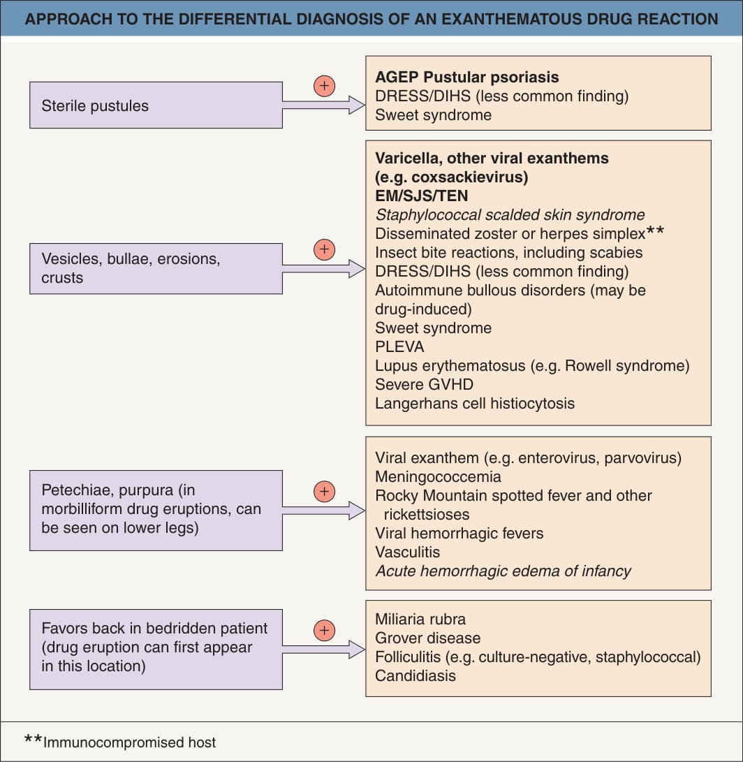
Fig. 21.4 Approach to the differential diagnosis of an exanthematous drug reaction. With a few exceptions (e.g. pityriasis rosea, drug-induced autoimmune bullous disorders), patients with these entities may be febrile. Entities in italics occur primarily in children and most common entities are in bold. Toxic shock syndrome can be staphylococcal or streptococcal (see Ch. 74). Drug-induced autoimmune bullous disorders: bullous pemphigoid or linear IgA bullous dermatosis > drug-induced pemphigus. Trimethoprim-sulfamethoxazoleassociated sudden conjunctivitis, lymphopenia, and rash combined with hemodynamic changes (SCoRCH) is a rare disorder that presents with generalized sunburn-like erythema, fever, hypotension, and mild facial and acral edema as well as kidney and/or liver involvement; it can appear within a day if prior exposure and between 4 to 11 days if no prior exposure.
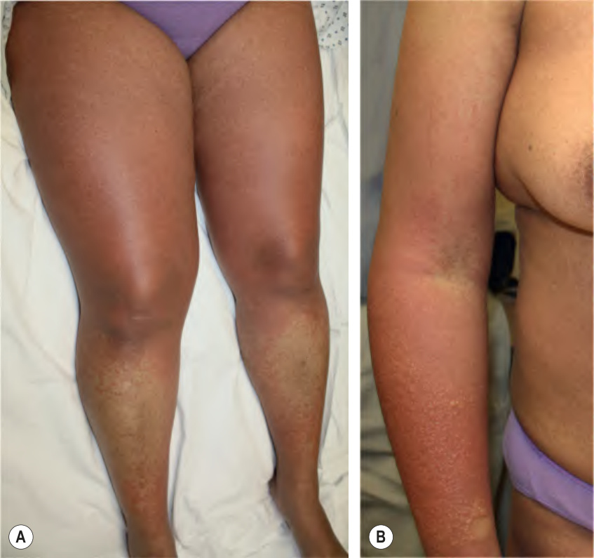
Fig. 21.5 Drug reaction with eosinophilia and systemic symptoms (DRESS)
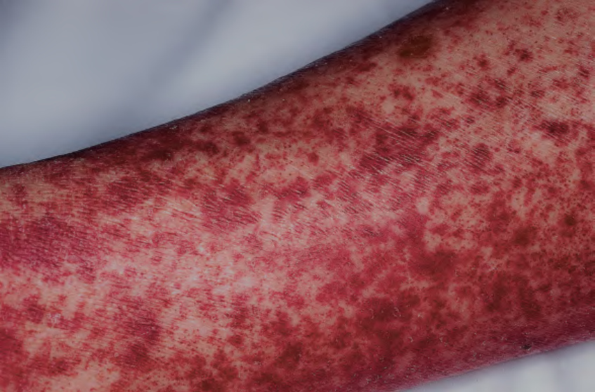
Fig. 21.6 Serum sickness due to antithymocyte globulin. The purpuric lesions are due to small vessel vasculitis in this patient with aplastic anemia. Courtesy Jean L. Bolognia, MD.
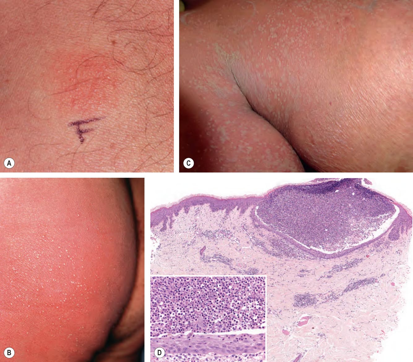
Fig. 21.7 Acute generalized exanthematous pustulosis (AGEP).
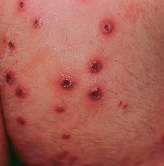
Fig. 21.8 Iododerma.
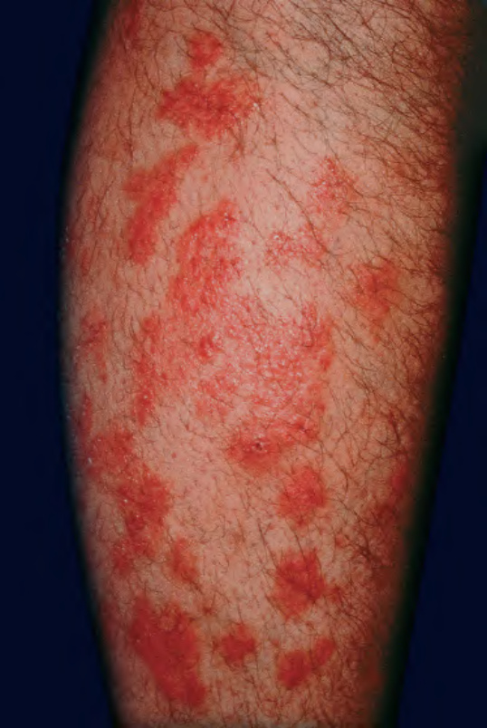
Fig. 21.9 Neutrophilic eccrine hidradenitis.
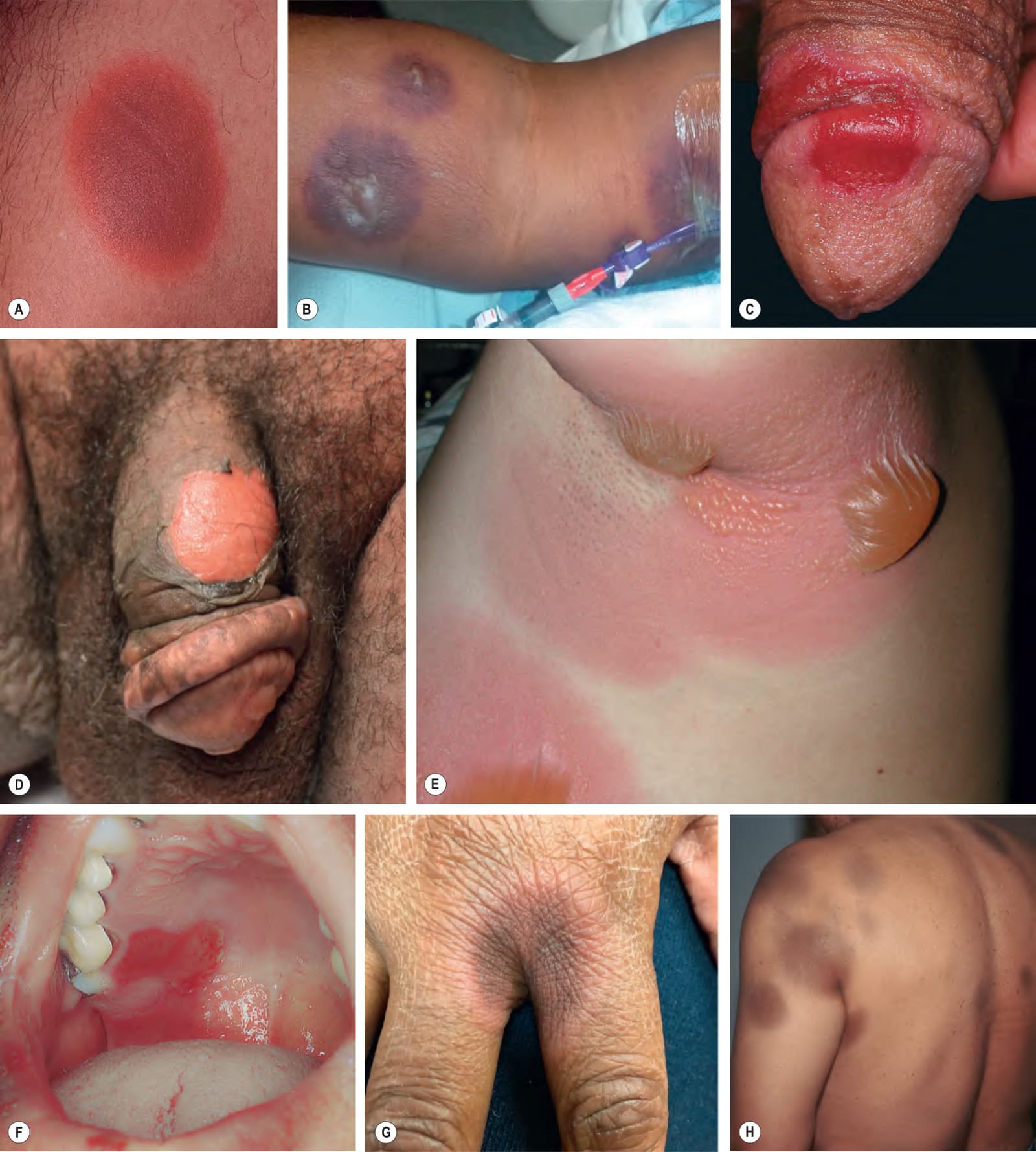
Fig. 21.10 Fixed drug eruptions.A An oval, well-demarcated red–brown plaque with a more erythematous border. B Multiple round violet-colored lesions with central bullae. C Erosive lesion of the penis due to epidermal detachment; this clinical presentation is sometimes misdiagnosed as recurrent HSV infection. D, E Generalized bullous FDE with involvement of the genitalia and intertriginous zones; erosions are seen following rupture of the bullae. Because of its more widespread distribution, this variant can be confused with TEN. F In the mouth, mucosal detachment leads to erosions. G As the inflammation resolves, the lesion acquires a brown hue. H Once inflammation is completely resolved, circular or oval areas of hyperpigmentation are commonly seen. Responsible drugs were phenolphthalein (A), acetaminophen (B), ciprofloxacin (C), naproxen (D), pseudoephedrine (E), allopurinol (F), metronidazole (G), and trimethoprim–sulfamethoxazole (H). B, Courtesy Jeffrey P. Callen, MD, and Tyler Geers, MD; C, F, G, Courtesy Kalman Watsky, MD; D, Courtesy Sara Perkins, MD; E, Courtesy Edward W. Cowen, MD; H, Courtesy Mary Stone, MD.
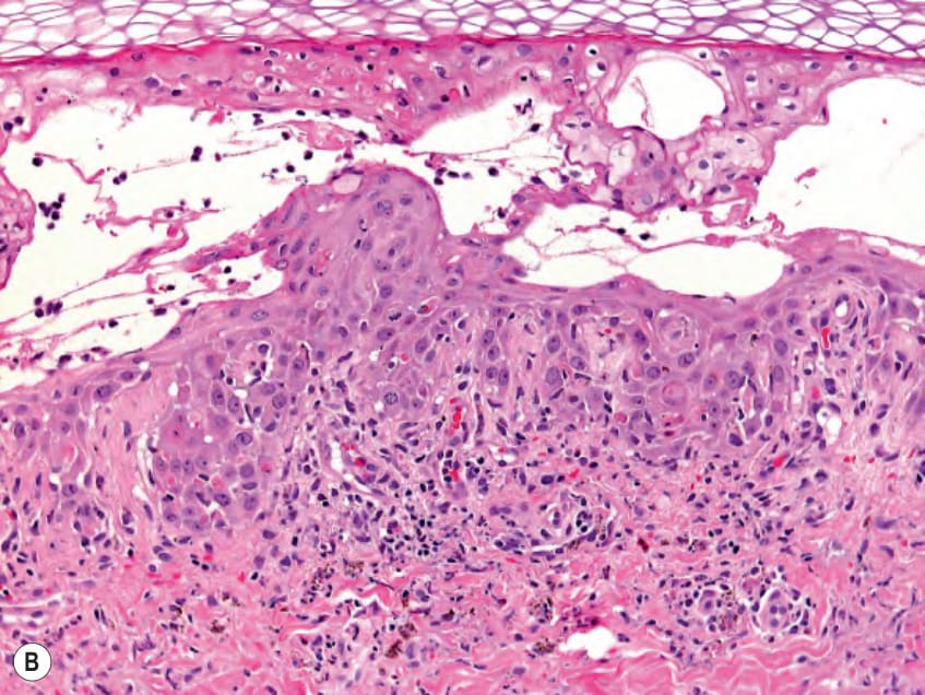
Fig. 21.11 Fixed drug eruptions – histopathologic features.A A few necrotic keratinocytes, mild vacuolar changes at the dermal–epidermal junction, papillary dermal edema, and melanophages can be seen as well as a mixed infiltrate with neutrophils and an occasional eosinophil. B Sometimes there is more extensive epidermal necrosis. Courtesy Lorenzo Cerroni, MD.
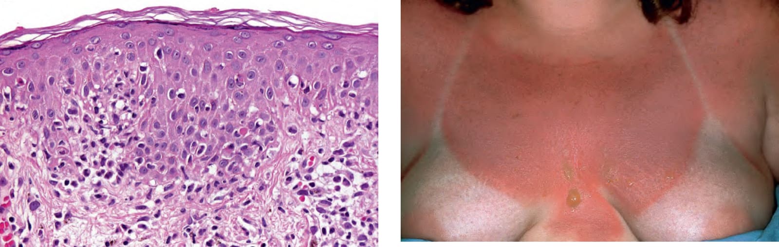
Fig. 21.12 Phototoxic reaction in a patient receiving methotrexate. The erythema and bullae are obviously limited to sun-exposed sites and resemble an exaggerated sunburn. Patients on methotrexate can also experience a “sunburn-recall” phenomenon.
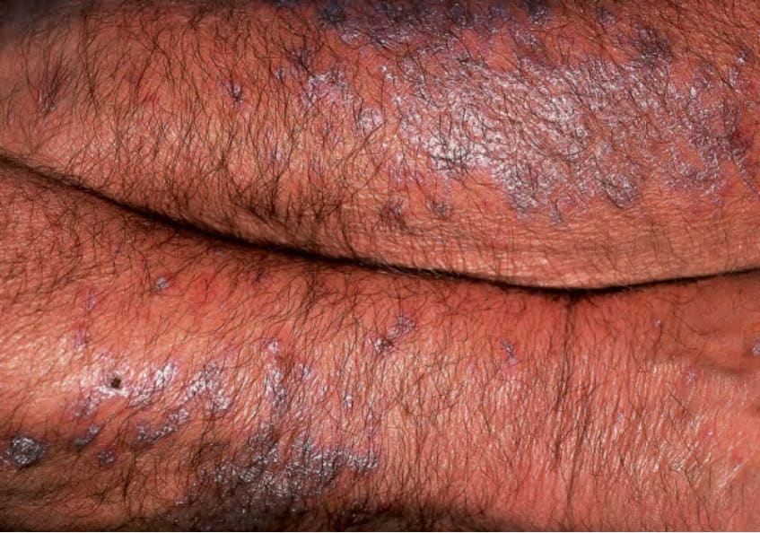
Fig. 21.13 Photolichenoid drug eruption due to hydrochlorothiazide. The lesions favored the extensor surfaces of the forearms.
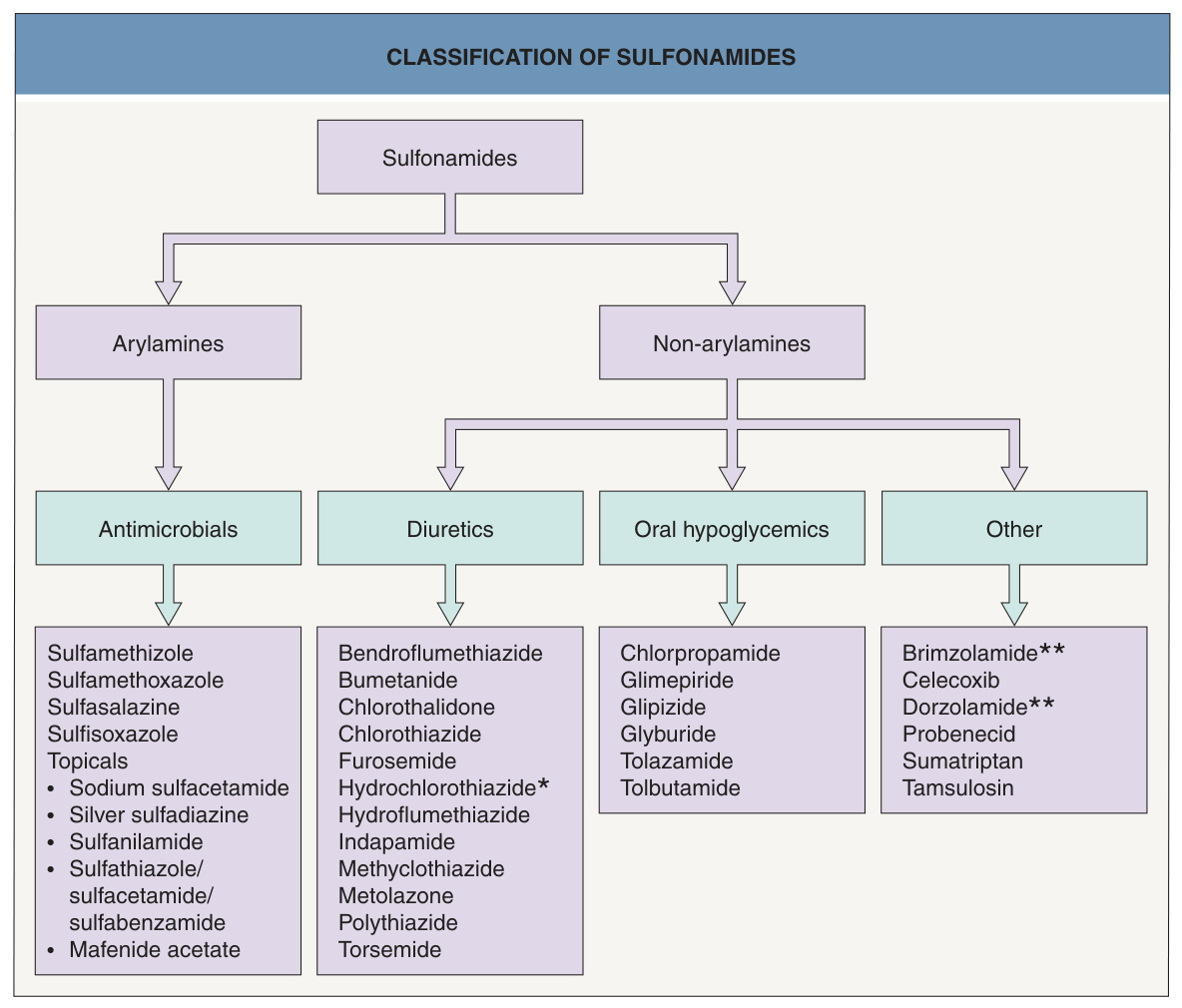
Fig. 21.14 Classification of sulfonamides.
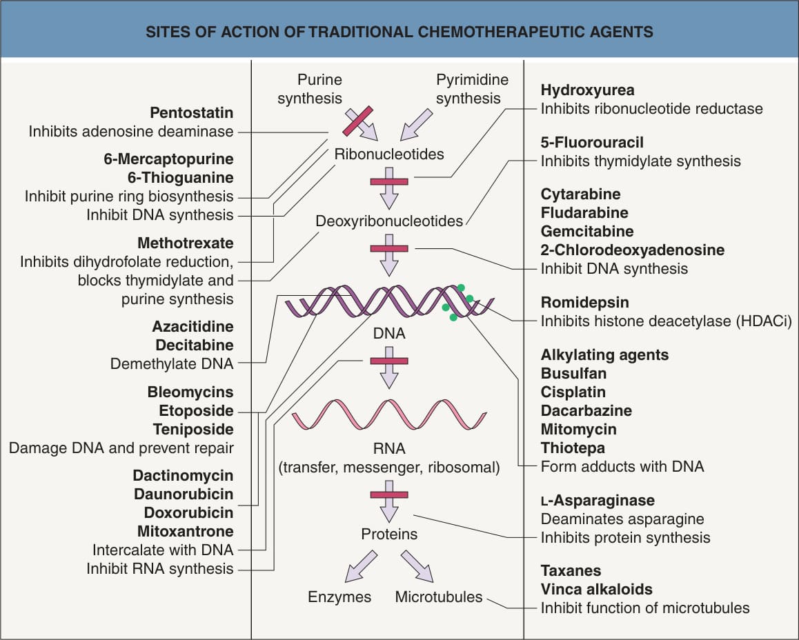
Fig. 21.15 Sites of action of chemotherapeutic agents.Adapted with permission from Brunton L, Lazo J, Parker K (eds.). Goodman & Gilman’s The Pharmacological Basis of Therapeutics, 11th edn. New York: McGraw-Hill Medical, 2005.
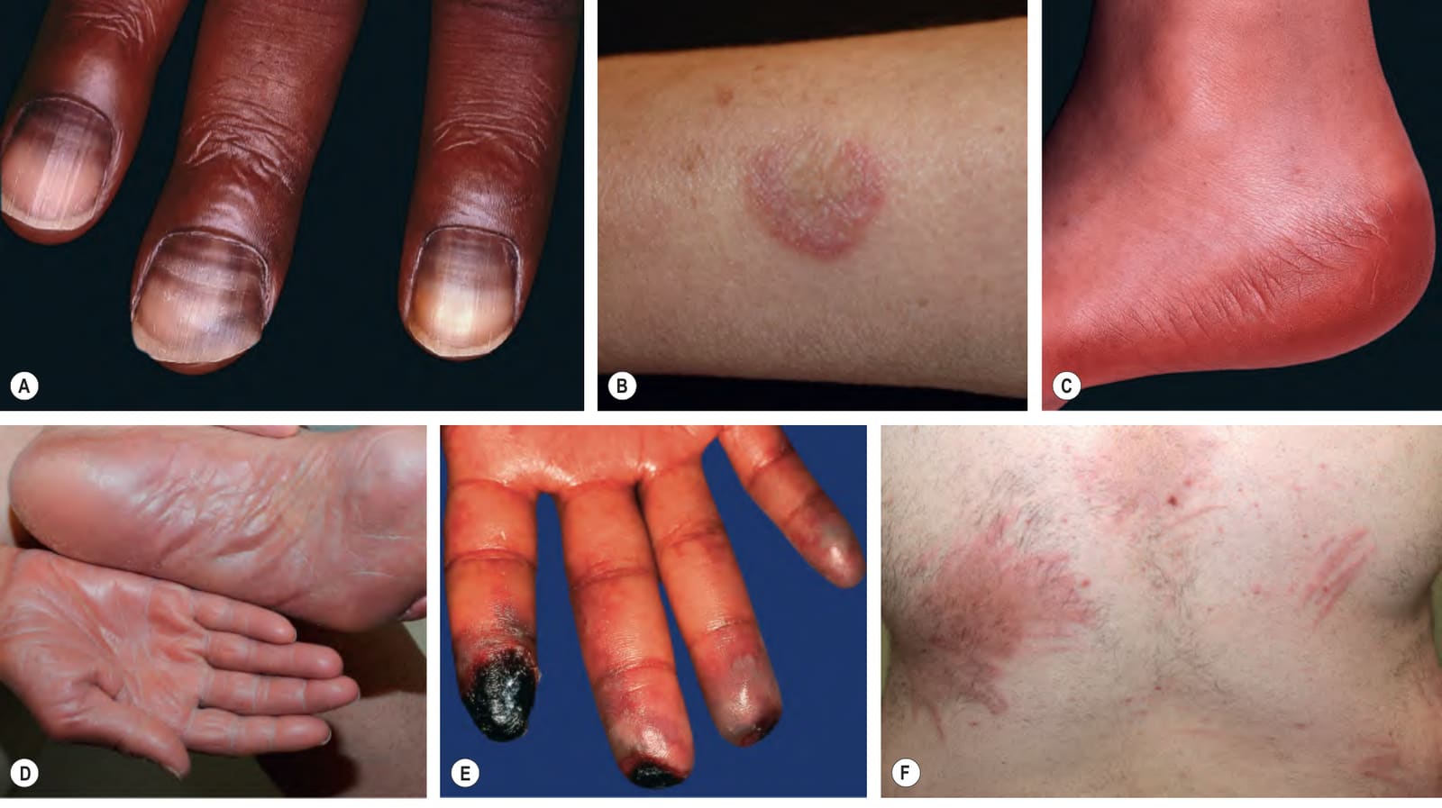
Fig. 21.16 Cutaneous side effects and complications of chemothera- peutic agents.A Horizontal melanonychia due to 5-fluorouracil. B Inflammation surrounding a seborrheic keratosis in a patient receiving paclitaxel. C Toxic erythema of chemotherapy (TEC) due to cytarabine, with obvious painful acral erythema involving the plantar surface. D TEC, also referred to as hand–foot syndrome when localized, due to capecitabine; diffuse glazed erythema and mild hyperkeratosis of palmoplantar skin. E Raynaud phenomenon and digital necrosis due to systemic bleomycin. F Multiple linear (flagellate) erythematous urticarial plaques in a patient receiving systemic bleomycin for Hodgkin lymphoma. G Chemical cellulitis due to extravasation of enfortumab vedotin (anti-Nectin-4 antibody attached to monomethyl auristatin, a cytotoxic microtubule inhibitor). A–C, Courtesy Jean L. Bolognia, MD; D, F, Courtesy Kalman Watsky, MD; G, Courtesy Katherine Given, MD.
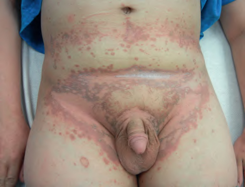
Fig. 21.17 Toxic erythema of chemotherapy. Symmetric involvement of inter-triginous zones and the scrotum. There is central desquamation and some of the lesions have a dusky color. The patient was receiving fludarabine plus intra-venous busulfan. Courtesy Leonard Kristal, MD.

Fig. 21.18 Toxic erythema of chemotherapy. Use of a number of terms (especially those based on histologic findings), including palmoplantar erythrodysesthesia, eccrine squamous syringometaplasia and epidermal dysmaturation, has created some confusion for clinicians. There is considerable overlap in the appearance of the symmetric erythematous to dusky patches which can develop edema, erosions, desquamation or purpura, whether they favor acral sites, intertriginous zones, or the elbows and knees. “Toxic erythema of chemotherapy” has been suggested as an encompassing term that allows simplification. In addition, there is no need to implicate additional diagnoses when lesions are not limited to the hands and feet. Insets: Erythema of the ears due to cytarabine (cytosine arabinoside), sometimes referred to as “Ara-C ears”; the petechiae are due to thrombocytopenia. Dusky edematous plaques of the palm, some of which have developed sterile bullae. Courtesy Jean L. Bolognia, MD and Boni Elewski, MD.
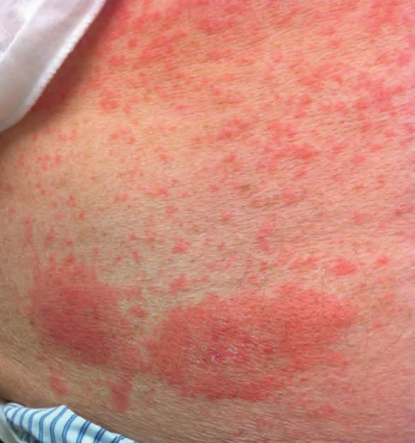
Fig. 21.19 Exanthematous (morbilliform) drug reaction to lenalidomide with accentuation at sites of bortezomib injections.Courtesy Dennis Cooper, MD.
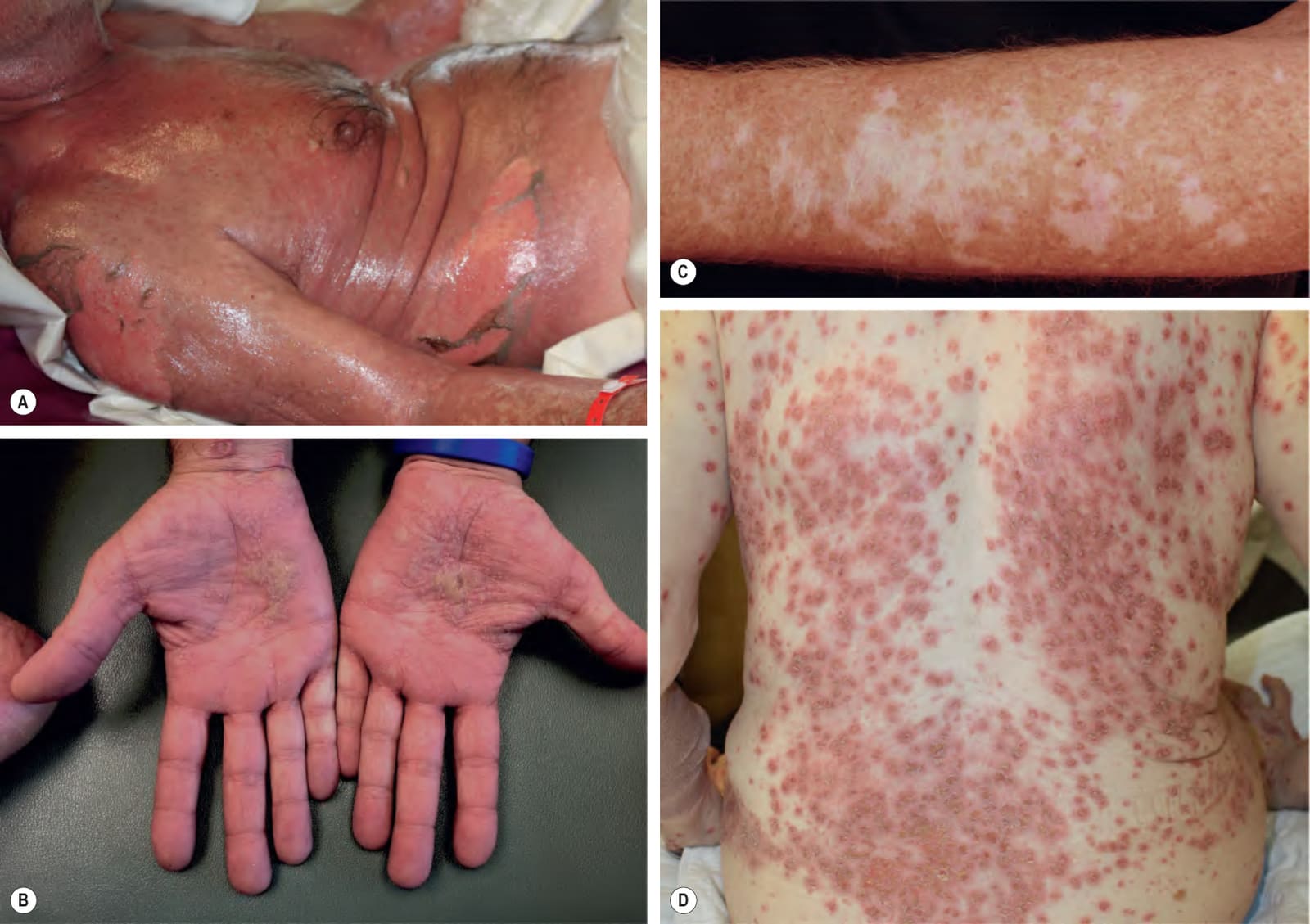
Fig. 21.20 Spectrum of cutaneous reactions to immune checkpoint inhibitors (immune-related adverse events).A SJS/TEN overlap due to ipilimumab. B Lichenoid dermatitis of the palms in a patient receiving pembrolizumab for metastatic colon cancer. C Vitiligo-like leukoderma in a patient with metastatic melanoma receiving nivolumab. D Widespread eruption of erythematous papules due to ipilimumab; many of the lesions are crusted. A, Courtesy Marianna Freudzon, MD; B, Courtesy Jonathan Leventhal, MD. C, Courtesy Jean L. Bolognia, MD; D, Courtesy Miriam Totonchy, MD.
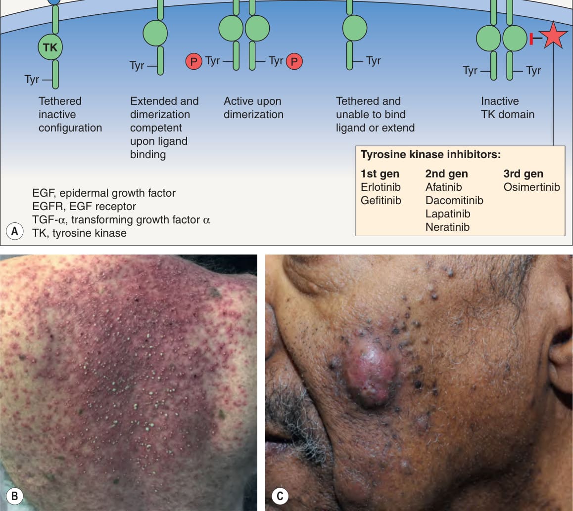
Fig. 21.21 Papulopustular (acneiform) reactions to epidermal growth factor receptor (EGFR) inhibitors.
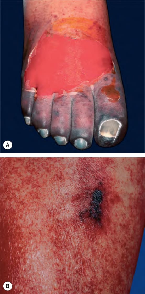
Fig. 21.22 Heparin-induced thrombocytopenia (HIT) syndrome.
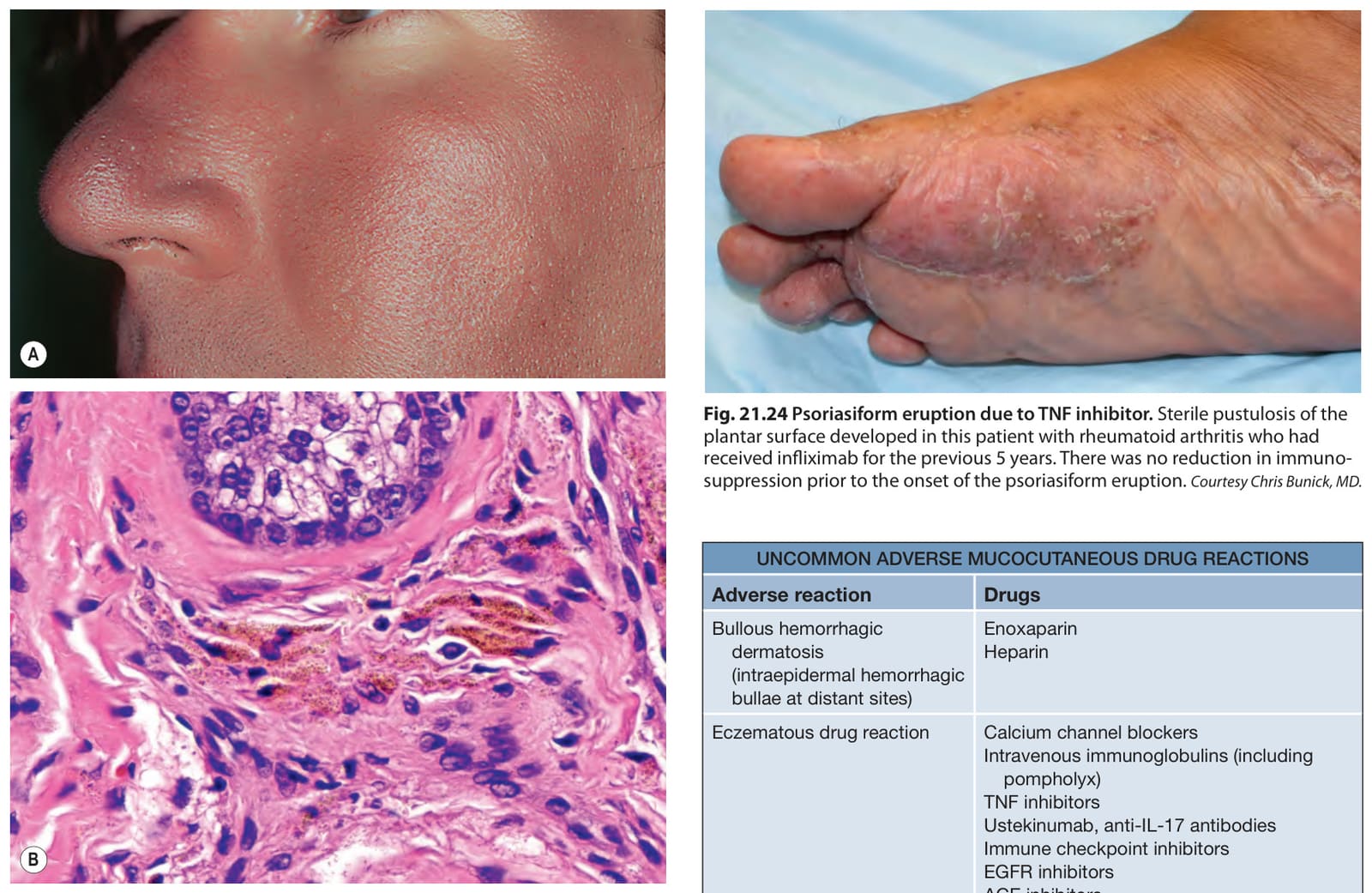
Fig. 21.23 Cutaneous discoloration due to amiodarone.A Gray–violet discoloration of the face. Note sparing of the lower eyelid. B Biopsy specimens demonstrate yellow–brown granules within dermal macrophages. A, Courtesy Jean L. Bolognia, MD; B, Courtesy Luis Requena, MD.
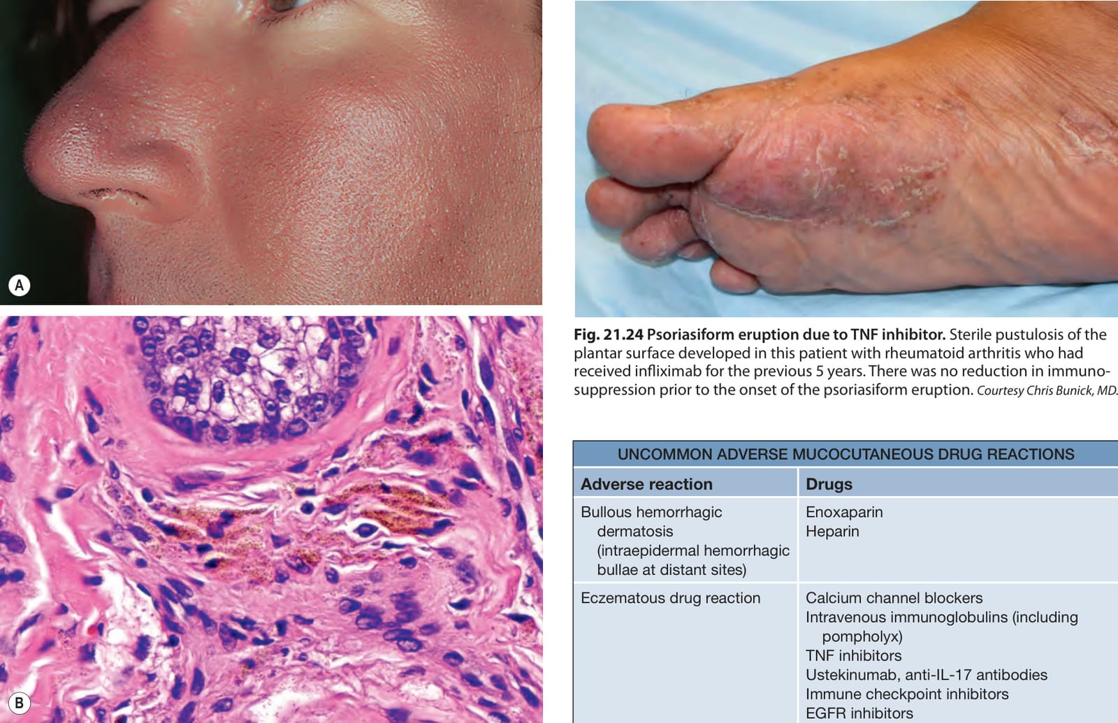
Fig. 21.24 Psoriasiform eruption due to TNF inhibitor. Sterile pustulosis of the plantar surface developed in this patient with rheumatoid arthritis who had received infliximab for the previous 5 years. There was no reduction in immunosuppression prior to the onset of the psoriasiform eruption. Courtesy Chris Bunick, MD.
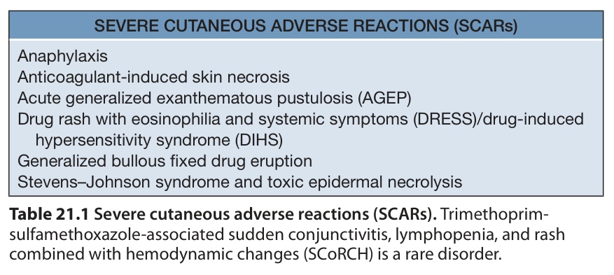
Table 21.1 Severe cutaneous adverse reactions (SCARs). Trimethoprim-sulfamethoxazole-associated sudden conjunctivitis, lymphopenia, and rash combined with hemodynamic changes (SCoRCH) is a rare disorder.
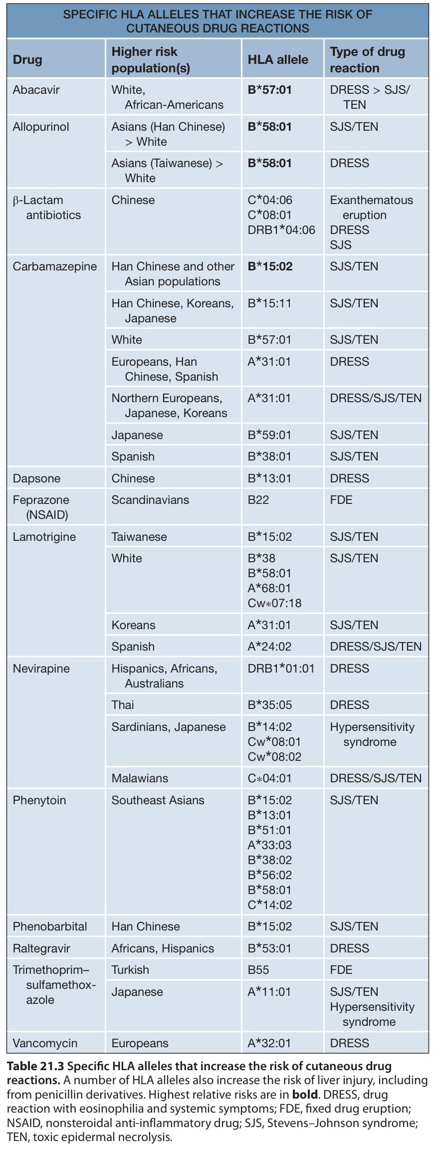
Table 21.3 Specific HLA alleles that increase the risk of cutaneous drug reactions. A number of HLA alleles also increase the risk of liver injury, including from penicillin derivatives. Highest relative risks are in bold. DRESS, drug reaction with eosinophilia and systemic symptoms; FDE, fixed drug eruption; NSAID, nonsteroidal anti-inflammatory drug; SJS, Stevens–Johnson syndrome; TEN, toxic epidermal necrolysis.
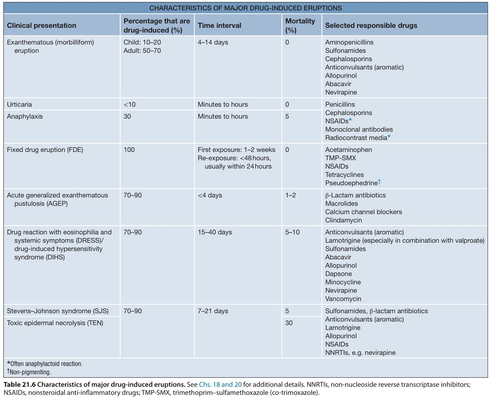
Table 21.6 Characteristics of major drug-induced eruptions. See Chs. 18 and 20 for additional details. NNRTIs, non-nucleoside reverse transcriptase inhibitors; NSAIDs, nonsteroidal anti-inflammatory drugs; TMP-SMX, trimethoprim–sulfamethoxazole (co-trimoxazole).
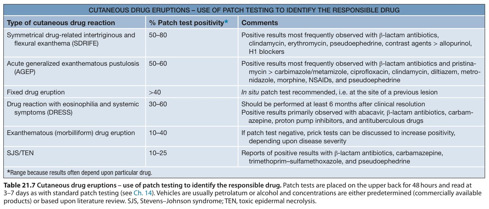
Table 21.7 Cutaneous drug eruptions – use of patch testing to identify the responsible drug. Patch tests are placed on the upper back for 48 hours and read at 3–7 days as with standard patch testing (see Ch. 14). Vehicles are usually petrolatum or alcohol and concentrations are either predetermined (commercially available products) or based upon literature review. SJS, Stevens–Johnson syndrome; TEN, toxic epidermal necrolysis.
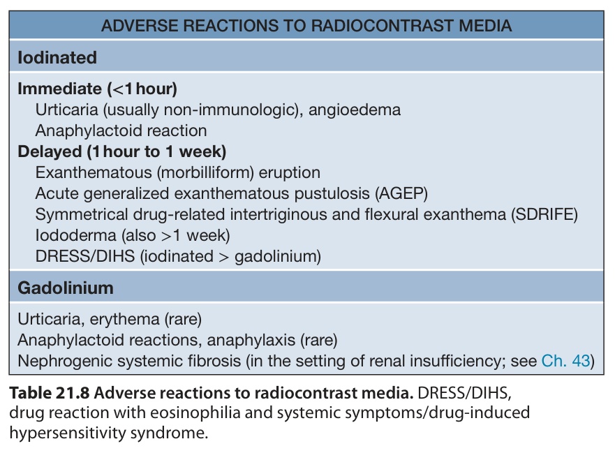
Table 21.8 Adverse reactions to radiocontrast media. DRESS/DIHS, drug reaction with eosinophilia and systemic symptoms/drug-induced hypersensitivity syndrome.
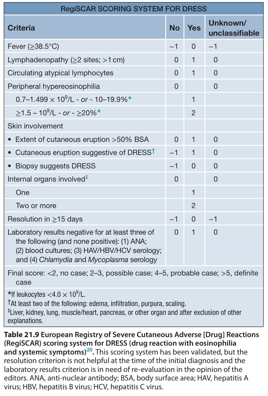
Table 21.9 European Registry of Severe Cutaneous Adverse [Drug] Reactions (RegiSCAR) scoring system for DRESS (drug reaction with eosinophilia and systemic symptoms). This scoring system has been validated, but the resolution criterion is not helpful at the time of the initial diagnosis and the laboratory results criterion is in need of re-evaluation in the opinion of the editors. ANA, anti-nuclear antibody; BSA, body surface area; HAV, hepatitis A virus; HBV, hepatitis B virus; HCV, hepatitis C virus.
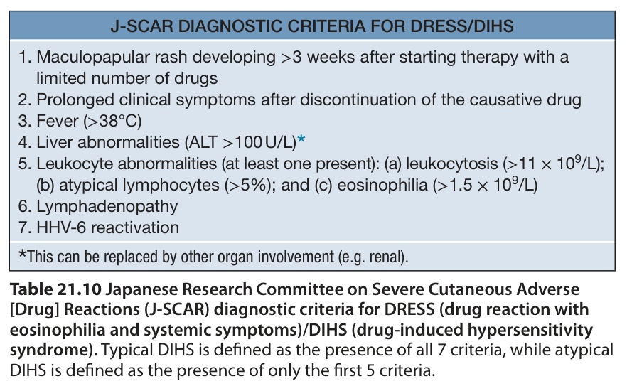
Table 21.10 Japanese Research Committee on Severe Cutaneous Adverse [Drug] Reactions (J-SCAR) diagnostic criteria for DRESS (drug reaction with eosinophilia and systemic symptoms)/DIHS (drug-induced hypersensitivity syndrome). Typical DIHS is defined as the presence of all 7 criteria, while atypical DIHS is defined as the presence of only the first 5 criteria.
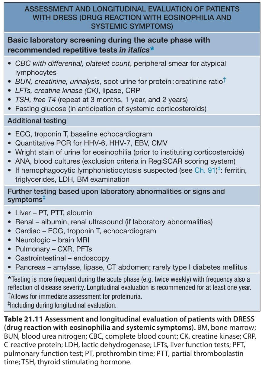
Table 21.11 Assessment and longitudinal evaluation of patients with DRESS
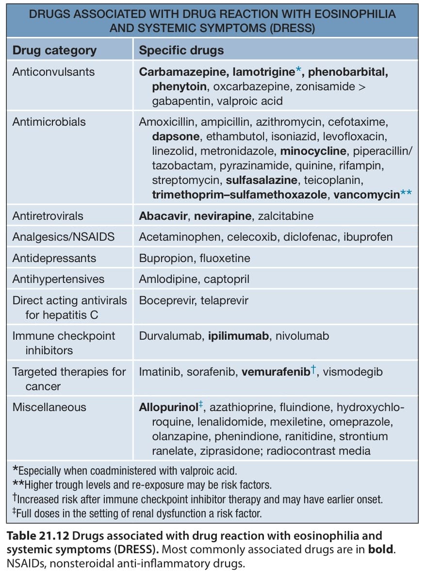
Table 21.12 Drugs associated with drug reaction with eosinophilia and systemic symptoms (DRESS). Most commonly associated drugs are in bold. NSAIDs, nonsteroidal anti-inflammatory drugs.
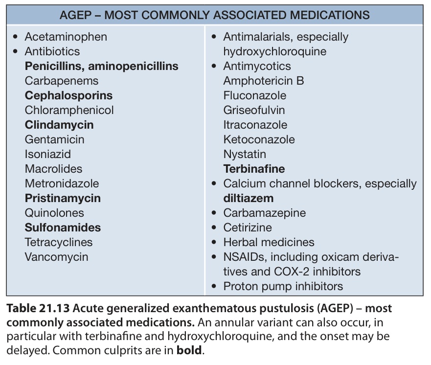
Table 21.13 Acute generalized exanthematous pustulosis (AGEP) – most commonly associated medications. An annular variant can also occur, in particular with terbinafine and hydroxychloroquine, and the onset may be delayed. Common culprits are in bold.
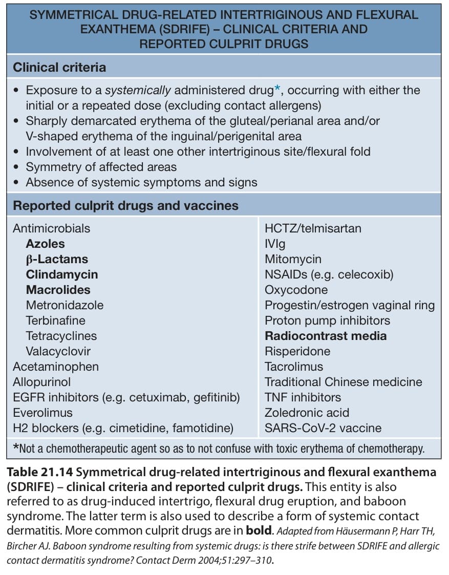
Table 21.14 Symmetrical drug-related intertriginous and flexural exanthema
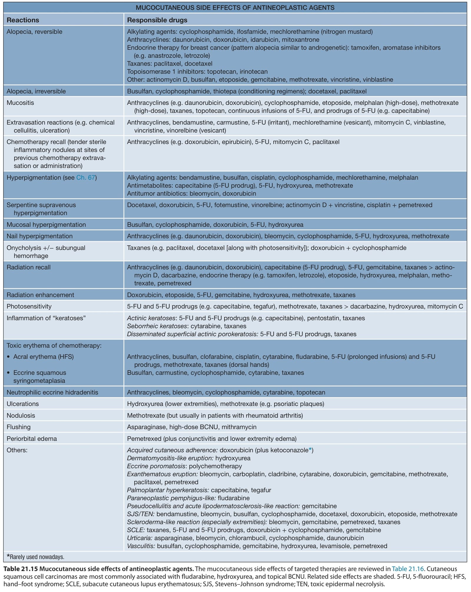
Table 21.15 Mucocutaneous side effects of antineoplastic agents. The mucocutaneous side effects of targeted therapies are reviewed in Table 21.16. Cutaneous squamous cell carcinomas are most commonly associated with fludarabine, hydroxyurea, and topical BCNU. Related side effects are shaded. 5-FU, 5-fluorouracil; HFS, hand–foot syndrome; SCLE, subacute cutaneous lupus erythematosus; SJS, Stevens–Johnson syndrome; TEN, toxic epidermal necrolysis.
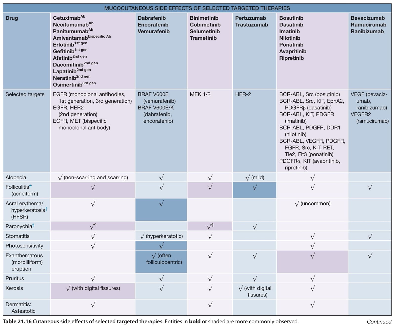
Table 21.16 Cutaneous side effects of selected targeted therapies. Entities in bold or shaded are more commonly observed.
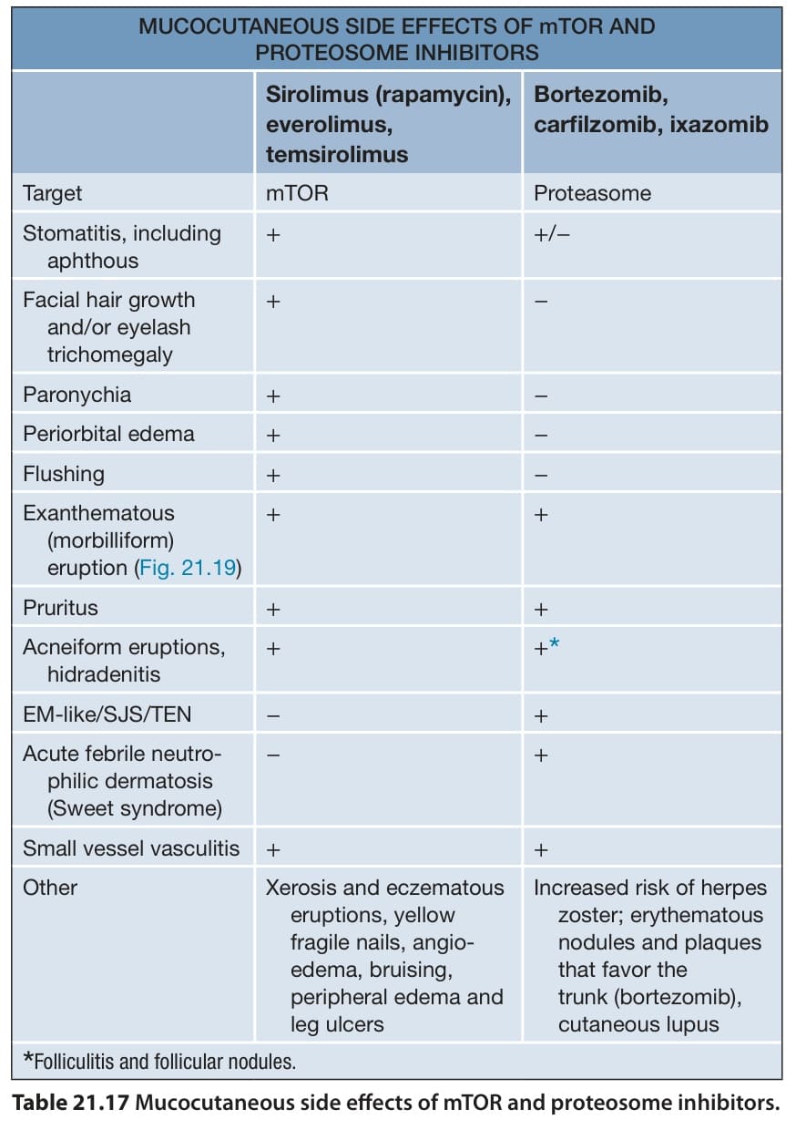
Table 21.17 Mucocutaneous side effects of mTOR and proteosome inhibitors.
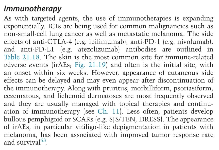
Table 21.18 Mucocutaneous and extracutaneous side effects of immune checkpoint inhibitors. These include anti-CTLA-4 (e.g. ipilimumab, tremelimumab), anti-PD-1 (e.g. nivolumab, pembrolizumab, cemiplimab), and anti-PD-L1 (e.g. atezolizumab, avelumab, durvalumab) antibodies. Certain reactions are more commonly associated with anti-PD-1/PD-L1 antibodies (e.g. lichenoid eruption, bullous pemphigoid, psoriasiform eruption), and combination anti-CTLA-4/-PD-1 therapy is associated with increased frequency and severity of immune-related adverse events. Rarely, subcutaneous fat necrosis, circumoral plasmacytosis, and repigmentation of gray hair have been observed. DRESS, drug reaction with eosinophilia and systemic symptoms; PLEVA, pityriasis lichenoides et varioliformis acuta; PLC, pityriasis lichenoides chronica; PRES, posterior reversible encephalopathy syndrome.
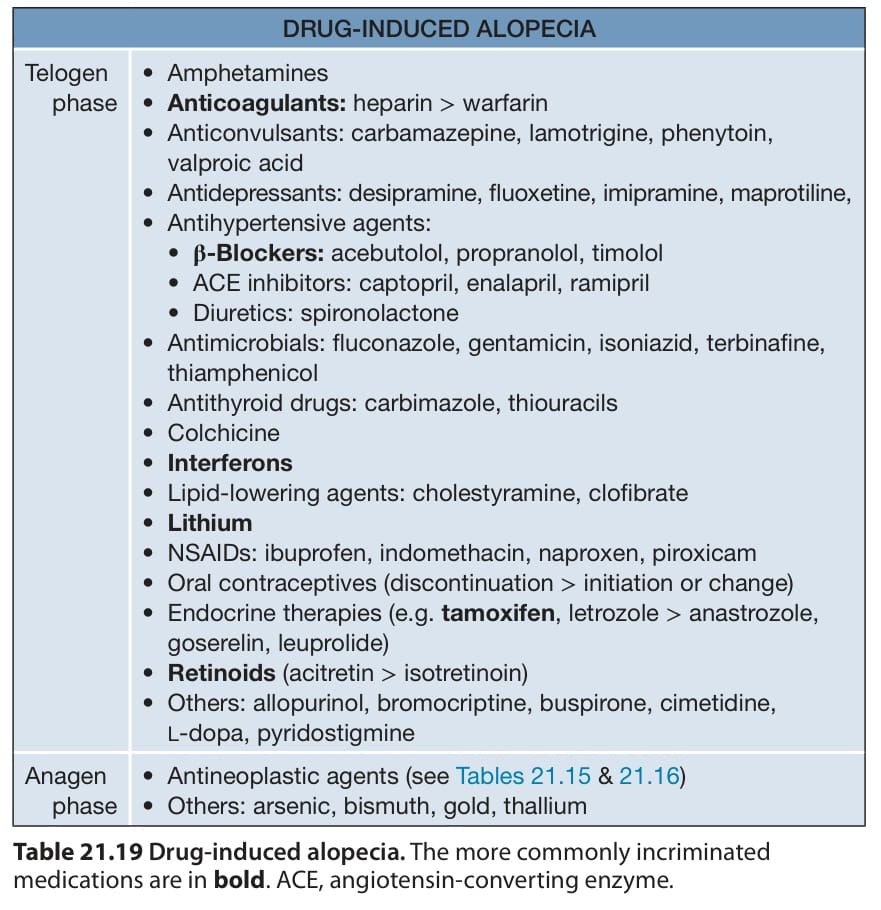
Table 21.19 Drug-induced alopecia. The more commonly incriminated medications are in bold. ACE, angiotensin-converting enzyme.
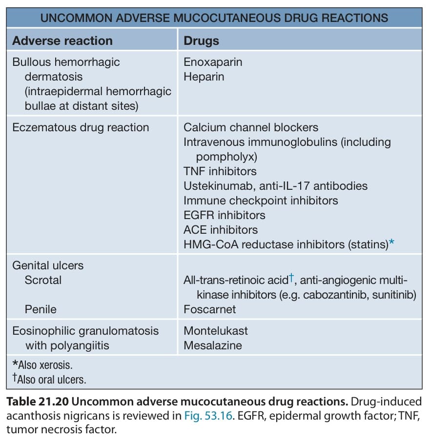
Table 21.20 Uncommon adverse mucocutaneous drug reactions. Drug-induced acanthosis nigricans is reviewed in Fig. 53.16. EGFR, epidermal growth factor; TNF, tumor necrosis factor.
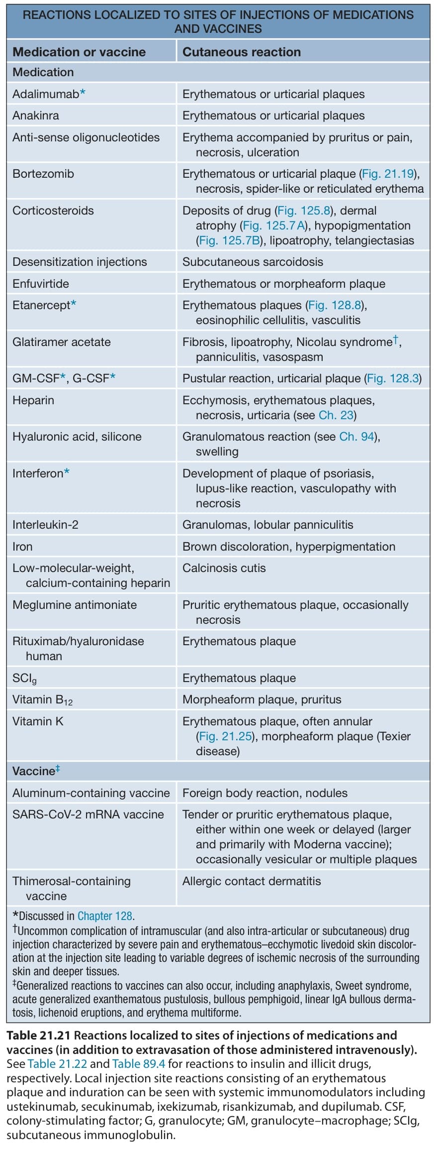
Table 21.21 Reactions localized to sites of injections of medications and vaccines (in addition to extravasation of those administered intravenously).
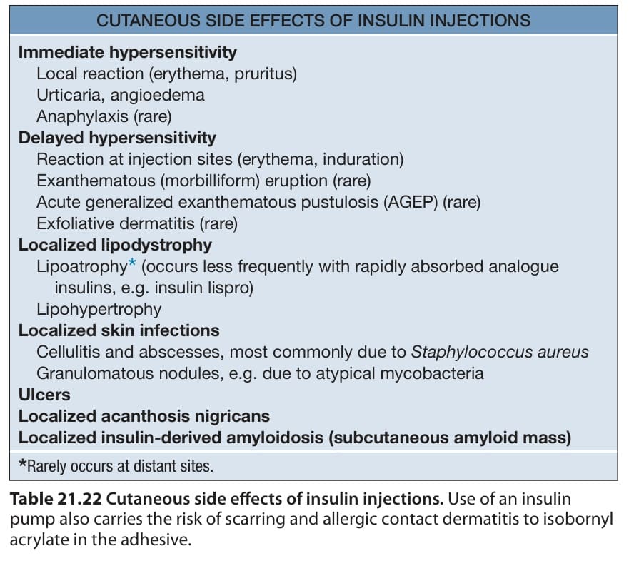
Table 21.22 Cutaneous side effects of insulin injections. Use of an insulin pump also carries the risk of scarring and allergic contact dermatitis to isobornyl acrylate in the adhesive.
K injections. A This patient was originally diagnosed as having erythema multiforme. B Large pink–violet plaque with areas of hemorrhage in an infant. B, Courtesy Julie V. Schaffer, MD.
Additional figures and tables on Skin reactions to “drugs” received by at least 1000 patients, Drugs received by more than 1000 patients with no skin reactions (rates estimated to be ≤3 per 1000), Features that suggest a severe cutaneous adverse reaction (SCAR) to a drug, Frequency of clinical and laboratory features in drug reaction with eosinophilia and systemic symptoms (DRESS)/drug-induced hypersensitivity syndrome (DIHS), Severity score of drug reaction with eosinophilia and systemic symptoms (DRESS)/drug-induced hypersensitivity syndrome (DIHS), and Management of immune checkpoint inhibitor cutaneous side effects, available in our eBook (see inside front cover for access code).
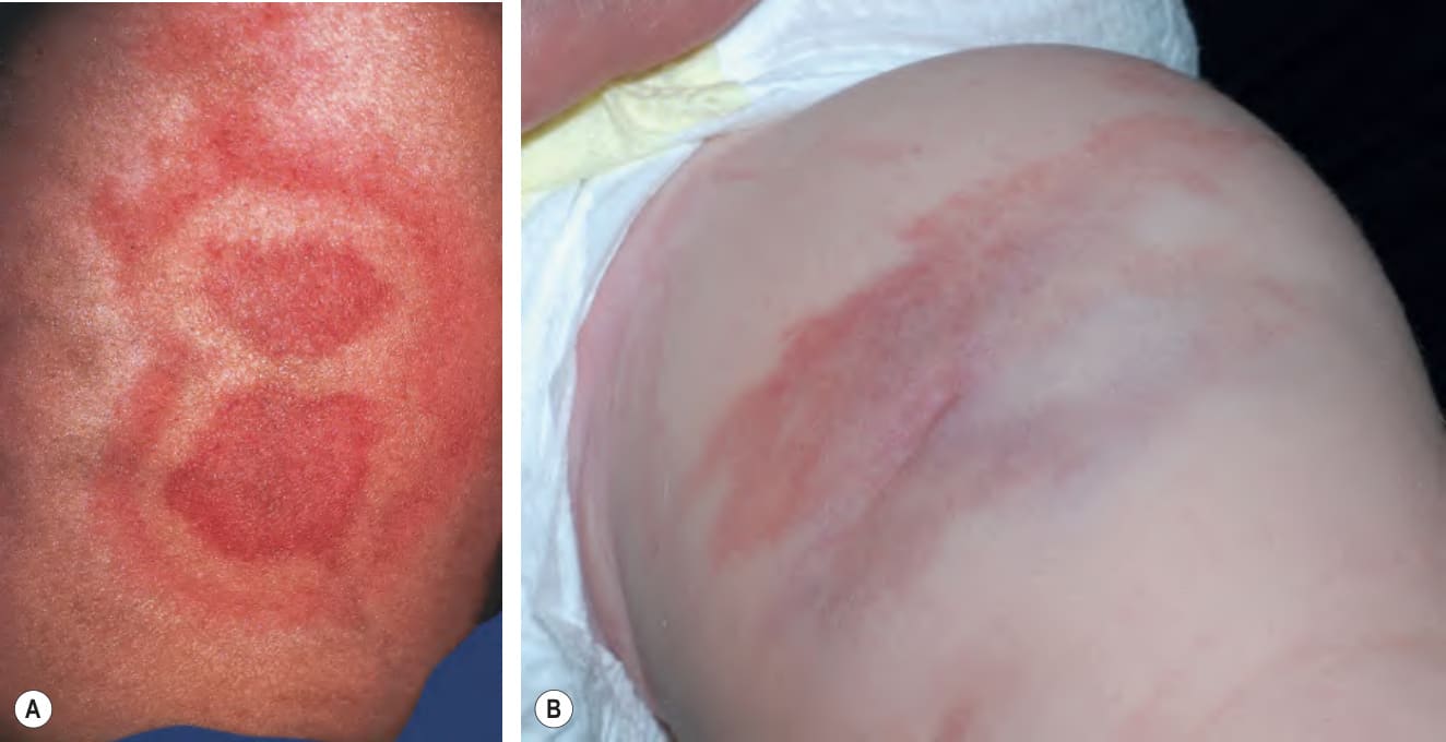
Fig. 21.25 Local reactions to vitamin