INTRODUCTION
This chapter addresses the evaluation and classification of patients pre-senting with purpura, defined as visible hemorrhage within the skin or mucous membranes. The differential diagnosis presented in this chapter is directed toward syndromes of primary purpura, where the hemorrhage is an integral part of lesion formation, rather than hemorrhage secondary to causes such as trauma and scratching or hemorrhage into established lesions due to dependency (e.g. stasis dermatitis, eczema craquelé).
This chapter outlines a diagnostic approach based upon lesion number and distribution pattern, with a further focus on the morphology of the purpuric lesions. The initial goal is to identify the most plausible of three broad categories: simple hemorrhage (vascular breach), inflammatory hemorrhage (vessel-directed inflammation), or microvascular occlusion/ischemia. Assigning the patient to one of these three major categories facilitates a more directed, efficient, and accurate evaluation to prove or disprove the suspected pathomechanism and it serves to narrow the list of possible diagnoses.
Warren W. Piette
Figure 22.1 illustrates this diagnostic approach based upon lesion number and distribution pattern. Interpretation of these clinical findings is then modified by lesional morphology, extracutaneous findings, and clinical context. For example, petechiae appearing in a generalized pattern requires exclusion of severe thrombocytopenia or perhaps thrombotic thrombocytopenic purpura, but they are most often due to a viral or drug eruption.
When widespread, purpuric lesions in a severely ill patient raise concern for serious drug eruptions (e.g. Stevens–Johnson syndrome) or some vasculitides whereas generalized retiform purpura in the setting of multi-organ disease suggests fulminant microvascular disease (e.g. catastrophic antiphospholipid syndrome) or sepsis-associated disseminated intravascular coagulation (DIC) with purpura fulminans. Predominantly acral lesions are divided into two subsets: (1) involvement of the ears and nose as well as the hands and feet, typically triggered by cold exposure; and (2) a more widespread distribution pattern with prominent involvement of the hands and feet, typically in the absence of cold exposure. A pauci-random distribution pattern is typical
of ANCA-associated vasculitides, various microvascular occlusive diseases, or lymphomatoid papulosis, and occasionally cutaneous polyarteritis nodosa (PAN). Multi-dependent patterns can result from severe thrombocytopenia but in the practice of dermatology is most commonly due to cutaneous small vessel vasculitis (see Ch. 24).
In 1989, the term retiform purpura was coined to describe a sign of certain vasculitic subsets, but nowadays it is generally used to denote a frequent marker of thrombotic or occlusive microvascular disease. The morphologies of purpuric lesions are divided into two major sub-sets: (1) exclusively macular, whose shapes can be round, oval, irregular, or geometric; and (2) palpable and/or retiform. The macular group consists of petechiae (Fig. 22.2A, Table 22.1), macular purpura (Table 22.2), and macular ecchymoses (Fig. 22.2B, Table 22.3). The palpable and/or retiform group consists of palpable purpura (Fig. 22.2C, Table 22.4), non-inflammatory retiform purpura (Fig. 22.2D; Table 22.5), and inflammatory retiform purpura (Table 22.6). In this chapter, lesions in the macular group are categorized on the basis of size and are defined as non-blanching and non-palpable; those in the palpable and/or retiform group can range in size from a few millimeters to several centimeters in diameter, may be palpable, and may partially blanch with pressure. The relevance of these morphologic subsets when sorting through the differential diagnosis is depicted in Figure 22.3.
Two major causes of purpura – microvascular occlusion syndromes and vasculitis – are discussed in Chapters 23 and 24. Occlusion is important to recognize because it may mimic vasculitis but requires a very different approach to determine its etiology and appropriate therapy. The most typical presentation of microvascular occlusion syndromes is retiform purpura (see Table 22.5).
Livedo reticularis is a reflection of the physiologic anatomy of slow flow states. The three-dimensional structure and flow regulation of the dermal and subcutaneous vasculature give rise to the net-like pattern of livedo reticularis (see Fig. 106.1). The diameter of the almost-circular to polygonal individual units within the net-like grid varies from 2 cm or larger on the back to 5 mm or less on the palms or soles.
Round to oval petechiae, <3 mm in diameter. B Solar (actinic) purpura in sites of actinic damage plus trauma. C Palpable purpura due to cutaneous small vessel vasculitis (inflammation plus hemorrhage). D Non-inflammatory (bland) retiform purpura as well as hemorrhagic bullae in a patient with disseminated intravascular coagulation (DIC). B, Courtesy Kalman Watsky, MD; D, Courtesy Judit Stenn, MD.
Retiform purpura morphology results from involvement of the same vessels that produce the livedo reticularis pattern. While the former was traditionally considered a component of livedo reticularis, the two should be distinguished based upon the presence or absence of purpura, hence the term “retiform purpura”. Given the size of dermal vessels, the clot within the vessel is too small to be seen grossly. What is actually observed is hemorrhage around the dermal vessels, presumably due to ischemia with hemorrhage prior to complete occlusion of the vessel. A complete reticulate pattern is seen infrequently. Instead, the morphology of retiform purpura is composed of “puzzle pieces” of the livedo reticularis pattern. While some lesions have a branching appearance, very few, if any, are truly stellate, i.e. a central area of necrosis or hemorrhage with a radiating star-like pattern composed of straight lines.
Retiform lesions can be inflammatory or non-inflammatory, but the age of the lesion may blur this morphologic distinction, so in practice both groups of diagnoses need to be considered (see Tables 22.5 & 22.6). Inflammatory retiform purpura is accompanied by peripheral erythema, but the wound healing response can eventually lead to erythema and leukocytoclasis in lesions initiated by non-inflammatory occlusion (Fig. 22.4). As a corollary, vasculitis sometimes produces lesions that have minimal early erythema and lesions lose erythema during resolution.
Lastly, the retiform lesions of immune complex vasculitis are invariably sparse in number and are accompanied by many round purpuric lesions. In addition, these retiform lesions are typically more finely branched than either occlusive or ANCA-associated retiform purpura. However, when within confluent areas of purpura, they may have more intense branching hemorrhage or necrosis. Of note, vasculitic retiform lesions do not simply represent the crowding or collision of more classic round purpuric lesions, and they may occur with or without cutaneous arteritis.
The presence of a multi-dependent round and retiform pattern (many round/few retiform lesions) is seen most commonly with three types of immune complex vasculitis: (1) IgA-predominant; (2) autoimmune connective tissue disease-associated; and (3) mixed cryoglobulinemia. This pattern of round and retiform purpuric lesions may occasionally
NSAIDs, nonsteroidal anti-inflammatory drugs.
be seen in microscopic polyangiitis or granulomatous polyangiitis, but the lesions are fewer in number and more randomly distributed. The combination of many round/few retiform lesions is not seen with simple hemorrhage or microvascular occlusive disease.
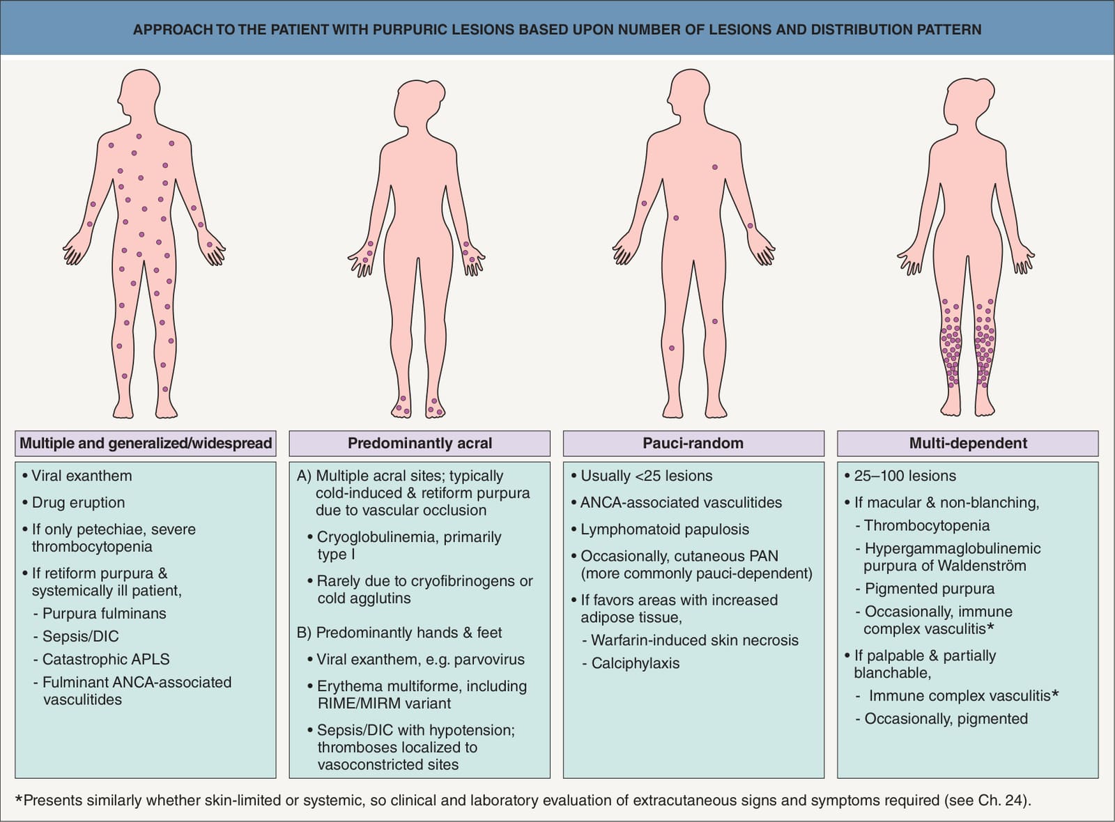
Fig. 22.1 Approach to the patient with purpuric lesions based upon number of lesions and distribution pattern. APLS, antiphospholipid antibody syndrome; DIC, disseminated intravascular coagulation; MIRM, Mycoplasma-induced rash and mucositis; RIME, reactive infectious mucocutaneous eruption; PAN, polyarteritis nodosa.
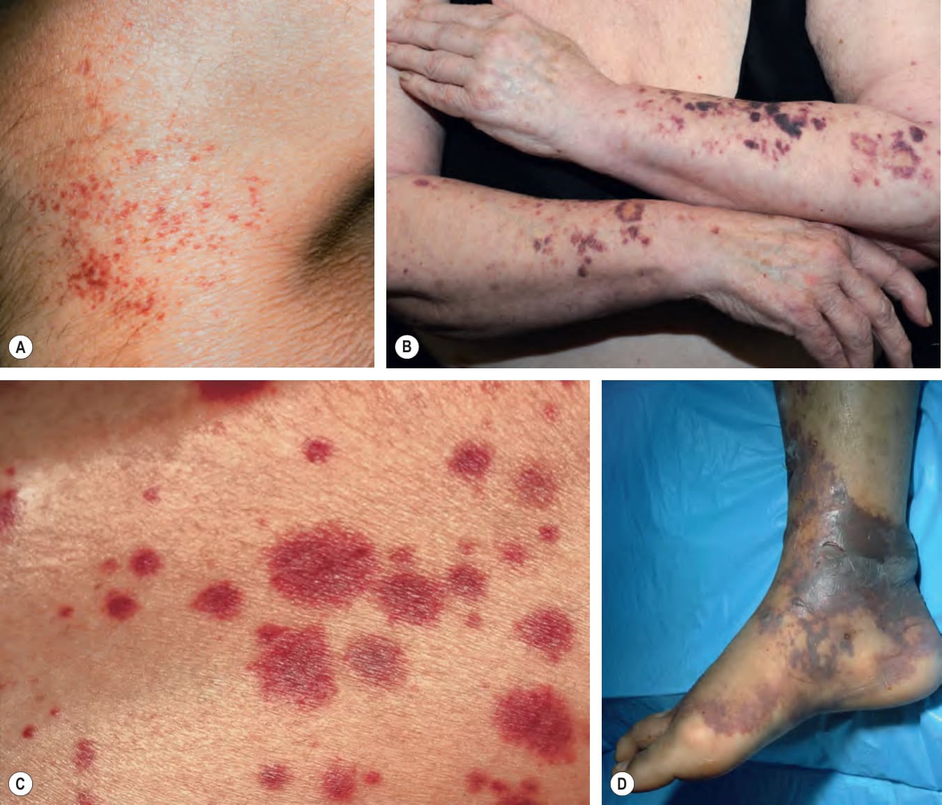
Fig. 22.2 Clinical examples of petechiae and purpura.A
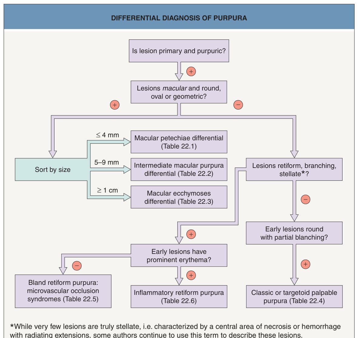
Fig. 22.3 Differential diagnosis of purpura.
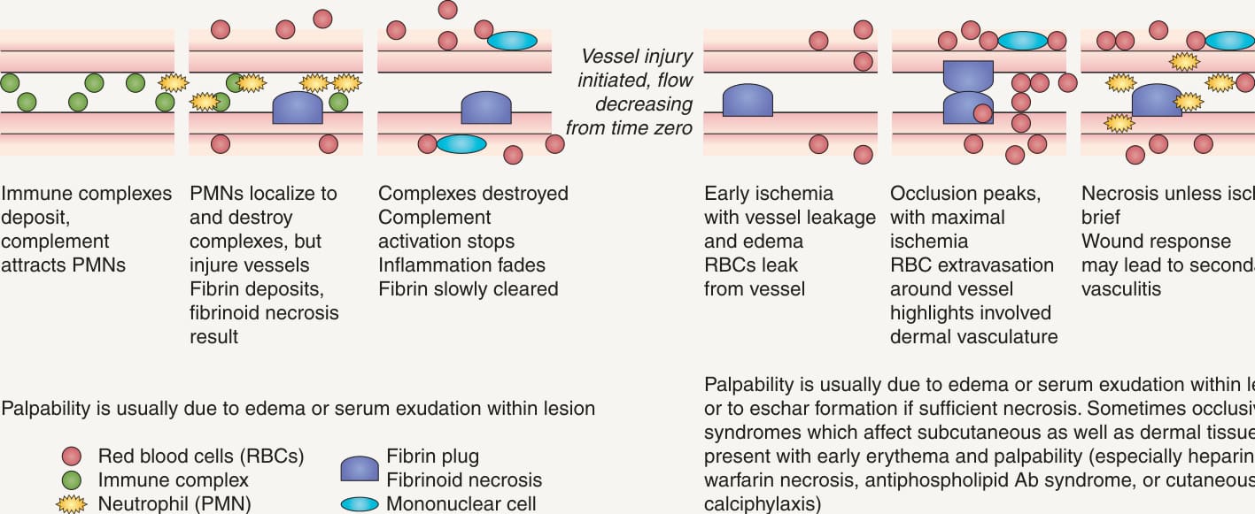
Fig. 22.4 Time course for skin lesions due to vasculitis versus microvascular occlusion.A Time course for lesions due to immune complex-mediated leukocytoclastic vasculitis. B Time course for lesions due to microvascular occlusion. Ab, antibody; tr, trace.
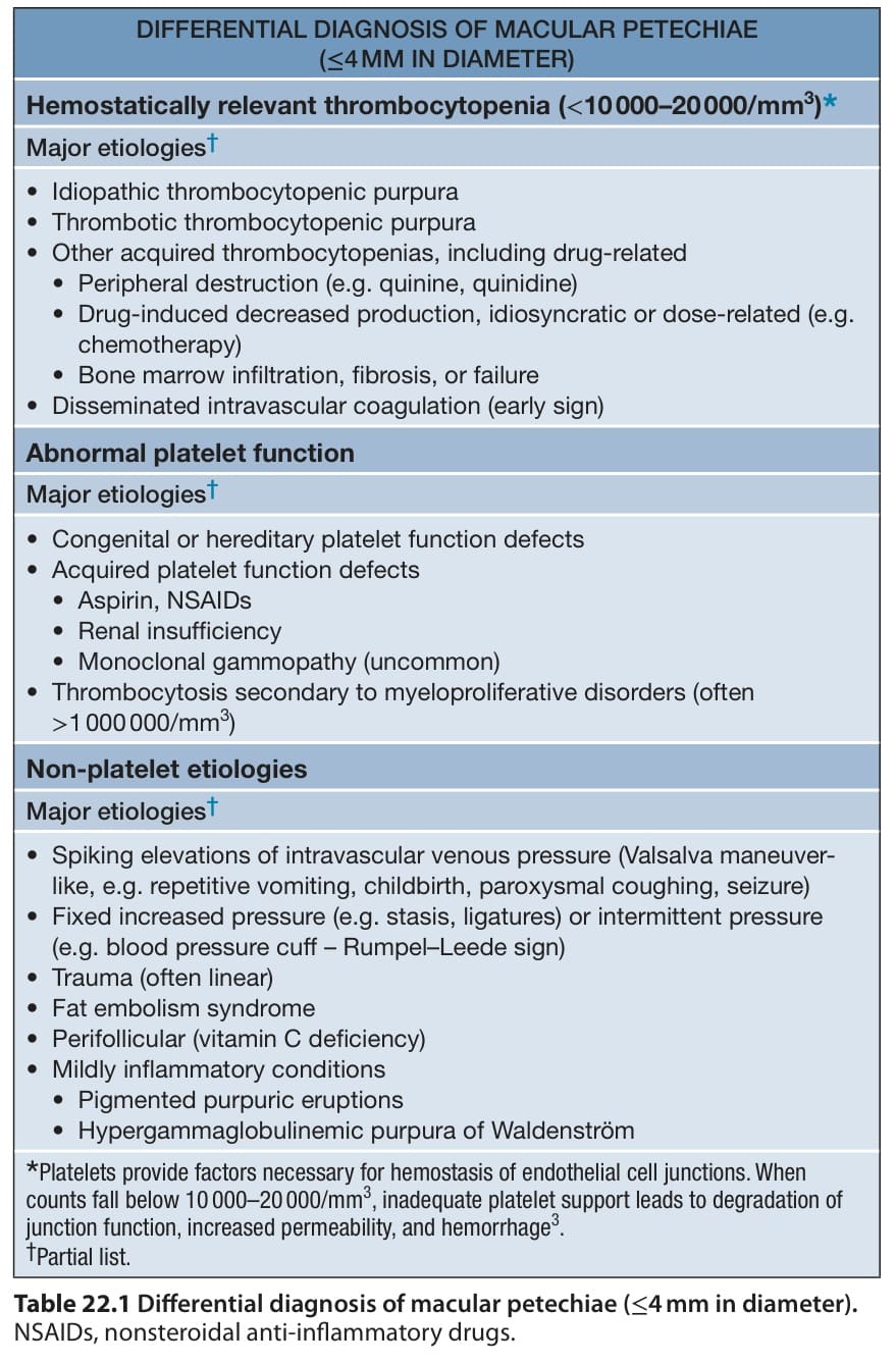
Table 22.1 Differential diagnosis of macular petechiae (≤4 mm in diameter).
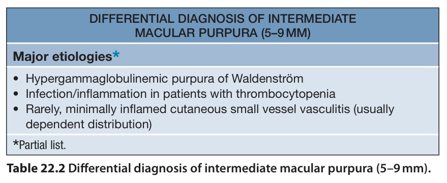
Table 22.2 Differential diagnosis of intermediate macular purpura (5–9 mm).
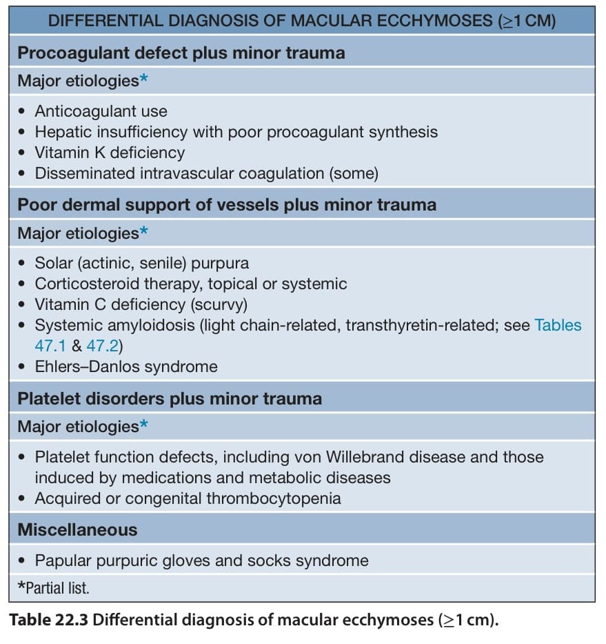
Table 22.3 Differential diagnosis of macular ecchymoses (≥1 cm).
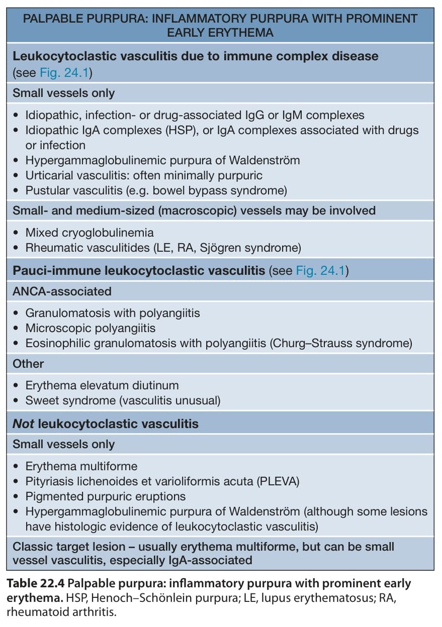
Table 22.4 Palpable purpura: inflammatory purpura with prominent early erythema. HSP, Henoch–Schönlein purpura; LE, lupus erythematosus; RA, rheumatoid arthritis.
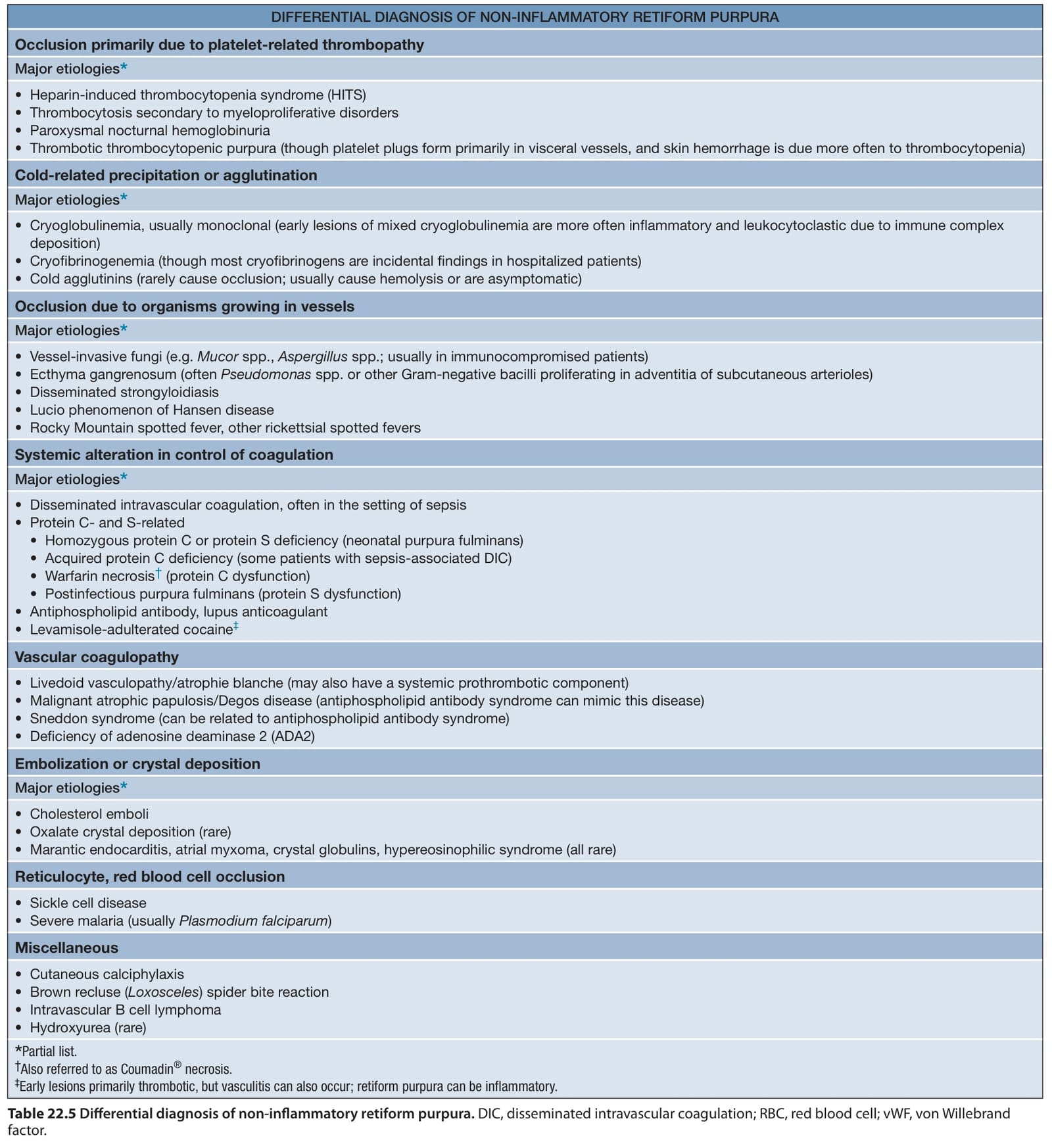
Table 22.5 Differential diagnosis of non-inflammatory retiform purpura. DIC, disseminated intravascular coagulation; RBC, red blood cell; vWF, von Willebrand factor.
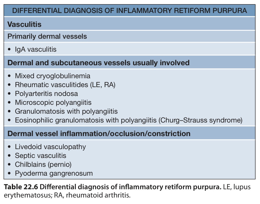
Table 22.6 Differential diagnosis of inflammatory retiform purpura. LE, lupus erythematosus; RA, rheumatoid arthritis.