HEMOSTASIS/COAGULATION
Primary hemostasis consists of adhesion, activation, and aggregation of platelets at the site of vascular injury. This leads to the formation of a platelet plug sufficient for routine minor injuries to the microvascular system. Platelets are also essential for maintaining the integrity of small blood vessels. There is molecular signaling crosstalk between megakaryocytes and endothelial cells, with platelets delivering megakaryocytic factors that are important for endothelial health.
Petechial hemorrhage due to severe thrombocytopenia (typically platelets <20 000/mcl) results from reduced delivery of these megakaryocyte/ platelet factors to the endothelium, thereby impairing endothelial cell function and widening of the gaps between endothelial cells. This probably also explains why platelet dysfunction does not typically result in petechial hemorrhage, since platelet number and factor delivery remain normal, but rather ecchymoses develop because platelet plugs fail to form at sites of injury.
When a platelet plug is inadequate due to the size of the vessel or the size of the injury, secondary hemostasis with clot formation is needed. Secondary hemostasis classically includes the intrinsic and extrinsic coagulation pathways that then converge into a common
pathway, driving the formation of fibrin from fibrinogen via stepwise amplifications (Fig. 22.5). The intrinsic (contact activation) pathway is the longer of the two; it begins with factor XII activation, typically due to injury-related exposure of endothelial collagen. This pathway is assessed by the activated partial thromboplastin time (aPTT). The extrinsic pathway is triggered by tissue factor released from endothelial and other cells, which converts factor VII to factor VIIa, and then as the extrinsic pathway converges with the intrinsic pathway to form the common pathway, factor X is converted to factor Xa. The extrinsic pathway is monitored by the prothrombin time (PT).
Whereas insufficient clotting can lead to death via hemorrhage, inappropriate clotting produces thrombosis, embolus, and/or necrosis. Uncontrolled clotting with fibrinolysis can produce both thrombosis and hemorrhage, as in DIC. Ideally, a thrombus should form rapidly at sites of injury, but should not extend beyond where it is needed. Clotting factors that escape the local injury site must be prevented from triggering clotting at distant sites. As might be expected for a system that requires exquisite regulation to perform properly, the control of coagulation is multifaceted. Basal coagulation encompasses the constant low-level activation of some components of the procoagulant, natural anticoagulant, and fibrinolytic pathways, so that these systems are primed for a rapid response when needed. While Figure 22.5 appears complicated enough for a dermatologist, there are additional modifiers of coagulation including prekallikrein, C1 esterase inhibitor (see Fig. 18.6), factor XIII, and protein Z/protein Z-dependent protease inhibitor.
The initiation phase of clotting is down-regulated by tissue factor pathway inhibitor (TFPI) and antithrombin III (ATIII). TFPI and ATIII are bound to heparan sulfate molecules on the surface of endothelial cells (see Fig. 22.5). Their presence in close proximity to sites of desired clot formation allows them to capture activated clotting factors and pre-vent them from leaving the area. ATIII can neutralize thrombin as well as factors IXa, Xa, and XIIa; this process is greatly enhanced by heparan sulfate or heparin binding to ATIII.
The thrombomodulin/protein C/protein S system is the other major natural anticoagulant pathway. While both ATIII and protein C pathways
Morphology can be used as a primary sorting method, but combining morphology with an initial pattern approach as depicted in Figure 22.1 can improve accuracy. When discrepancies between the two arise, additional investigation is warranted.
are important in large vessels, the protein C system seems to be critical for normal function of the microvasculature. Thrombin that escapes the clot site can bind to thrombomodulin on the endothelial cell surface. Once bound to thrombomodulin, thrombin no longer activates procoagulant factors, but instead avidly binds and activates protein C, a vitamin K-dependent anticoagulant protein (see Fig. 22.5). The anticoagulant function of activated protein C is enhanced by binding to a phospholipid surface, a high-density lipoprotein (HDL), and protein S (another vitamin K-dependent anticoagulant). This complex then inactivates factors Va and VIIIa. The factor V Leiden mutation is a single amino acid change at the site of cleavage by activated protein C, and this change renders it much more resistant to inactivation, thus explaining the associated prothrombotic state. In other patients, a polymorphism in the prothrombin gene, specifically 20210A within an untranslated region, increases the level of prothrombin and can lead to a prothrombotic state.
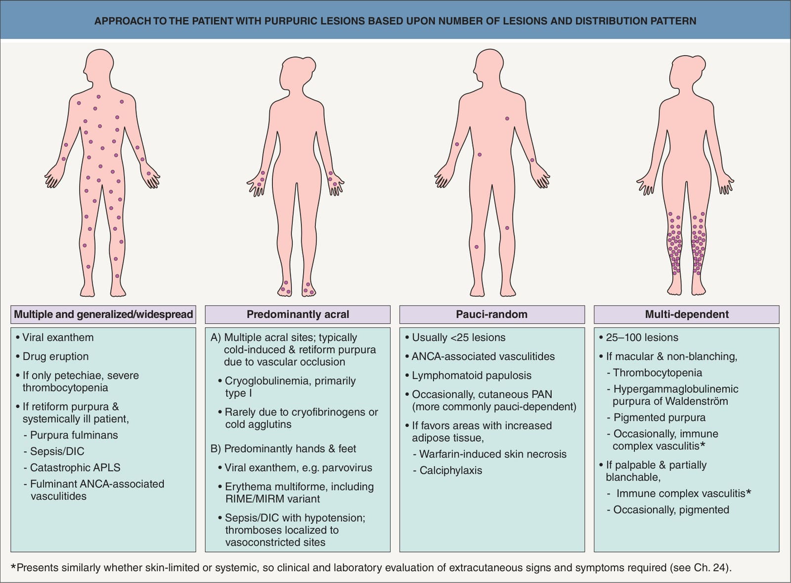
Fig. 22.1 Approach to the patient with purpuric lesions based upon number of lesions and distribution pattern. APLS, antiphospholipid antibody syndrome; DIC, disseminated intravascular coagulation; MIRM, Mycoplasma-induced rash and mucositis; RIME, reactive infectious mucocutaneous eruption; PAN, polyarteritis nodosa.
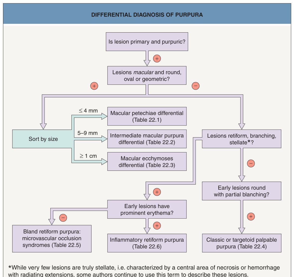
Fig. 22.3 Differential diagnosis of purpura.
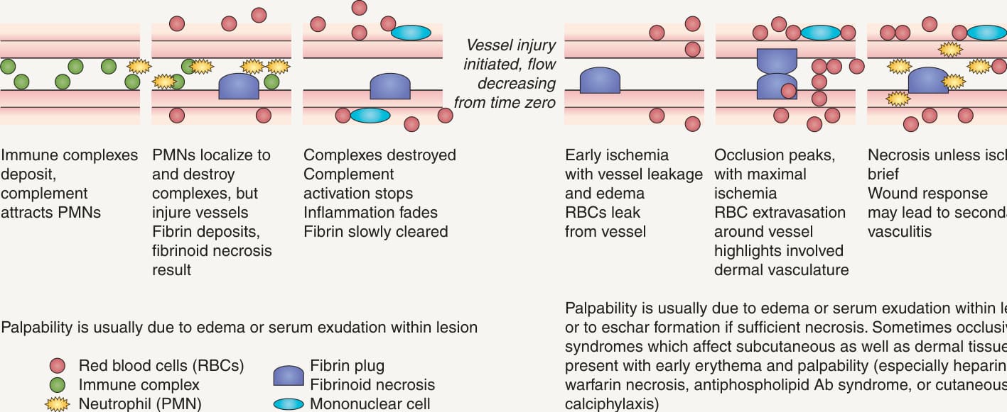
Fig. 22.4 Time course for skin lesions due to vasculitis versus microvascular occlusion.A Time course for lesions due to immune complex-mediated leukocytoclastic vasculitis. B Time course for lesions due to microvascular occlusion. Ab, antibody; tr, trace.
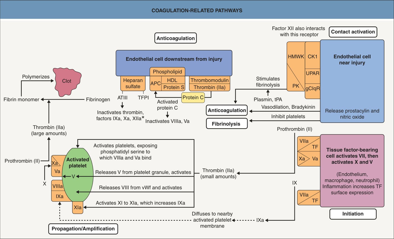
*Fig. 22.5 Coagulation-related pathways. TFPI can inactivate factor Xa, and the TFPI–factor Xa complex can inhibit VIIa. Enhanced by binding of heparin to ATIII. APC, activated protein C; ATIII, antithrombin III; CK1, cytokeratin 1; gC1qR, globular head of the C1q receptor; HDL, high-density lipoprotein; HMWK, high-molecularweight kininogen; PK, prekallikrein; TF, tissue factor; TFPI, tissue factor pathway inhibitor; tPA, tissue plasminogen activator; UPAR, urokinase plasminogen activator receptor; vWF, von Willebrand factor.
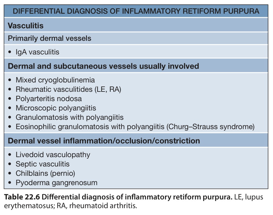
Table 22.6 Differential diagnosis of inflammatory retiform purpura. LE, lupus erythematosus; RA, rheumatoid arthritis.