INTRODUCTION
The differential diagnosis of microvascular syndromes involving the skin is extensive. It is critical to distinguish between inflammatory injury and non-inflammatory occlusive injury to vessels. The diagnosis of occlusive syndromes in the skin is greatly facilitated by recognizing the telltale lesions of retiform purpura or non-inflammatory (bland) necrosis.
Diseases that produce skin lesions secondary to microvascular occlusion have often been lumped with cutaneous vasculitic syndromes or discussed primarily as systemic diseases with little attention paid to cutaneous findings. While there is some overlap in the pathogenesis of deep venous thrombosis and pulmonary emboli and the pathogenesis of cutaneous microvascular occlusion syndromes, the two can differ substantially with respect to disease etiology and treatment.
The differential diagnosis for occlusive syndromes is best approached through major pathophysiologic categories (Table 23.1). These categories frequently have sufficiently distinctive clinical settings that a minimal history and physical examination plus laboratory findings quickly allows for an efficient sorting of the most likely diagnoses (Fig. 23.1). This is especially important in life-threatening situations when making timely decisions is crucial. A reasonable panel of initial laboratory tests is listed in Table 23.2.
It must be emphasized that some inflammatory syndromes capable of producing retiform purpura may occasionally present with lesions that have minimal inflammation (see Ch. 22). In particular, this may occur with the anti-neutrophil cytoplasmic antibody (ANCA)-positive syndromes of granulomatosis with polyangiitis (GPA) and microscopic polyangiitis (MPA), as well as ANCA-negative cutaneous polyarteritis nodosa.
Because treatment depends on pathogenesis (see Fig. 22.4), accurate diagnosis is critical prior to instituting appropriate therapy. The use of anti-inflammatory therapy for occlusive disease may not only be useless but actually harmful, and the same may be true for anticoagulant therapy in most vasculitides.
Warren W. Piette
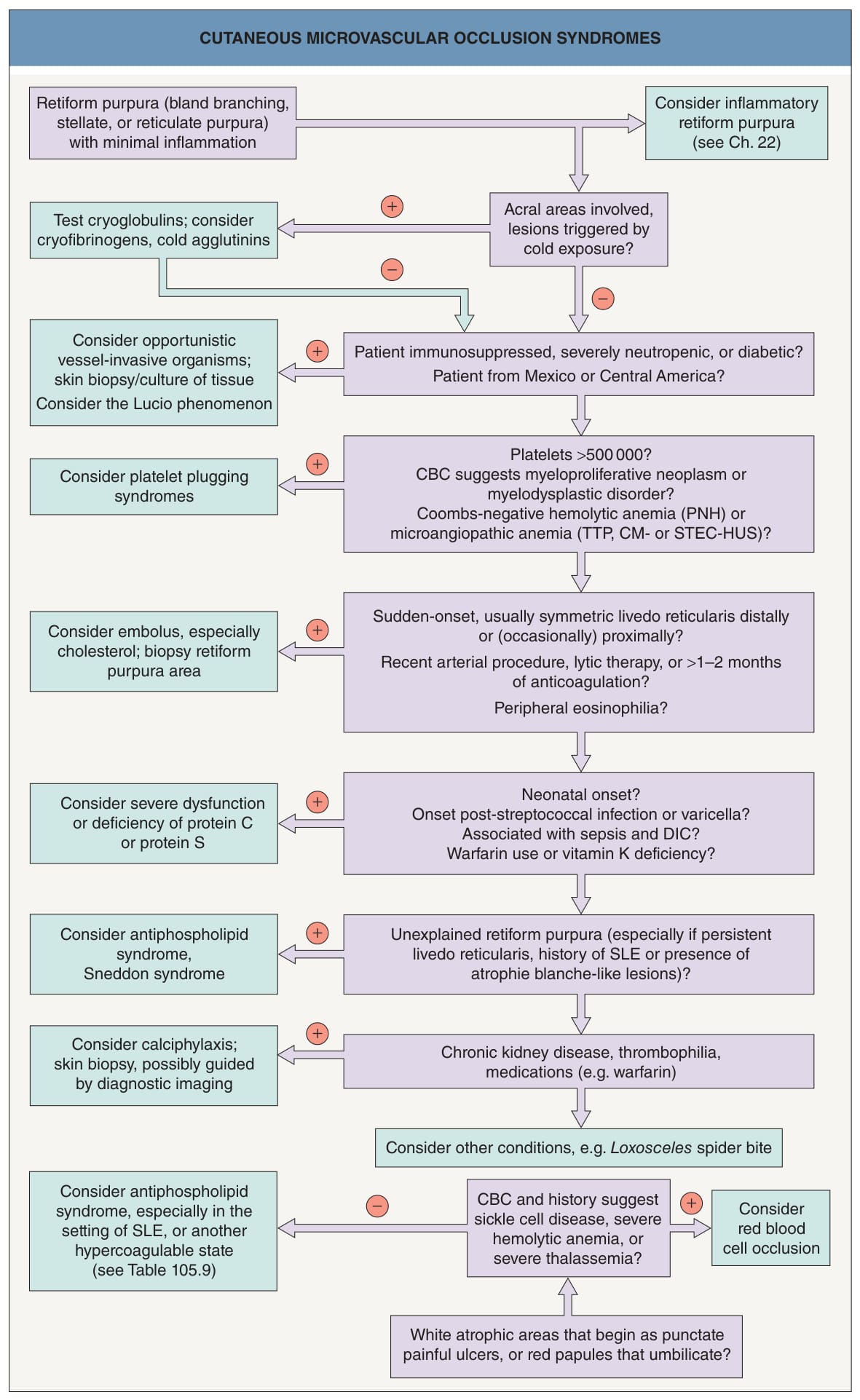
Fig. 23.1 Cutaneous microvascular occlusion syndromes. CM-HUS, complement-mediated hemolytic uremic syndrome; DIC, disseminated intra-vascular coagulation; PNH, paroxysmal nocturnal hemoglobinuria; SLE, systemic lupus erythematosus; STEC-HUS, Shiga toxin-producing Escherichia coli hemolytic uremic syndrome; TTP, thrombotic thrombocytopenic purpura.
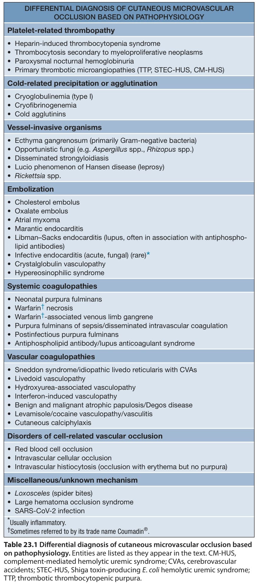
Table 23.1 Differential diagnosis of cutaneous microvascular occlusion based on pathophysiology. Entities are listed as they appear in the text. CM-HUS, complement-mediated hemolytic uremic syndrome; CVAs, cerebrovascular accidents; STEC-HUS, Shiga toxin-producing E. coli hemolytic uremic syndrome; TTP, thrombotic thrombocytopenic purpura.
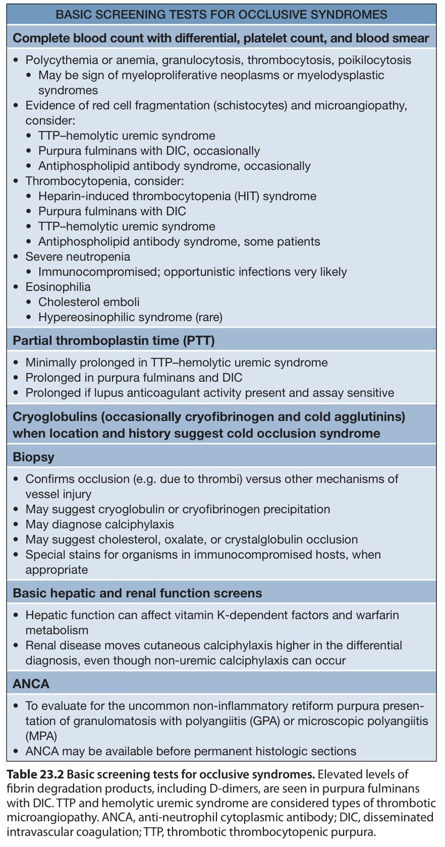
Table 23.2 Basic screening tests for occlusive syndromes. Elevated levels of fibrin degradation products, including D-dimers, are seen in purpura fulminans with DIC. TTP and hemolytic uremic syndrome are considered types of thrombotic microangiopathy. ANCA, anti-neutrophil cytoplasmic antibody; DIC, disseminated intravascular coagulation; TTP, thrombotic thrombocytopenic purpura.