GENERAL CLINICAL FEATURES
The skin lesions of CSVV usually appear 7–10 days after the inciting event; rarely, drug-induced CSVV may appear within hours of administration of the responsible drug. In systemic vasculitis, systemic symptoms often precede the appearance of associated cutaneous lesions (average, 6 months), but the interval is variable and cutaneous findings can certainly be a presenting manifestation. As noted, the cutaneous findings of vasculitis depend upon the predominant size of the vessels involved. CSVV typically presents with palpable or macular purpura,
but urticarial papules, pustules, vesicles, petechiae, or targetoid lesions can be seen (Fig. 24.2 & 24.3). The lesions favor dependent sites, as well as areas under tight-fitting clothing, reflecting the influence of hydrostatic pressure and stasis on immune complex deposition. In general, the lesions are asymptomatic, but they may itch, burn, or sting.
In medium-sized vessel vasculitis, the affected blood vessels reside within the reticular dermis or subcutis. As a result, it typically presents with livedo racemosa, retiform purpura, ulcers, subcutaneous nodules, and/or digital necrosis. In general, the presence of larger ulcerations or necrosis suggests deeper arterial involvement. The combination of purpuric macules and papules plus features of medium-sized vessel disease points to a “mixed” pattern of vasculitis with involvement of both small and medium-sized vessels, as in cryoglobulinemic vasculitis and ANCA-associated vasculitis (see Table 24.1).
Common manifestations of systemic vasculitis include inflammatory arthritis as well as constitutional symptoms, such as fever, weight loss, and malaise. These common findings can be seen in virtually any type of systemic vasculitis, whereas other signs and symptoms (e.g. motor neuropathy, sinusitis) are more often associated with specific disease states. As in the skin, systemic manifestations tend to correlate with the size of the affected blood vessels; for example, glomerulonephritis is a manifestation of small vessel disease, while renal artery aneurysms and renovascular hypertension are features of medium-sized vessel vasculitis. In a population-based study of 84 patients with biopsyproven LCV, 39 patients (46%) had systemic manifestations, with renal involvement most commonly observed (17 of 39 patients).
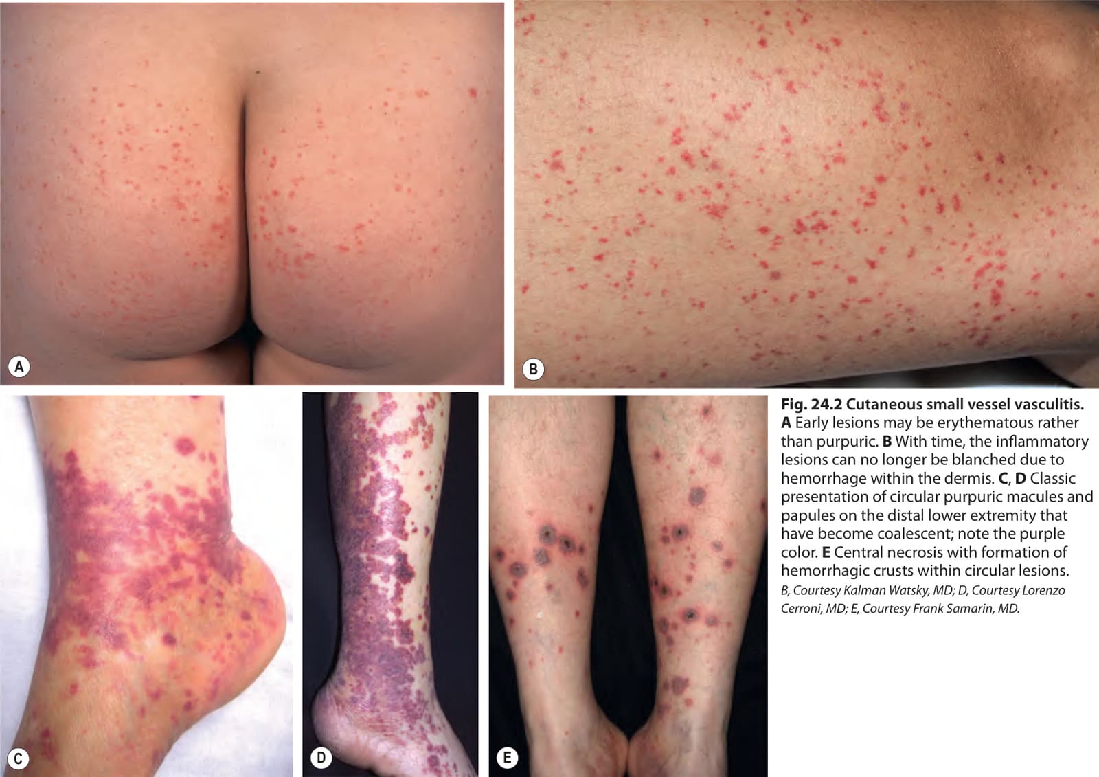
Fig. 24.2 Cutaneous small vessel vasculitis.
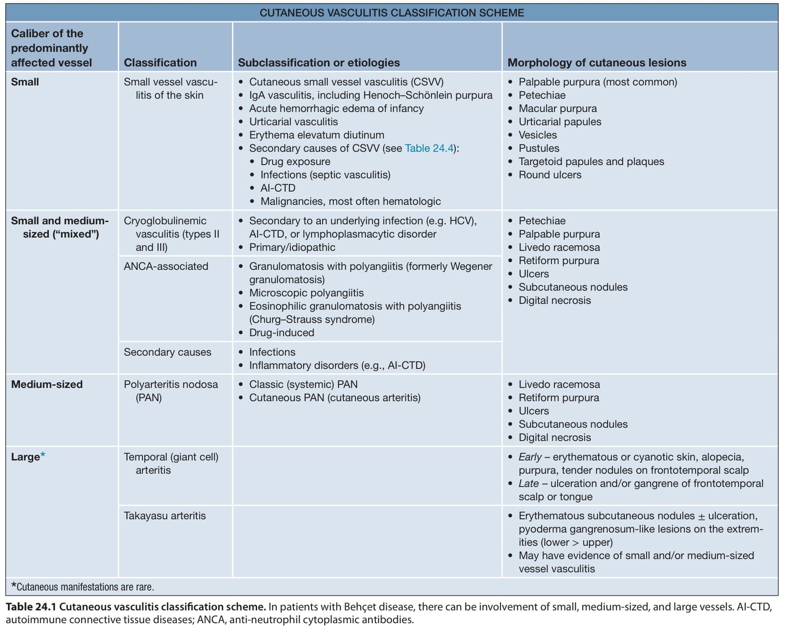
Table 24.1 Cutaneous vasculitis classification scheme. In patients with Behçet disease, there can be involvement of small, medium-sized, and large vessels. AI-CTD, autoimmune connective tissue diseases; ANCA, anti-neutrophil cytoplasmic antibodies.