PATHOLOGY
Timing and appropriate sampling of the most likely involved vessels increase the diagnostic yield of a skin biopsy. Ideally, lesions should be biopsied for routine processing within the first 24 to 48 hours of appearance. When small vessel vasculitis is suspected, a biopsy specimen for direct immunofluorescence (DIF) should be obtained, as the presence of certain immunoglobulins (e.g. IgA) may suggest a particular diagnosis and prognosis. Biopsies for DIF should also be obtained from lesions of recent onset, ideally those that are only hours old. In general, punch biopsies are adequate for the diagnosis of small vessel vasculitis, while an incisional deep biopsy may be required to diagnose vasculitis of larger vessels.
In cutaneous vasculitis, the histologic findings vary depending upon the type and age of the sampled lesion in addition to the size of the affected vessel. The classic histopathologic features of CSVV are referred to as LCV and consist of transmural infiltration of the walls of small vessels (primarily postcapillary venules) by neutrophils undergoing karyorrhexis of their nuclei, as well as fibrinoid necrosis of the vessel walls (Fig. 24.4). Other findings include leukocytoclasia (degranulation and fragmentation of neutrophils, leading to the production of nuclear dust), extravasated erythrocytes, and signs of endothelial cell damage. However, lesions present for greater than 48 to 72 hours may have a predominantly mononuclear rather than neutrophilic infiltrate. In the case of palpable purpura, the most common clinical lesion of CSVV,
the small vessel involvement accounts for the small size of the lesions, the inflammatory response for the palpability and symptomatology (e.g. burning pain, pruritus), the red blood cell extravasation from damaged blood vessels for the non-blanching purpura, and the effect of gravity on immune complex deposition for the distribution of lesions on the lower legs and other dependent areas.
Some studies suggest that additional histologic findings may provide clues to the underlying etiology, e.g. eosinophils in drug-induced CSVV, thrombi in patients exposed to levamisole-tainted cocaine, and thrombi plus dense dermal inflammation in septic vasculitis. Involvement of deeper dermal vessels can alert the clinician to look for an underlying etiology rather than presuming the CSVV is idiopathic.
Vasculitis of medium-sized blood vessels is characterized by similar changes involving the vessels (e.g. small arteries) of the deep reticular dermis and subcutaneous fat. Neovascularization of the adventitia, in the form of small capillaries, is commonly seen in older lesions of medium-sized vessel vasculitis.
In ~80% of cases of small vessel vasculitis of the skin, DIF demonstrates deposition of C3, IgM, IgA, and/or IgG (generally in that order of frequency) in a granular pattern within the vessel walls. Immunoglobulin deposition is highest (up to 100%) in skin lesions present for ≤48 hours. On the other hand, in 30% of samples obtained 48–72 hours after lesion onset, the DIF will be negative for immunoglobulins, and only C3 will be detected in lesions present for >72 hours. In patients with ANCA-associated vasculitis, the DIF of lesional skin is usually negative. After controlling for duration, it is preferable to biopsy more proximal lesions for DIF in order to avoid nonspecific vascular fluorescence that can occur at sites of increased hydrostatic pressure.
As stated previously, the term LCV is a histologic designation. Terms used to describe additional histologic forms of cutaneous vasculitis include “lymphocytic” and “granulomatous” (see below). Use of the term lymphocytic vasculitis requires clarification, as it does not represent a conventional type of vasculitis but rather a histologic
A Early lesions may be erythematous rather than purpuric. B With time, the inflammatory lesions can no longer be blanched due to hemorrhage within the dermis. C, D Classic presentation of circular purpuric macules and papules on the distal lower extremity that have become coalescent; note the purple color. E Central necrosis with formation of hemorrhagic crusts within circular lesions. B, Courtesy Kalman Watsky, MD; D, Courtesy Lorenzo Cerroni, MD; E, Courtesy Frank Samarin, MD.
finding seen in a number of non-vasculitic disorders such as pernio and pityriasis lichenoides et varioliformis acuta (Table 24.2).
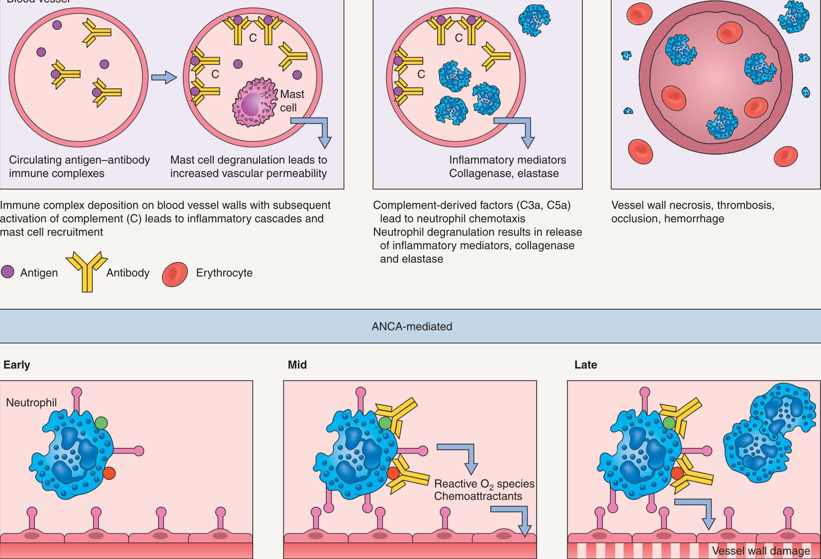
Fig. 24.1 Pathogenesis of cutaneous vasculitis – immune complex- versus anti-neutrophil cytoplasmic antibody (ANCA)-mediated.A In immune complex-mediated vasculitis, circulating antigens (e.g. infectious agents, medications, neoplasms) induce antibody formation. Binding of antibodies to circulating antigens creates immune complexes. Immune complex deposition within postcapillary venules activates complement and subsequently leads to an increase in adhesion molecule expression on the endothelium. Complement split products (C3a and C5a) induce mast cell degranulation and neutrophil chemotaxis. Mast cell degranulation leads to increased vascular dilation and permeability, enhancing immune complex deposition and leukocyte tethering to endothelium. Increased adhesion between inflammatory cells (especially neutrophils) and the endothelium is mediated by elevated expression of selectins (E-selectin, P-selectin) and members of the immunoglobulin superfamily (ICAM-1, VCAM-1, PECAM-1) on endothelial cells in concert with the upregulation of their corresponding ligands and receptors/adhesion molecules on leukocytes (e.g. P-selectin glycoprotein ligand-1, LFA-1, Mac-1) (see Ch. 102 for details). Neutrophils release proteolytic enzymes (such as collagenases and elastases) and free oxygen radicals that damage the vessel wall. In addition, formation of the membrane attack complex (C5–C9) on the endothelium leads to the activation of the clotting cascade and the release of cytokines and growth factors with ensuing thrombosis, inflammation, and angiogenesis. B In ANCA-mediated vasculitis, following primary activation by cytokines such as tumor necrosis factor (TNF), intracellular proteins from neutrophils (e.g., proteinase 3 [PR3], myeloperoxidase [MPO]) become expressed on the cell surface. After formation of ANCAs that recognize these antigens, binding of the autoantibodies to neutrophils leads to increased neutrophil adhesion to vessel walls and subsequent cellular activation. Neutrophils then release reactive oxygen species and other toxic mediators that result in vessel wall damage (see A). Because the vessel damage in ANCA-positive vasculitides is directly mediated by neutrophils rather than by immune complexes, they are referred to as “pauci-immune” vasculitides. Formation of ANCAs may be related to an impairment in neutrophil apoptosis which results in a prolonged opportunity for autoantibody development.
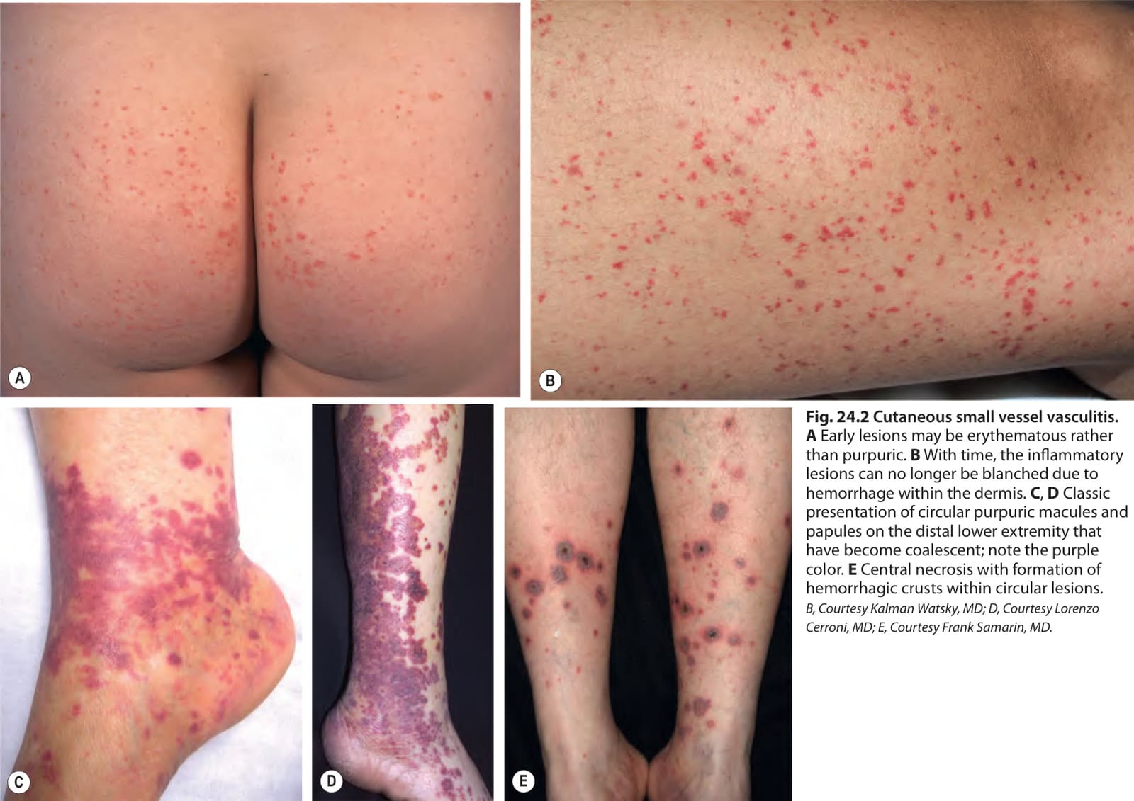
Fig. 24.2 Cutaneous small vessel vasculitis.
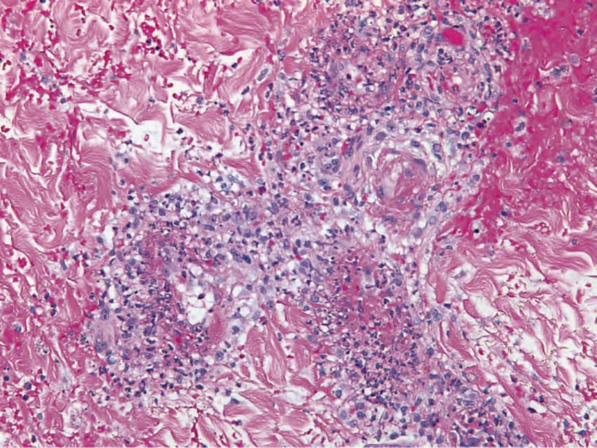
Fig. 24.4 Cutaneous small vessel vasculitis – histopathologic features.
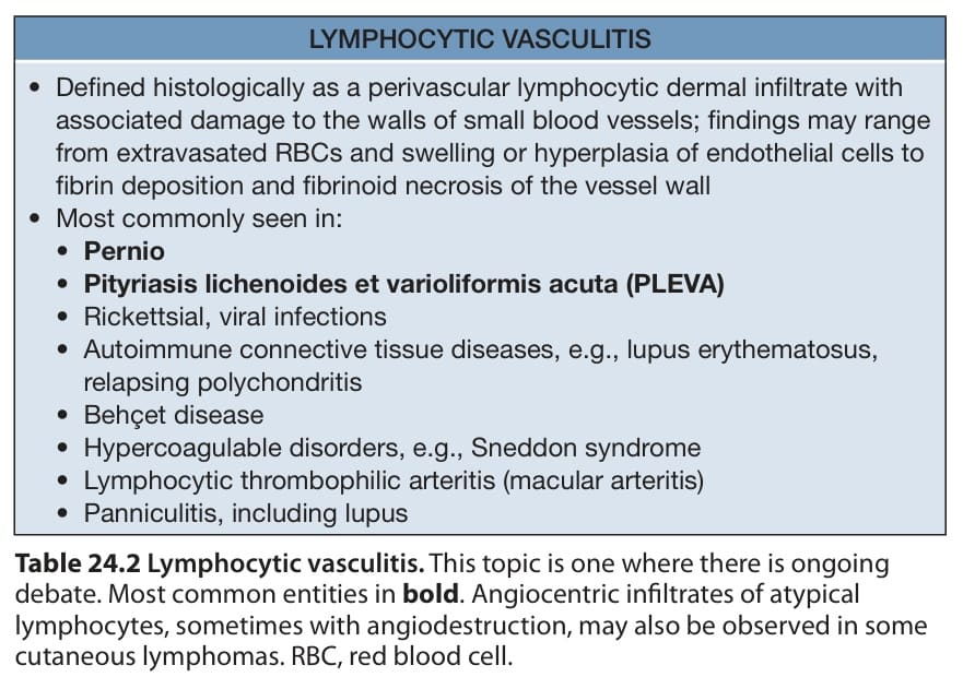
Table 24.2 Lymphocytic vasculitis. This topic is one where there is ongoing debate. Most common entities in bold. Angiocentric infiltrates of atypical lymphocytes, sometimes with angiodestruction, may also be observed in some cutaneous lymphomas. RBC, red blood cell.