SMALL VESSEL VASCULITIS
Synonyms: IgM/IgG immune complex vasculitis Cutaneous leukocytoclastic vasculitis Cutaneous leukocytoclastic angiitis Hypersensitivity vasculitis Cutaneous necrotizing venulitis
Key features
A single-organ, skin-limited vasculitis involving small blood vessels
Presents with palpable purpura, urticarial lesions, and/or hemor- rhagic macules or vesicles; less common findings include targetoid lesions, pustules, and round ulcerations
Lesions favor the lower extremities and other dependent areas or pressure points
Histologically, there is leukocytoclastic vasculitis involving small vessels, primarily postcapillary venules
Must be differentiated from vasculitis with extracutaneous involvement
Angiocentric segmental inflammation with endothelial swelling, a neutrophilic infiltrate with leukocytoclasia, red blood cell extravasation, and fibrinoid necrosis of blood vessel walls. The neutrophils are present around and within the walls of the small dermal blood vessels. Courtesy David F. Fiorentino, MD.
Introduction
CSVV is a type of single-organ, skin-limited vasculitis that involves primarily the dermal postcapillary venules and is characterized histologically by LCV. The dermatologic addendum to the Chapel Hill Consensus Conference Nomenclature proposes the term “IgM/ IgG immune complex vasculitis” to further characterize this entity. Because clinical manifestations of small vessel vasculitis of the skin (e.g. palpable purpura) and histologic findings of LCV may be seen in other disease entities (e.g. IgA vasculitis), systematic evaluation and consideration of other types of vasculitis involving small cutaneous vessels is necessary before establishing the diagnosis of CSVV (see Table 24.1). CSVV is often idiopathic in nature, but it may be secondary to an underlying cause such as an infection, medication, or autoimmune connective tissue disease (Table 24.4). Within the spectrum of small vessel vasculitis of the skin are several entities whose rather unique epidemiologic and clinical features warrant subclassification (see Table 24.1) and separate discussion (following sections).
Epidemiology
CSVV occurs in both sexes and at all ages, but it is more common in the adult population. It is estimated that ~10% of those affected are children. The annual population-based incidence of CSVV (excluding those with a distinct clinical subtype of small vessel vasculitis) is 21 cases per million.
Pathogenesis
CSVV is mediated by immune complex deposition (see Fig. 24.1A).
Clinical features
CSVV typically presents spontaneously, or after exposure to an inciting agent, with a crop of lesions consisting of non-blanching purpuric macules and papules, petechiae, urticarial lesions, or hemorrhagic vesicles ranging in size from 1 mm to several centimeters (see Fig. 24.2 & 24.3). Lesions often begin with erythema and then rapidly become purpuric; larger lesions arise via coalescence of papules. Occasionally, pustules, targetoid lesions, and round ulcerations are seen. CSVV favors dependent areas, as well as areas affected by trauma (Koebner phenomenon) or under tight-fitting clothing. Prolonged sitting or standing, as well as exercise, in particular walking or hiking in hot weather, can induce CSVV on the lower extremities. Although they may be asymptomatic, lesions can be associated with burning pain or pruritus and may be psychologically distressing. Residual postinflammatory hyperpigmentation can persist for months after the primary process resolves.
By definition, CSVV is skin-limited, or skin-predominant, so it is appropriate to apply this label to patients with small vessel vasculitis of the skin in whom there is no evidence of systemic vasculitis. That said, patients with CSVV commonly have low-grade constitutional symptoms and arthralgias which may accompany flares of skin vasculitis. Because of a lack of clarity regarding terminology and disease definitions in the literature, estimates of the prevalence of systemic symptoms in CSVV vary. Available data suggest that systemic symptoms occur in 5%–25% of patients with CSVV, with arthralgias and arthritis occurring most commonly (15%–65%), followed by genitourinary signs or symptoms (3%–7%), and gastrointestinal involvement (3%–5%). A population-based study demonstrated systemic symptoms in 11 of 38 patients (29%) with CSVV. In general, more overt signs or symptoms of systemic disease, such as marked constitutional symptoms, inflammatory arthritis, and gastrointestinal, renal, or neurologic involvement should increase the clinical suspicion for a systemic vasculitis. In one study, the presence of paresthesias or fever were identified as risk factors for an associated systemic disease. A stepwise approach to evaluation, guided by the presenting signs and symptoms, is recommended to help identify those patients with systemic manifestations of vasculitis or important underlying disorders (see Fig. 24.19).
Approximately 90% of patients with CSVV will have spontaneous resolution of cutaneous lesions within several weeks or a few months, while another 10% will have chronic or recurrent disease at intervals of months to years. In the latter group, the average duration of disease activity is reported to be ~24–28 months. The presence of arthralgias or cryoglobulinemia and an absence of fever may portend chronicity. An underlying cause for the CSVV, such as an autoimmune connective tissue disease or neoplasm, also affects prognosis.
Differential diagnosis
In addition to determining if the patient has a particular subtype of small vessel vasculitis (see Table 24.1) or has cutaneous vasculitis as a manifestation of a systemic vasculitic syndrome, causes of secondary cutaneous vasculitis also need to be considered (see Table 24.4; Fig. 24.5 & 24.6). Entities listed in Table 24.3, especially the first three categories, may mimic cutaneous vasculitis and need to be included in the clinical differential diagnosis.
Treatment
The initial intervention in CSVV consists of supportive care and elimination of potential triggers. The need for and choice of pharmacotherapy depends on the chronicity and severity of the cutaneous involvement and any potential underlying disease state. In cases of small vessel vasculitis of the skin where systemic involvement is identified, the choice of therapy must appropriately address those manifestations. Specific therapeutic options are outlined in Table 24.10.
CSVV often resolves without any specific treatment. Because it is usually self-limited as well as skin-limited, supportive measures (e.g. rest, leg elevation, compression) or symptomatic therapy (e.g. antihistamines, NSAIDs) may be sufficient.
Chronic (lasting >4 weeks) or severe cutaneous disease generally requires more aggressive systemic therapy. There is a dearth of high-quality data to guide management, but a number of agents, including colchicine, dapsone and azathioprine, appear to be effective for skin and joint manifestations in some patients and are usually well tolerated (see Table 24.10). A randomized trial is currently underway to assess the efficacy of these agents for skin-limited vasculitis.
Patients with severe, ulcerating, or progressive cutaneous disease who require rapid control of symptoms can be treated with a short course of high-dose oral corticosteroids (e.g. up to 1 mg/kg/day of prednisone). Because of adverse effects associated with long-term corticosteroid use, a taper over 4 to 6 weeks should be attempted. If the patient develops recurrent CSVV as the dosage is decreased, addition of a steroid-sparing agent is warranted. However, corticosteroids do not represent an appropriate long-term plan for chronic or recurrent CSVV. In addition to the oral medications listed previously, methotrexate (<25 mg weekly), mycophenolate mofetil, and rituximab have been reported to be useful in recalcitrant CSVV.
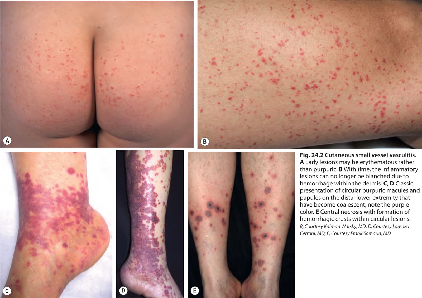
Fig. 24.2 Cutaneous small vessel vasculitis.
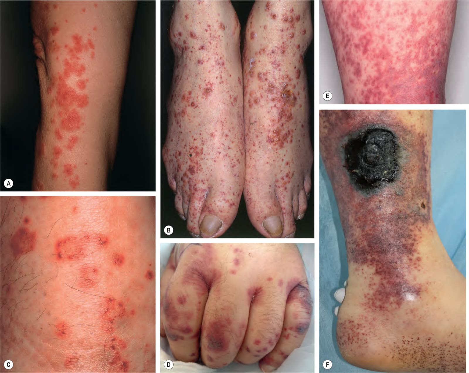
Fig. 24.3 Clinical variants of cutaneous small vessel vasculitis.A Targetoid appearance that can resemble erythema multiforme. B Hemorrhagic vesicles in addition to palpable purpura. C Hemorrhagic crusts in an annular configuration. D Lesions involving the upper extremities. E Purpuric macules and papules with areas of confluence that resemble a purpuric morbilliform drug eruption. F Coalescence of palpable purpura as well as edema and an ulcer with a thick black eschar; petechiae are also present and in the instep resemble pinpricks. B, Courtesy Kalman Watsky, MD; D, Courtesy Lindy Fox, MD, and Kanade Shinkai MD; E, Courtesy David A. Wetter, MD; F, Courtesy Jean L. Bolognia, MD.
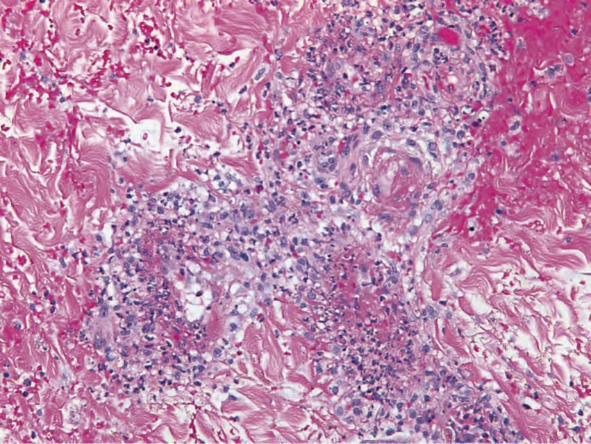
Fig. 24.4 Cutaneous small vessel vasculitis – histopathologic features.
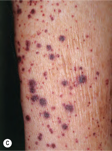
*Fig. 24.5 Etiologies of cutaneous small vessel vasculitis (CSVV). CSVV was idiopathic in 76% of cases in a population-based study from Olmsted County, Minnesota.
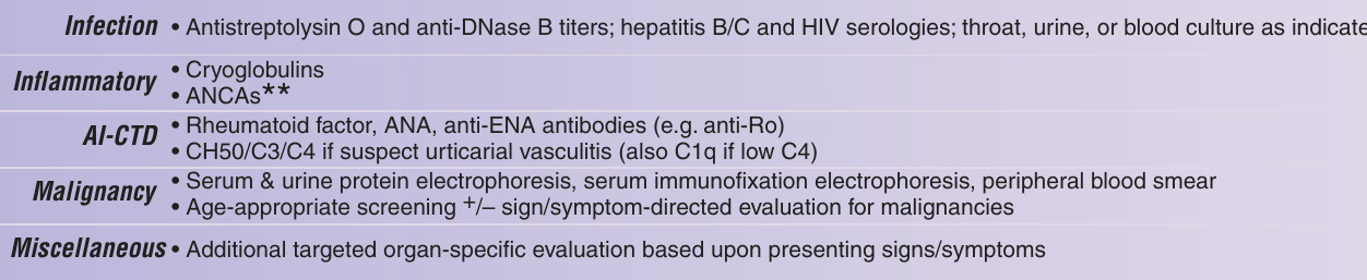
Fig. 24.19 Approach to the patient with suspected cutaneous vasculitis. AI-CTD, autoimmune connective tissue disease; ANA, anti-nuclear antibody; ANCA, anti-neutrophil cytoplasmic antibody; BMZ, basement membrane zone; BUN, blood urea nitrogen; CBC, complete blood count; CT, computerized tomography; DIF, direct immunofluorescence; ELISA, enzyme-linked immunosorbent assay; ENA, extractable nuclear antigen; ESR, erythrocyte sedimentation rate; H&E, hematoxylin and eosin; HIV, human immunodeficiency virus; Ig, immunoglobulin; IgAV, IgA vasculitis (Henoch– Schönlein purpura); SLE, systemic lupus erythematosus. Adapted from Goeser MR, et al. Am J Clin Dermatol 2014;15:299–306.
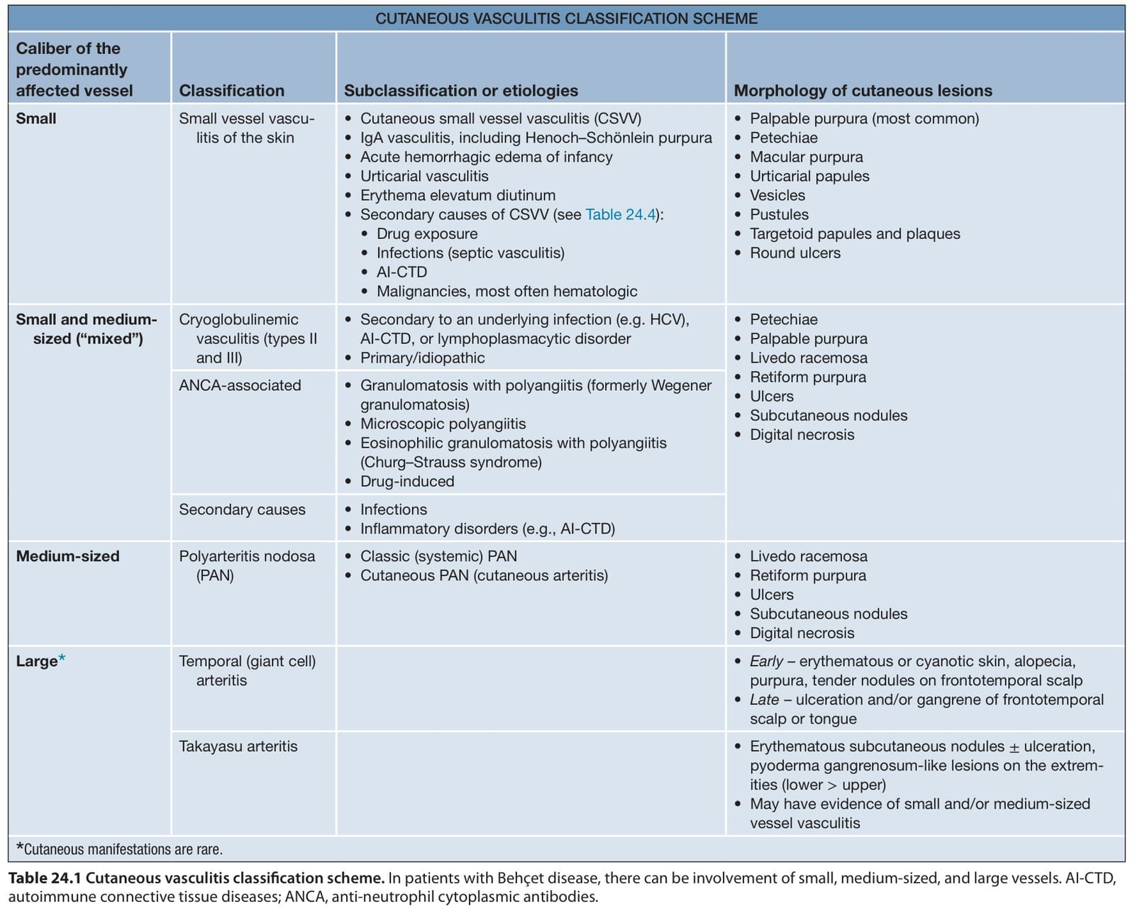
Table 24.1 Cutaneous vasculitis classification scheme. In patients with Behçet disease, there can be involvement of small, medium-sized, and large vessels. AI-CTD, autoimmune connective tissue diseases; ANCA, anti-neutrophil cytoplasmic antibodies.
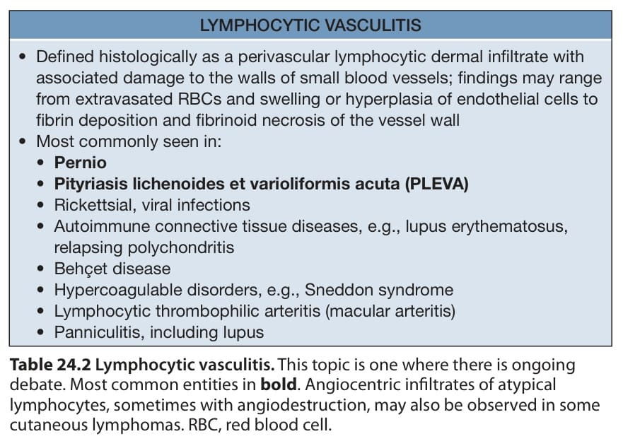
Table 24.2 Lymphocytic vasculitis. This topic is one where there is ongoing debate. Most common entities in bold. Angiocentric infiltrates of atypical lymphocytes, sometimes with angiodestruction, may also be observed in some cutaneous lymphomas. RBC, red blood cell.
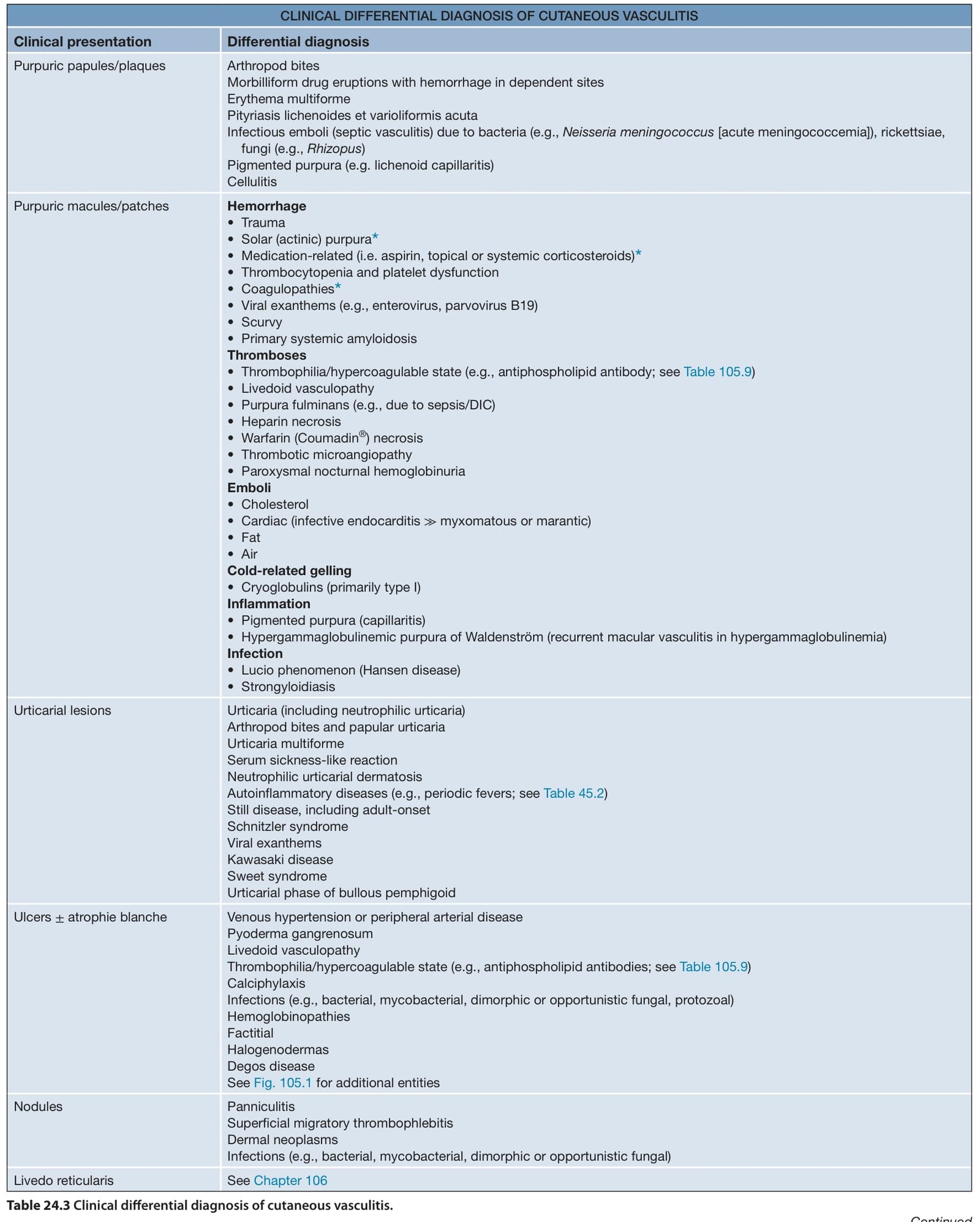
Table 24.3 Clinical differential diagnosis of cutaneous vasculitis.
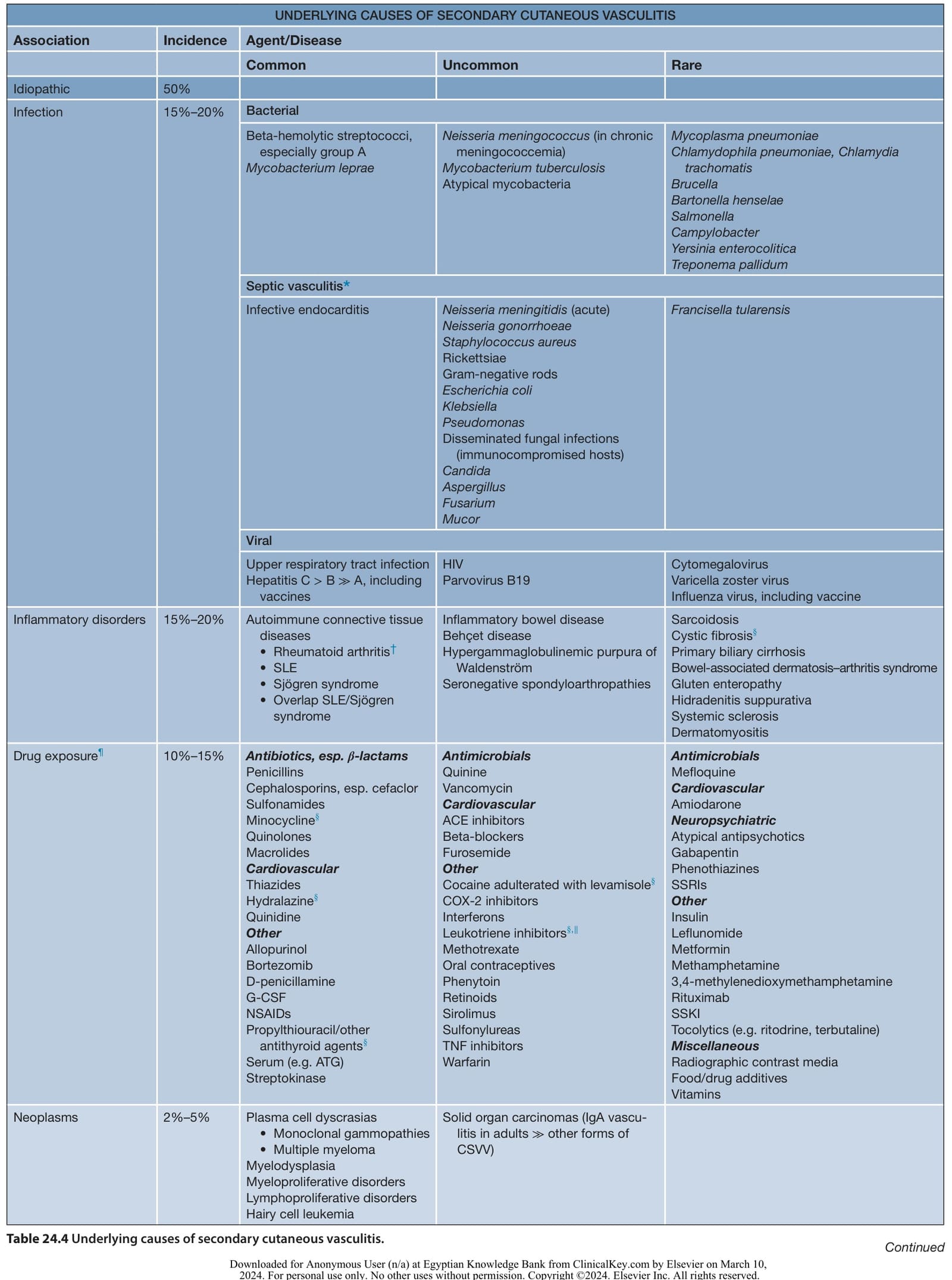
Table 24.4 Underlying causes of secondary cutaneous vasculitis. Continued

Table 24.10 Therapeutic ladder for patients with vasculitis.