IgA Vasculitis (Henoch–Schönlein Purpura)
Synonyms: Schönlein–Henoch purpura Anaphylactoid purpura Purpura rheumatica Cutaneous small vessel vasculitis secondary to IgA immune complexes
Key features
Most common form of vasculitis in children <10 years of age
Frequently preceded by an upper respiratory tract infection
Intermittent palpable purpura on the extremities with
IgA-dominant immune deposits in the walls of small blood vessels
Systemic involvement, in particular gastrointestinal, joint, and renal, is often observed, but IgA vasculitis may be skin-limited
In adults (men > women), rarely associated with an underlying malignancy
Introduction
IgA vasculitis (IgAV), also referred to as Henoch–Schönlein purpura (HSP), is a type of vasculitis affecting small blood vessels in which vascular IgA deposition is a key diagnostic and pathophysiologic feature. This particular type of vasculitis occurs more commonly in children than in adults. The classic tetrad consists of palpable purpura, arthritis, abdominal pain, and hematuria. However, IgAV may be skinlimited or skin-predominant, with either no associated symptoms or mild arthralgias and/or constitutional symptoms.
The underlying diseases were Sjögren syndrome (A), rheumatoid arthritis (B), relapsing polychondritis with myelodysplasia (C), and endocarditis due to Streptococcus mitis (D). Note the Koebner phenomenon in A and rheumatoid nodules in B. Cutaneous vasculitis in association with relapsing polychondritis and myelodysplasia points to a likely diagnosis of VEXAS syndrome. C, Courtesy Jean L. Bolognia, MD; D, Courtesy David A. Wetter, MD.
Epidemiology
IgAV is the most common form of vasculitis in children, with an incidence of 30 to 270 cases per million children per year. The average age of onset is 6 years, and 90% of cases occur in children <10 years of age. In adults, the incidence of HSP is 8 to 18 cases per million, and the population-based incidence of biopsy-proven cutaneous IgA vasculitis is 13 cases per million per year. The disorder follows a seasonal pattern, with a peak occurrence during the winter. HSP has a slight male predominance in both children and adults.
Pathogenesis
IgAV frequently presents 1 to 2 weeks following an upper respiratory tract infection, especially in children. Although several studies have reported that 20%–50% of HSP patients have positive antistreptolysin O (ASO) titers, no causal role for group A β-hemolytic streptococci has been demonstrated. IgAV may be triggered by a medication or rarely it is a paraneoplastic phenomenon.
IgA (specifically IgA1) is thought to play a fundamental role in the pathogenesis of IgAV. IgA deposits in the postcapillary venules of the skin and mesangium and, less commonly, circulating IgA-containing immune complexes and increased serum levels of IgA have been demonstrated in patients with IgAV. In a case series of over 400 patients with IgAV, increased serum IgA levels were found in ~30%. Altered glycosylation of the hinge region of IgA1 may promote the formation of macromolecular complexes that lodge within the mesangium and activate the alternate complement pathway.
Certain genetic polymorphisms may predispose to more severe disease. For example, HLA-B35 positivity may predispose to renal disease, while patients who do not have the ICAM-1 469 K/E variant have less severe gastrointestinal involvement.
Clinical features
IgAV classically presents with palpable purpura in association with arthritis, abdominal pain, and/or nephritis. Cutaneous lesions begin as erythematous macules or urticarial papules and evolve into palpable purpura ranging in size from pinpoint to several millimeters (Fig. 24.7). Urticaria, vesicles, bullae, targetoid lesions, and foci of necrosis can also be seen as well as lower extremity edema. Typically, lesions are symme trically distributed on the lower extremities and buttocks but may also involve the trunk, upper extremities, and face. Individual lesions usually regress within 10 to 14 days, with resolution of cutaneous involvement over a period of several weeks to months. Recurrences of skin disease are observed in 5%–10% of patients.
Extracutaneous manifestations of IgAV are common. Arthritis occurs in up to 75% of patients with IgAV and most commonly affects the joints of the lower extremities (knees and ankles). Gastrointestinal involvement (50%–75% of patients) may precede the purpura and presents with colicky abdominal pain (65%), gastrointestinal bleeding (30%), and/or vomiting. Intussusception and bowel perforation are rare complications.
Renal involvement occurs in 40%–50% of patients and typically presents with microscopic hematuria (40%), often accompanied by proteinuria (25%). The appearance of cutaneous lesions generally precedes the development of nephritis. When it occurs, renal involvement usually appears within the first month of diagnosis (85%) and develops within six months in >95% of patients. Therefore, close observation, with frequent urinalyses, is recommended during this time period. In pediatric patients, risk factors for the development of nephritis include male sex, age >10 years at onset, severe gastrointestinal symptoms, arthritis/arthralgia, persistent or recurrent purpura, and several laboratory abnormalities (leukocytosis, thrombocytosis, elevated ASO titer, low C3). Depending upon the series, persistent renal disease has been observed in 8%–50% of patients, emphasizing the need for
longitudinal monitoring until all abnormalities resolve. Fortunately, only 1%–3% of children develop long-term renal impairment. Poor prognostic factors include renal failure at the time of onset, nephrotic syndrome, hypertension, and decreased factor XIII activity.
In young boys, orchitis is a rare manifestation of systemic disease. The lung is also a rare site of involvement, presenting as hemoptysis and/or pulmonary infiltrates due to diffuse alveolar hemorrhage.
Adult patients with IgAV present with clinical signs and symptoms similar to those in children. However, adults are more likely than children to: develop chronic renal insufficiency (up to 30%); have diarrhea and leukocytosis; require more aggressive therapy; and have a longer hospital stay. Compared to younger adults, those >65 years of age are more likely to develop a 50% increase in serum creatinine and less likely to achieve clinical remission. Lastly, adult patients with IgAV may have an underlying malignancy, most often cancer of a solid organ, in particular the lung. Because 90% of the latter group are men, older male patients should be current with cancer screening guidelines and undergo symptom-based screening. However, there is no consensus regarding the recommended extent of the screening or the need for repeated screening.
Pathology
Histologically, LCV of the small dermal blood vessels is seen. In one series of adults with IgA vasculitis (age >40 years), an absence of dermal eosinophils was associated with an increased risk of renal involvement. The presence of perivascular IgA deposition by DIF is the defining pathologic feature of IgAV (Fig. 24.8). Other immunoglobulins (C3, IgM, IgG) may be seen but are generally not as prominent as IgA and are considered nonspecific deposits. A few small studies have suggested that the presence of other findings, such as perivascular C3 or IgM deposits and papillary dermal edema, may be associated with an increased risk of renal involvement. Of note, endothelial deposits of IgA are ~80% sensitive and specific for IgAV. Therefore, patients with signs and symptoms concerning for IgAV should be followed closely and treated appropriately for suspected systemic complications even if the DIF is negative for IgA.
Differential diagnosis
The clinical differential diagnosis includes any of the vasculitides involving small blood vessels (see Table 24.1), particularly CSVV, since the cutaneous findings in IgAV and CSVV can be indistinguishable. Several entities listed in Table 24.3, especially the first three categories, are also in the clinical differential diagnosis. Because vascular IgA deposition is not 100% sensitive or specific, the diagnosis of IgAV is supported by the presence of IgA in the appropriate clinical setting.
Treatment
As with other types of vasculitis, management of IgAV is dictated by the extent and severity of systemic and cutaneous manifestations. Because IgAV is generally self-limited, resolving over the course of weeks to months, treatment may be primarily supportive. For skin-limited IgAV, dapsone or colchicine may decrease the duration of cutaneous lesions and frequency of recurrences (see Table 24.10). Systemic corticosteroids are effective in treating associated arthritis and abdominal pain, as well as reducing the gastrointestinal complications and duration of skin lesions, but they do not prevent recurrences of purpura and are not suitable as a long-term therapy. In adults, the following factors may predict relapsing disease: age >30 years, persistent purpura >1 month, abdominal pain, hematuria, an underlying systemic disorder, and absence of IgM on DIF.
In patients with renal involvement (e.g. microscopic hematuria, proteinuria), referral to a nephrologist is indicated. Corticosteroids have not been shown to be effective in preventing renal disease due to IgAV and are therefore not recommended. For example, a Cochrane review of interventions for preventing and treating IgAV nephritis found no difference in the risk of persistent kidney disease (up to 12 months following treatment) in children who received prednisone for 2–4 weeks at the time of presentation, as compared to placebo or supportive therapy. In a randomized, doubleblind, placebo-controlled trial involving 352 children with new-onset IgAV, early treatment with 14 days of prednisolone did not reduce the prevalence of proteinuria 12 months after disease onset. For those who do develop evidence of IgAV nephritis, the treatment approach depends on disease severity and whether the patient is a child or adult. Individuals with limited evidence of renal involvement, mild proteinuria (<1 g/day), and normal serum creatinine warrant close monitoring and consideration for treatment
Schönlein purpura).A, B Strong staining for IgA within the walls of dermal blood vessels. Courtesy Laura Najera, MD.
with an angiotensin-converting enzyme inhibitor (ACEi) or angiotensin receptor blocker (ARB). Those with more significant proteinuria (≥1 g/day), elevated serum creatinine, or crescentic glomerulonephritis on kidney biopsy should initially receive, in addition to an ACEi or ARB, high-dose systemic corticosteroids. In sum, the current consensus is that corticosteroids do not prevent renal disease but should be used to treat severe nephritis.
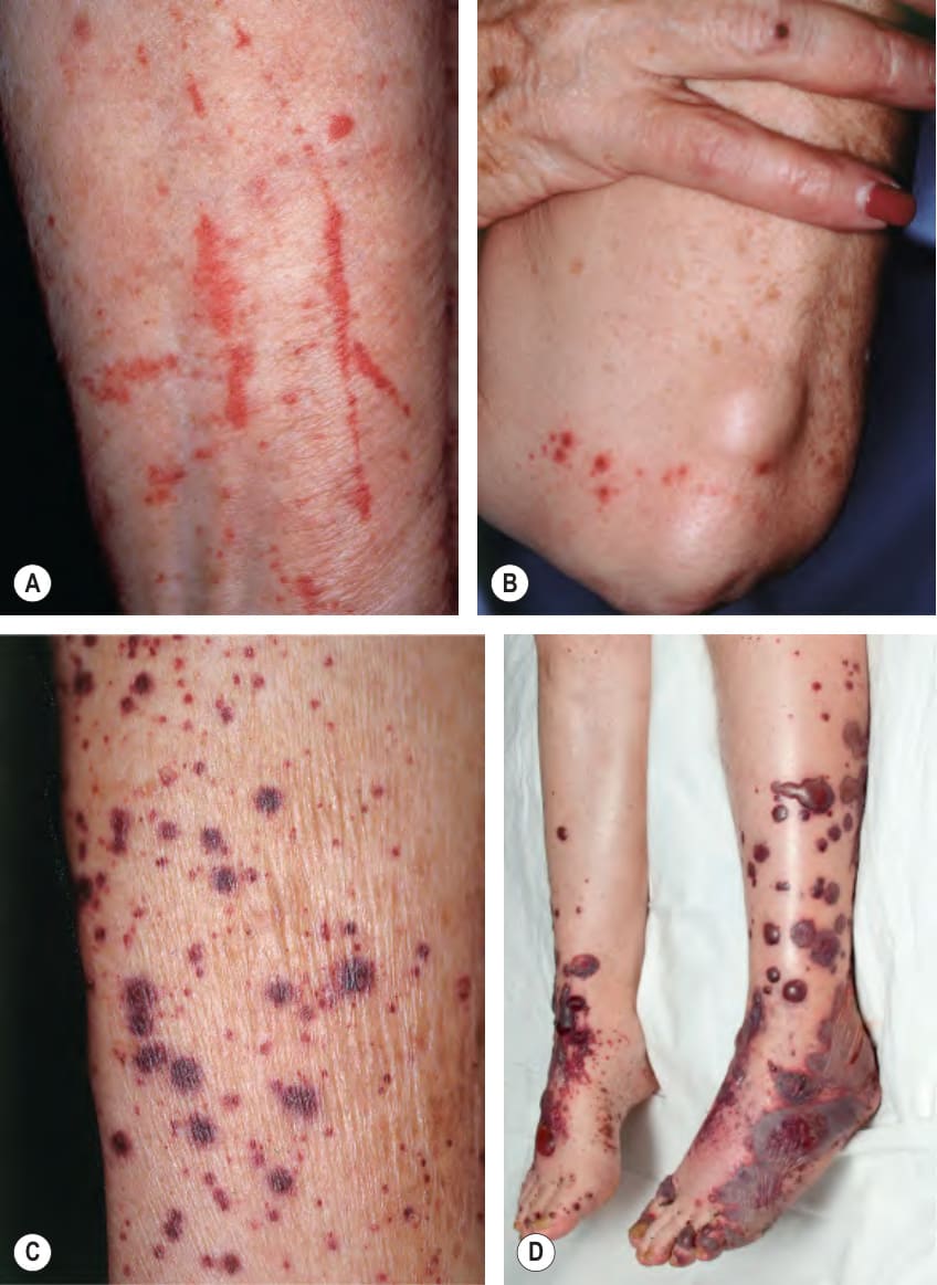
Fig. 24.6 Cutaneous small vessel vasculitis associated with systemic disorders.
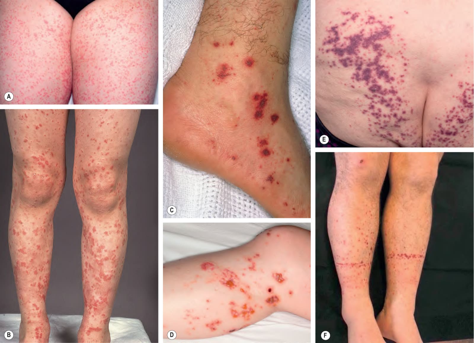
Fig. 24.7 IgA vasculitis (Henoch–Schönlein purpura).A, B Early inflammatory lesions that are beginning to develop a hemorrhagic component. Sites of previous involvement on the shins are now dull pink patches. C More developed lesions with central necrosis. D Less common vesiculobullous variant. E Purpuric macules and papules of the buttocks with coalescence into a retiform configuration on the upper left; inflammatory retiform purpura can be seen in some forms of vasculitis, especially IgA vasculitis (see Table 22.6). F Accentuation of lesions at the sock lines due to increased pressure. A, D, E, Courtesy David A. Wetter, MD; F, Courtesy Carlo Mainetti, MD.
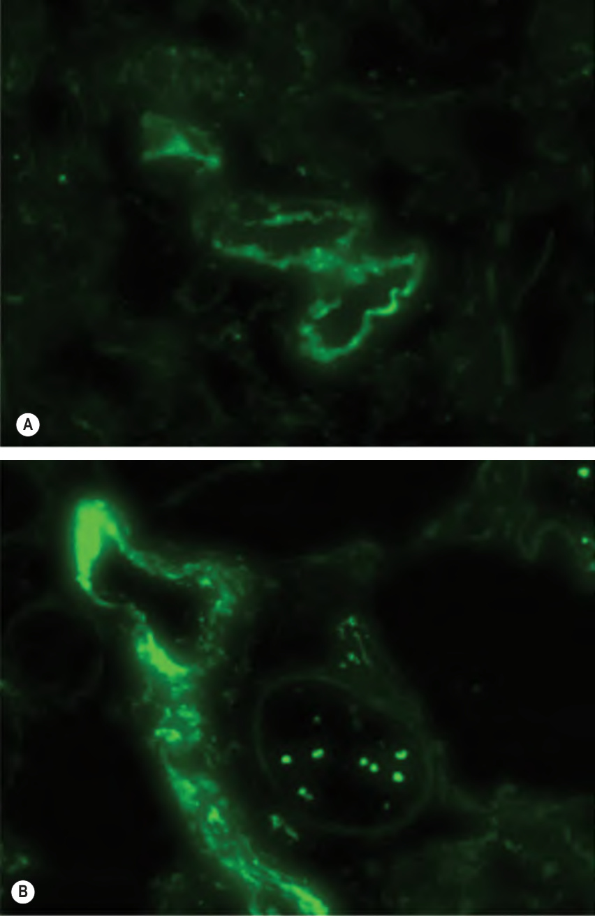
Fig. 24.8 Direct immunofluorescence microscopy of IgA vasculitis (Henoch–
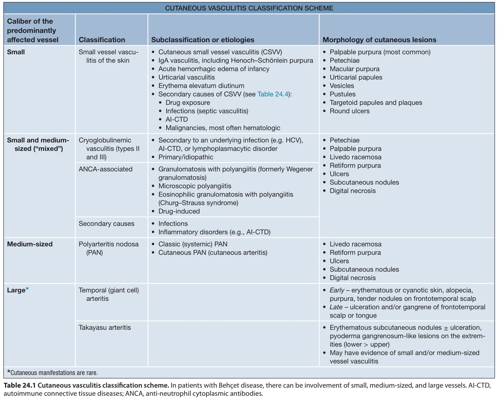
Table 24.1 Cutaneous vasculitis classification scheme. In patients with Behçet disease, there can be involvement of small, medium-sized, and large vessels. AI-CTD, autoimmune connective tissue diseases; ANCA, anti-neutrophil cytoplasmic antibodies.
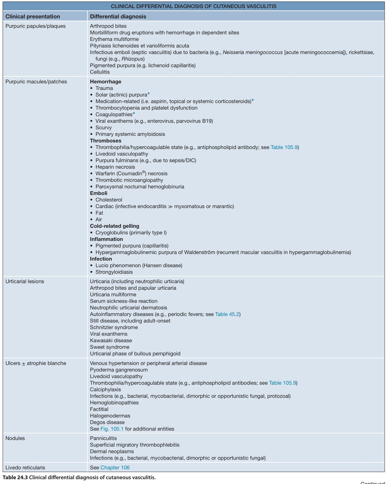
Table 24.3 Clinical differential diagnosis of cutaneous vasculitis.

Table 24.10 Therapeutic ladder for patients with vasculitis.