Urticarial Vasculitis
Synonyms/Variants: Normocomplementemic urticarial vasculitis Hypocomplementemic urticarial vasculitis Hypocomplementemic urticarial vasculitis syndrome Anti-C1q vasculitis McDuffie syndrome
Key features
Recurrent urticarial lesions lasting >24 hours and resolving with bruise-like discoloration
Two major subtypes: normocomplementemic and hypocomple- mentemic; low serum complement levels suggest systemic disease
Clinical manifestations include constitutional symptoms, arthritis, and obstructive lung disease
Associated with autoimmune connective tissue disease, especially systemic lupus erythematosus
Introduction
Urticarial vasculitis is a clinicopathologic entity consisting of persistent urticarial lesions that demonstrate the histopathologic features of LCV. Its association with autoimmune connective tissue disease and potential overlap with systemic lupus erythematosus (SLE) distinguish this disorder from typical CSVV. Although the relationship between urticarial vasculitis and neutrophilic urticaria is an area of debate, these and other related conditions (e.g. Still disease) may occur within a
spectrum (see below). For this chapter, the definition of urticarial vasculitis is as follows: urticarial lesions that histopathologically demonstrate vasculitis, as defined by a minimum of leukocytoclasia with vessel wall necrosis, with or without fibrinoid deposits, perivascular inflammation, or red blood cell extravasation.
Epidemiology
The population-based incidence of urticarial vasculitis is 5 cases per million per year. In patients with chronic urticaria in whom a strict definition of urticarial vasculitis (see above) is applied, the prevalence of urticarial vasculitis is said to be ~5%, although this is likely an overestimation. The peak incidence is during the fifth decade, and 60%–80% of patients with urticarial vasculitis are female. The hypocomplementemic form occurs almost exclusively in women. Approximately 70%–80% of cases of urticarial vasculitis are normocomplementemic, and they follow a benign course with an average duration of 3 years.
Pathogenesis
The pathogenesis of urticarial vasculitis is similar to that of typical CSVV. In urticarial vasculitis, it is thought that complement activation triggers mast cell release of inflammatory mediators such as TNF that in turn increase the expression of ICAM on mast cells (important for eosinophil transmigration) and E-selectin on endothelial cells.
Although urticarial vasculitis is most often idiopathic, it can be associated with autoimmune connective tissue diseases (especially SLE and Sjögren syndrome), serum sickness, cryoglobulinemia, infections, medications, and hematologic malignancies (see Table 24.5).
Clinical features
Lesions of urticarial vasculitis consist of erythematous, indurated wheals (Fig. 24.10), with or without angioedema, that favor the trunk
and proximal extremities. Urticarial vasculitis is distinguished from chronic urticaria by the persistence of individual lesions beyond 24 hours, burning pain rather than pruritus, and the presence of purpura and/or postinflammatory hyperpigmentation. Blanching of the skin via diascopy may reveal a central red–purple or brown macule, suggesting vasculitis. However, these features are not always present. Rarely, bullae, erythema multiforme-like lesions, livedo reticularis, Raynaud phenomenon, and laryngeal edema are clinical manifestations of urticarial vasculitis.
When evaluating patients with urticarial lesions that histologically demonstrate features of LCV, the most important prognostic feature is the presence or absence of hypocomplementemia. Patients with normal complement levels tend to have skin-limited disease and are perhaps best considered to have a subset of CSVV with hive-like lesions. Those with hypocomplementemia are much more likely to have systemic manifestations, including gastrointestinal, joint, or renal involvement, and to meet diagnostic criteria for SLE. The hypocomplementemic urticarial vasculitis syndrome (HUVS) is typically a more severe syndrome defined by specific diagnostic criteria:
●Two major criteria: (1) urticaria for 6 months; and (2) hypocomplementemia – plus –
●Two or more minor criteria: (1) vasculitis on skin biopsy; (2) arthralgia or arthritis; (3) uveitis or episcleritis; (4) glomerulo nephritis; (5) recurrent abdominal pain; or (6) positive C1q precipitin test with a low C1q level. Patients with hypocomplementemia who do not meet the criteria for HUVS are considered to have hypocomplementemic urticarial vasculitis but not HUVS.
Musculoskeletal involvement is the most common extracutaneous manifestation of urticarial vasculitis. Arthralgias of the hands, elbows, knees, ankles, and feet occur in half of all patients with urticarial vasculitis, but up to 50% of patients with HUVS have frank arthritis. Up to 20% of patients with HUVS have pulmonary symptoms which can resemble asthma or chronic obstructive pulmonary disease (COPD) and include cough, dyspnea, hemoptysis, laryngeal edema, and pleural effusions. COPD is especially severe in smokers with urticarial vasculitis, and it is worse than would be expected due to smoking alone; therefore, patients should be told to avoid smoking. Renal involvement, manifesting as proteinuria or microscopic hematuria, occurs in up to 15% of patients with HUVS. Gastrointestinal manifestations (abdominal pain, nausea, vomiting, diarrhea) occur in up to 30% of patients. Cardiac and central nervous system involvement are rare but reported. HUVS shares features with SLE, but distinctive clinical findings in HUVS include ocular inflammation (30%; conjunctivitis, episcleritis, iritis, uveitis), angioedema (50%), and COPD-like symptoms (20%). In a study of 57 patients with hypocomplementemic urticarial vasculitis, various systemic manifestations were observed, including ocular (56%), pulmonary (19%), gastrointestinal (18%), and renal (14%).
In patients with urticarial vasculitis, the most common abnormal laboratory studies are an elevated ESR, low serum C3 and C4 levels, and a positive ANA. HUVS is usually marked by low serum complement levels (which may vary from non-detectable to normal, even during flares) plus the presence of anti-C1q precipitin and depressed C1q levels. Although up to a third of patients with SLE have circulating anti-C1q antibodies, and up to half of patients with HUVS have a positive ANA, patients with HUVS rarely have anti-dsDNA or anti-Sm antibodies.
Pathology
Histologically, urticarial vasculitis is defined as the presence of leukocytoclasia with vessel wall necrosis, with or without fibrinoid deposits, perivascular inflammation, or red blood cell extravasation. In one series, an interstitial neutrophilic infiltrate was more commonly observed in patients with the hypocomplementemic form of urticarial vasculitis. Eosinophils may also be noted. Although findings can be subtle and consist only of interstitial neutrophils or perivascular lymphocytes with extravasated erythrocytes (especially in older lesions), these findings alone do not fulfill the histologic criteria for the diagnosis of urticarial vasculitis.
By DIF, 70% of lesions demonstrate deposits of immunoglobulin, C3, or fibrinogen around blood vessels. A granular pattern of immunoreactants along the basement membrane zone occurs in ~80% of lesions and, when accompanied by hypocomplementemia, suggests the diagnosis of SLE. Basement membrane immunoreactants have also been associated with renal disease.
Differential diagnosis
The main entity in the differential diagnosis is urticaria (including delayed pressure urticaria, in which lesions can last longer than 24 hours). Urticarial vasculitis should also be distinguished from neutrophilic urticaria, which, although considered by some to be within the spectrum of urticarial vasculitis, is not associated with hypocomplementemia or autoimmune disease and is better thought of as a subset of urticaria that histologically demonstrates a neutrophilic infiltrate without vasculitis. The differential diagnosis also includes disorders that clinicopathologically present with urticarial lesions and an inter-stitial neutrophilic infiltrate (i.e. neutrophilic urticarial dermatosis), including Schnitzler syndrome, adult-onset Still disease, and cryopyrinassociated periodic syndromes (see Ch. 45). Additional entities to consider are the urticarial phase of bullous pemphigoid, atypical erythema multiforme, urticaria multiforme, Sweet syndrome, tumid lupus erythematosus, SLE (see above), and rheumatoid neutrophilic dermatitis. For lesions of angioedema, the differential diagnosis includes acquired and inherited forms of angioedema (see Ch. 18).
Treatment
No randomized clinical trials have been performed to evaluate possible therapeutic options for urticarial vasculitis. Antihistamines may reduce the swelling and pain associated with cutaneous lesions but do not alter the course of the disease. Oral corticosteroids are effective, but the duration of use should be kept to a minimum (see Ch. 125). Indomethacin, dapsone (with or without pentoxifylline), colchicine, hydroxychloroquine, and mycophenolate mofetil have all been reported to be beneficial (see Table 24.10). Rituximab, omalizumab, anakinra, and canakinumab have been reported to improve recalcitrant hypocomplementemic urticarial vasculitis.
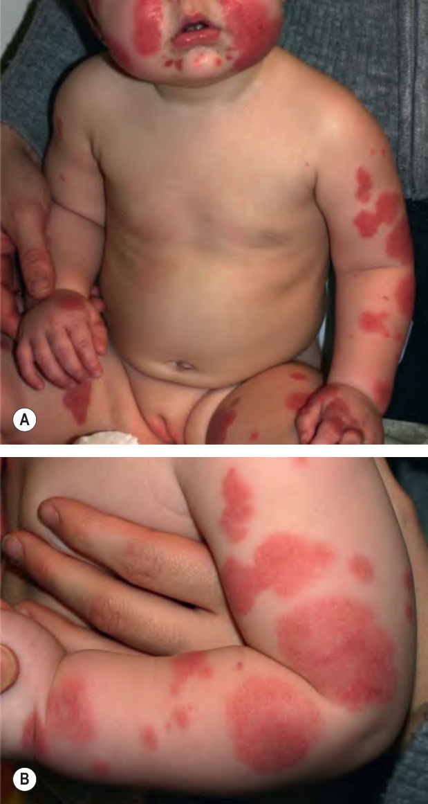
Fig. 24.9 Acute hemorrhagic edema of infancy.A, B Multiple edematous erythematous plaques on the face and extremities of a toddler. Some of the lesions have begun to become dusky. Courtesy Ilona J. Frieden, MD.
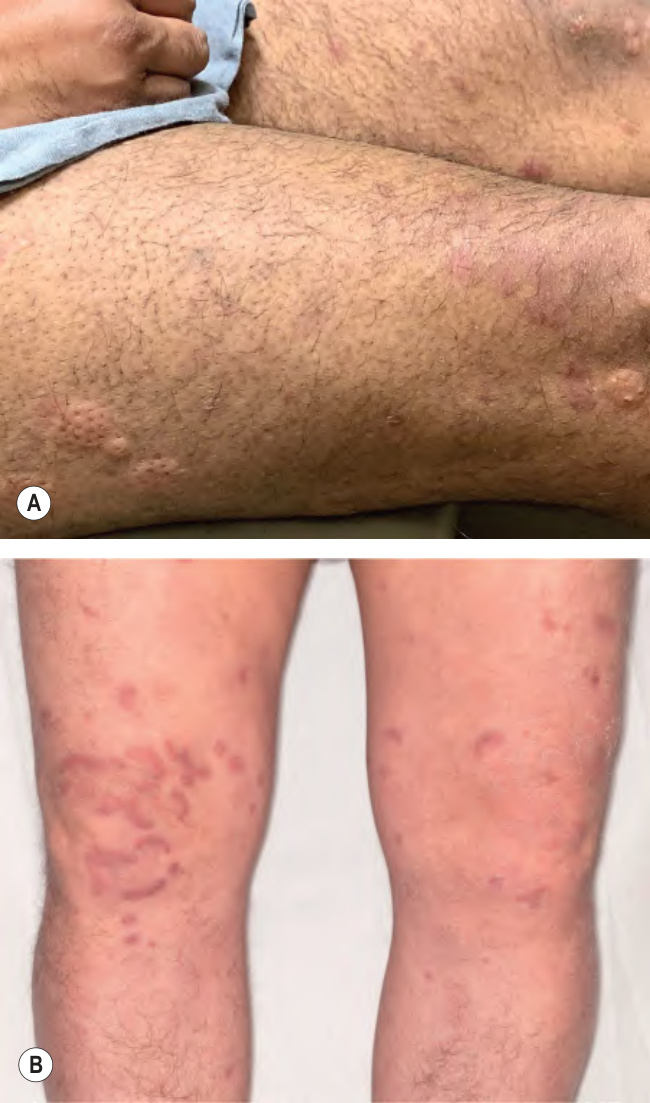
Fig. 24.10 Urticarial vasculitis.A Urticarial papules and purpuric macules in a patient with hypocomplementemic urticarial vasculitis. B Arcuate, dull violet plaques of the lower extremities. B, Courtesy David A. Wetter, MD.
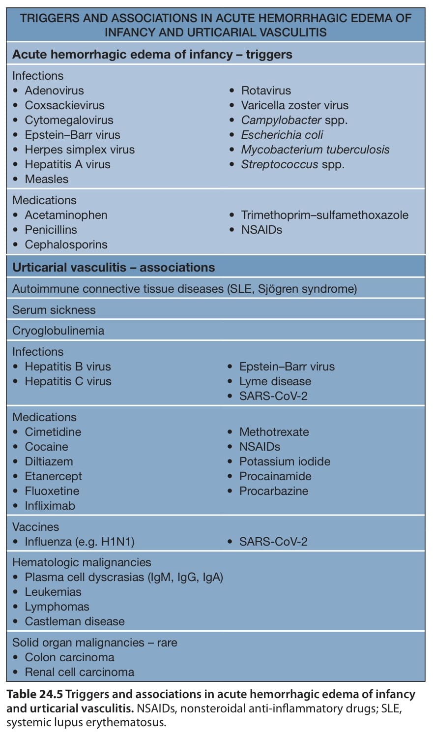
Table 24.5 Triggers and associations in acute hemorrhagic edema of infancy and urticarial vasculitis. NSAIDs, nonsteroidal anti-inflammatory drugs; SLE, systemic lupus erythematosus.

Table 24.10 Therapeutic ladder for patients with vasculitis.