Erythema Elevatum Diutinum
Key features
Symmetric, red–violet to red–brown papules and plaques
Persistent lesions develop on extensor surfaces
Fibrosing leukocytoclastic vasculitis
Introduction
Erythema elevatum diutinum (EED) is a rare, chronic dermatosis characterized by red–violet to red–brown papules, plaques, and nodules that favor extensor surfaces. Histopathologic features consist of LCV in early lesions, followed by fibrotic replacement of the dermis in older lesions.
Epidemiology
EED is rare, with descriptions of several hundred cases in the literature. The disease can develop at any age but is more common in middle-aged and older adults (ages 30–60 years). An earlier onset occurs more often in the setting of HIV infection. The male-to-female ratio is approximately equal, and no racial predilection has been observed.
Pathogenesis
Although the etiology of EED is unknown, circulating immune complexes, with repeated deposition, associated inflammation and partial healing, are thought to represent the underlying pathogenesis. Immune complex deposition results in complement activation, neutrophilic infiltration, and the release of destructive enzymes. The latter results in fibrin deposition in and around small dermal vessels during the later stages of the disease.
EED has been described in association with a number of systemic diseases, including infections, autoimmune diseases, and both benign and malignant hematologic disorders, especially IgA monoclonal gammopathy. Associated infections include β-hemolytic streptococci, HBV, HIV, tuberculosis, and syphilis. Lesions identical to EED, both clinically and histologically, have been reproduced by the intradermal injection of streptococcal antigen into non-lesional skin of patients with EED. In patients with HIV infection, EED may be due to either HIV-related antigen–antibody complexes directly damaging small blood vessels or HIV-induced immunosuppression allowing other infectious agents to serve as the antigenic stimulus.
Autoimmune or inflammatory conditions associated with EED include granulomatosis with polyangiitis (formerly Wegener granulomatosis), inflammatory bowel disease, celiac disease, relapsing polychondritis, SLE, and rheumatoid arthritis. In addition to plasma cell dyscrasias (especially IgA monoclonal gammopathy), associated hematologic disorders include myelodysplasia, myeloproliferative disorders, and hairy cell leukemia.
Clinical features
The typical cutaneous lesions of EED are violaceous, red–brown or yellowish papules, plaques, or nodules that are symmetrically distributed. They favor acral and periarticular sites, in particular the extensor surfaces of the elbows, knees, ankles, hands, and fingers (Fig. 24.11). Additional sites of involvement include the face, retroauricular area, trunk, axillae, buttocks, and genitalia. Initially, the lesions are erythematous, but with time they become red–brown or violaceous in color as well as doughy or firm to palpation due to fibrosis.
In general, the lesions are asymptomatic, but they can be associated with a burning sensation or pruritus, especially early on. Annular plaques with a raised border and verrucous plaques on the soles have also been described. Nodular lesions, primarily palmoplantar, that progress to form bulky masses are characteristic of EED in the setting of HIV infection. Arthralgias may develop in underlying joints, but extracutaneous involvement, with the exception of ocular disease, is extremely rare. There have been reports of associated peripheral keratitis, nodular scleritis, panuveitis, and blindness. The disease is chronic and has a relapsing and remitting course; the majority of cases resolve spontaneously over a period of 5 to 10 years, but the disease can last up to 40 years.
In patients presenting with EED, evaluation for an associated infection (e.g. streptococcal, viral hepatitis, HIV, syphilis), monoclonal
gammopathy (serum immunofixation electrophoresis), or autoimmune disorder (see above) should be considered, based upon the clinical context.
Pathology
Early lesions of EED demonstrate the changes of LCV, with a neutrophilic infiltrate within the upper and mid dermis (Fig. 24.12A), admixed with some eosinophils. Involvement of the papillary and perifollicular adventitial dermis (without a grenz zone) is observed in more mature lesions. The latter are also characterized by the presence of granulation tissue and perivascular, concentric, or storiform fibrosis plus mixed inflammation that is composed predominantly of neutrophils. In the late stage, capillary walls may have fibrinoid necrosis or fibrosis (Fig. 24.12B); vertically oriented capillaries can also be seen, and it may be difficult to discern the features of vasculitis. Intracellular lipidosis, previously termed “extracellular cholesterolosis”, is a classic finding in late-stage lesions.
Differential diagnosis
The clinical differential diagnosis depends upon the stage of the lesion. For earlier lesions, one can consider neutrophilic dermatoses (e.g. Sweet syndrome, rheumatoid neutrophilic dermatitis) and palisaded neutrophilic and granulomatous dermatitis. Late-stage lesions may be confused primarily with tuberous xanthomas, granuloma annulare, rheumatoid nodules, fibroid nodules of Borrelia, and multi-centric reticulohistiocytosis. Other granulomatous disorders (e.g. sarcoidosis, Hansen disease [leprosy], necrobiotic xanthogranuloma) are sometimes in the differential diagnosis, and for an isolated lesion, dermatofibroma or dermatofibrosarcoma protuberans (DFSP). Occasionally, lesions with a prominent vascular component, especially in an HIV-infected patient, might be mistaken for Kaposi sarcoma or bacillary angiomatosis.
Histologically, the earliest lesions may share features with the neutrophilic dermatoses, but foci of LCV help to distinguish EED. Granuloma faciale may also be considered, but a predominance of eosinophils and plasma cells and the presence of a grenz zone are distinguishing features, as is the localization of lesions to the face. The differential diagnosis of later lesions includes tuberous xanthomas, fibrotic disorders or tumors (e.g. DFSP; see Chs. 98 & 116), and, occasionally, fibroid nodules of Borrelia, Kaposi sarcoma, and bacillary angiomatosis.
Treatment
Dapsone is the mainstay therapeutically, but relapses frequently occur upon discontinuation. If present, treatment of any associated infection or underlying systemic disorder is also recommended. Other therapies include NSAIDs, niacinamide, tetracyclines, chloroquine, colchicine, and plasmapheresis (see Table 24.10). Intralesional corticosteroids may be helpful for mild cases, but systemic corticosteroids are rarely indicated.
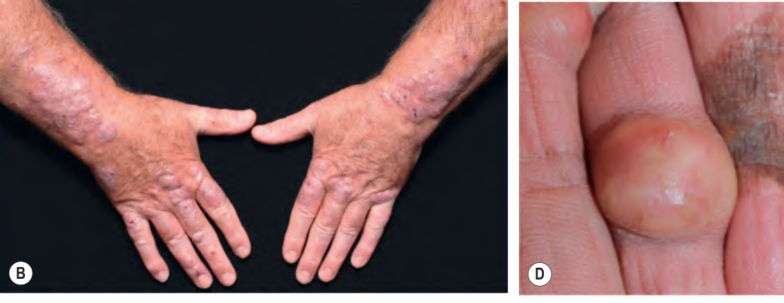
Fig. 24.11 Erythema elevatum diutinum.A Erythematous papulonodules on the upper knee, representing acute lesions, admixed with older red–brown nodules. B Symmetrically distributed, dull pink to violet–brown nodules and plaques on the extensor forearms and dorsal hands. C Firm nodule on the dorsum of the hand in a patient with HIV infection, representing a late-stage lesion. D These yellow nodules are sometimes misdiagnosed as xanthomas. The hyperpigmented patches represent skin grafts at sites of excision. A, Courtesy Kenneth Greer, MD; B, Courtesy David A. Wetter, MD; C, Courtesy Rachel Moore, MD; D, Courtesy, Kalman Watsky, MD.
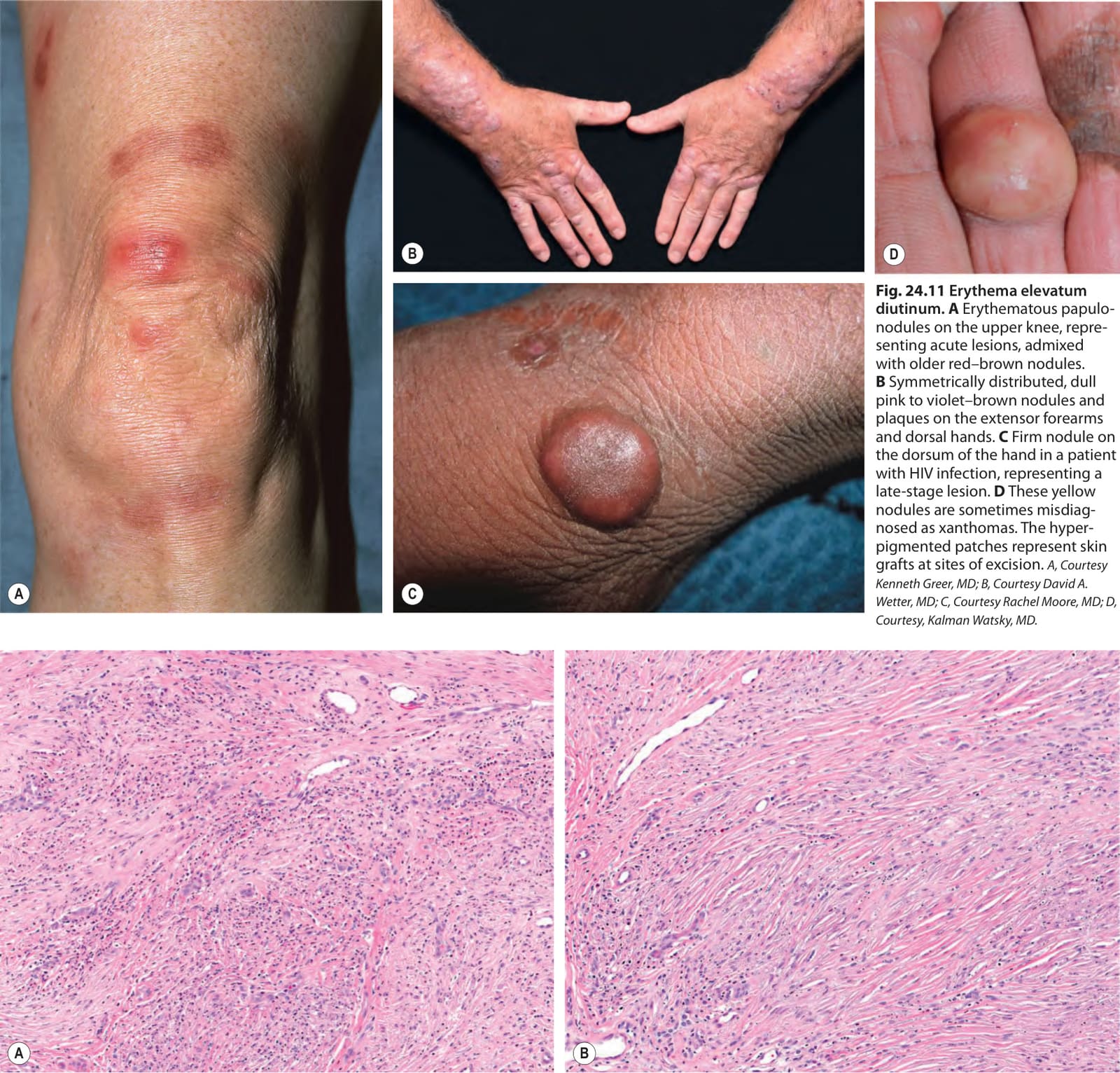
Fig. 24.12 Erythema elevatum diutinum – histopathologic features.A An early-stage lesion: dense neutrophilic infiltrates with focal leukocytoclastic vasculitis. B A late-stage lesion: a sparser inflammatory infiltrate and marked fibrosis. Courtesy Lawrence E. Gibson, MD.
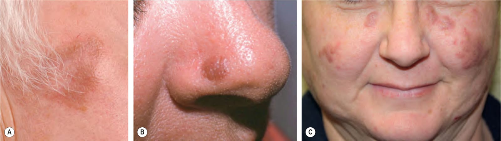
Fig. 24.13 Granuloma faciale.A Single light brown plaque of the lateral malar cheek. B Single red–brown papule of the nose. Clues to the diagnosis include prominent follicular openings and a lack of secondary changes such as scale. C Less commonly, patients present with multiple lesions. A, Courtesy Department of Dermatology, Medical University of Graz; B, Courtesy Cloyce L. Stetson, MD; C, Courtesy Jeffrey P. Callen, MD.

Table 24.10 Therapeutic ladder for patients with vasculitis.