Granuloma Faciale
Key features
Firm red–brown or violaceous plaques or nodules, often solitary and most commonly on the face
Histologic features of small vessel vasculitis, with dense dermal infil- trates of neutrophils, lymphocytes and plasma cells, admixed with numerous eosinophils; the inflammation spares the upper papillary dermis (grenz zone), and fibrosis may be present
Introduction
Granuloma faciale is a chronic inflammatory dermatosis characterized by red–brown to violaceous plaques or nodules. The face is the most common site of involvement and lesions are more often solitary than multiple. Because histologic features of vasculitis are frequently present, the disease can be classified as a subtype of skin-limited vasculitis.
Epidemiology
Granuloma faciale occurs predominantly in middle-aged White men but has been observed in Black and Asian men as well as women.
Pathogenesis
While the precise pathogenesis remains unknown, granuloma faciale is thought to be a localized form of skin-limited small vessel vasculitis. Direct immunofluorescence (DIF) microscopy demonstrates granular deposition of IgG, IgA, IgM, and/or C3 in blood vessel walls, a nonspecific finding that suggests a role for immune complex-mediated vascular damage. Interferon-γ and increased IL-5 production have been implicated as potentially important disease mediators. An increased ratio of IgG4- : IgG-bearing circulating plasma cells and/or relative increases of IgG4-positive plasma cells in skin lesions has been observed, but patients do not typically meet consensus criteria for IgG4-related disease.
Clinical features
Granuloma faciale usually presents as a solitary, asymptomatic, red– brown or violaceous plaque on the face, with a predilection for the forehead, cheek, and preauricular area (Fig. 24.13A,B). Less often, multiple papules or plaques may be present (Fig. 24.13C); in one retrospective analysis of 66 patients, one-third had more than one lesion. Uncommon locations for granuloma faciale include the ears, scalp, trunk, and extremities; in the previously cited study, 7% of patients had extrafacial involvement. The individual lesions tend to persist and only occasionally resolve spontaneously. Granuloma faciale has not been associated with systemic disease.
Pathology
In the dermis, there are perivascular and interstitial infiltrates of neutrophils, lymphocytes, and plasma cells, admixed with numerous eosinophils. Characteristically, the inflammation spares the upper papillary dermis, resulting in a “grenz zone” (Fig. 24.14). Features of leukocytoclastic vasculitis are most prominent early on, with older lesions tending to have fewer neutrophils and more eosinophils and plasma cells, as well as fibrosis. Because of the presence of eosinophils, IgG4-bearing plasma cells, and lamellar fibrosis, granuloma faciale can resemble IgG4-related disease histologically (see Table 26.2).
Differential diagnosis
The clinical differential diagnosis of granuloma faciale includes cutaneous lymphoma, persistent arthropod bite reactions, angiolymphoid hyperplasia with eosinophilia, tumid lupus erythematosus, and several granulomatous disorders (e.g. sarcoidosis, Hansen disease, granulomatous rosacea). The histopathology of granuloma faciale may have features in common with rheumatoid neutrophilic dermatitis, neutrophilic dermatosis associated with lupus erythematosus, epithelioid hemangioma (angiolymphoid hyperplasia with eosinophilia), or arthropod bite reaction. Granuloma faciale may also resemble erythema elevatum diutinum (EED), both clinically and histopathologically. However, EED presents as multiple red–brown papules, plaques, or nodules in a symmetric distribution on the extensor aspects of the extremities, with a predilection for skin that overlies joints. Fibrosis also tends to be more pronounced, and lipid-laden macrophages may be seen in EED, but not in granuloma faciale.
Treatment
Because of the facial location, treatment is often desired. Unfortunately, granuloma faciale is frequently resistant to therapy. Topical or intra-lesional corticosteroids (e.g. triamcinolone suspension 2.5–5 mg/ml) and topical calcineurin inhibitors (pimecrolimus, tacrolimus) are firstline therapeutic options. Prolonged therapy (e.g. 3–6 months) may be necessary for improvement. As with other recalcitrant dermatoses, there are several alternatives with reported efficacy, including topical and oral dapsone (50–150 mg daily), topical ruxolitinib, hydroxychloroquine, clofazimine (300 mg daily), topical PUVA, and TNF inhibitors.
Cryosurgery, electrosurgery, surgical excision, dermabrasion, and CO or pulsed dye laser therapy may be effective, but each carries a risk of scarring given the depth of inflammation. In addition, recurrence after excision has been reported. Laser therapy targeting the prominent vascular component (e.g. 595 nm pulsed dye laser, 532 nm potassium titanyl phosphate laser) has led to improvement in a number of patients.
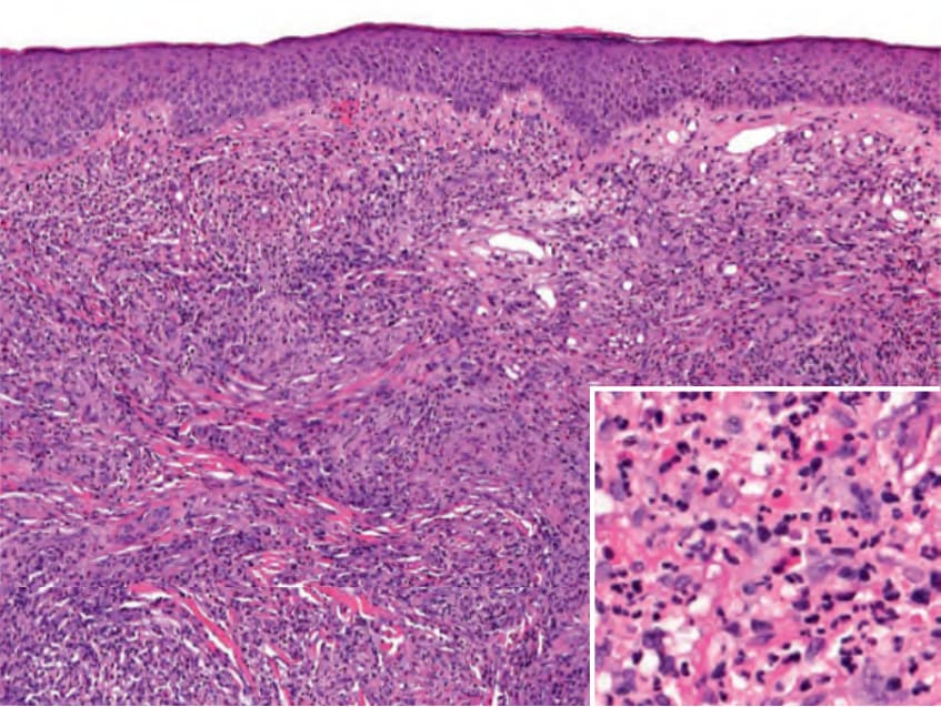
Fig. 24.14 Granuloma faciale – histopathologic features. Dense diffuse dermal inflammation with a grenz zone. Inset shows mixed infiltrate of lymphocytes, eosinophils, neutrophils, and plasma cells. Courtesy Lorenzo Cerroni, MD.