Cryoglobulinemic Vasculitis
Synonyms: Type II or III cryoglobulinemia Mixed cryoglobulinemia Cryoglobulinemic purpura Essential cryoglobulinemic vasculitis
Key features
Associated with mixed cryoglobulinemia (IgM and IgG), most commonly in the setting of hepatitis C virus (HCV) infection
Palpable purpura, typically on the lower extremities
Myalgias and arthralgias
Peripheral neuropathy and glomerulonephritis
Introduction
Cryoglobulins are cold-precipitable immunoglobulins that can be divided into three subtypes, all of which have cutaneous manifestations (Table 24.6). Type I cryoglobulinemia, composed primarily of monoclonal IgM (and less frequently IgG > IgA), results in microvascular occlusion without vasculitis (see Ch. 23). In contrast, vasculitis is seen with type II and III cryoglobulins, also referred to as “mixed cryoglobulins,” because they are composed of polyclonal IgGs – the autoantigens – and monoclonal or polyclonal IgMs, respectively. The IgM autoantibodies bind other antibodies and therefore have rheumatoid factor activity. Approximately 15% of patients with circulating mixed cryoglobulins present with symptoms due to cryoglobulinemic vasculitis. Cryoglobulinemic vasculitis typically affects the skin, joints, peripheral nervous system, and kidneys.
Epidemiology
The frequency of mixed cryoglobulinemia varies according to geographic location; e.g. there is a higher prevalence in Southern Europe than in Northern Europe or North America. In northwest Spain, the annual incidence was determined to be 4.8 cases per million. Geographic variations in the frequency of mixed cryoglobulinemia likely reflect differences in the prevalence of HCV infection. While 40%–60% of patients with HCV infection have cryoglobulinemia, overt cryoglobulinemic vasculitis develops in ~5%–30% of these individuals.
Pathogenesis
Mixed cryoglobulinemia occurs in the setting of specific infections, autoimmune connective tissue diseases, and hematologic malignancies (see Table 24.6). When the association between HCV infection and mixed cryoglobulinemia was discovered in 1989, a cause for 80%–90% of “essential” cryoglobulinemia became apparent. Only 5% of cases are associated with HBV infection, and occasionally, HIV, EBV, cytomegalovirus, Leishmania, and Treponema spp. have been implicated. In a minority of patients, mixed cryoglobulinemia has been associated with autoimmune connective tissue diseases, in particular Sjögren syndrome, SLE, and rheumatoid arthritis. Approximately 5% of the patients with mixed cryoglobulinemia have a lymphoproliferative disorder (e.g. B cell non-Hodgkin lymphoma, chronic lymphocytic leukemia, macroglobulinemia).
It is thought the underlying infectious, autoimmune, or lymphoproliferative disease causes B cell stimulation and proliferation, resulting in expansion of cryoglobulin-producing B cell clones. A significant percentage of lymphocytes in HCV-infected individuals possess a t(14;18) translocation that results in a rearrangement of bcl-2, whose protein product has an anti-apoptotic function; this can lead to B lymphocyte proliferation and may contribute to production of cryoglobulins. In HCV-infected patients who do not possess this bcl-2 rearrangement, B cell activation and subsequent cryoglobulin and autoantibody production is believed to result from chronic immune stimulation by the virus. For these reasons, patients with mixed cryglobulinemia are at increased risk of developing B cell non-Hodgkin lymphoma.
Cryoglobulinemic vasculitis occurs when immune complexes form from circulating cryoglobulins and deposit within the walls of small blood vessels. As with other forms of immune complex-mediated vasculitis, this immune complex deposition initiates complement activation and vascular inflammation. The presence of the hepatitis C virion (in association with IgM and IgG antibodies) within vessel walls raises the possibility that viral particles are a component of the cryoprecipitate.
Clinical features
Cutaneous involvement occurs in nearly all patients with cryoglobulinemic vasculitis, most commonly manifesting as palpable purpura of the lower extremities (Fig. 24.15). Other cutaneous findings include livedo reticularis, retiform purpura, necrosis, and ulceration, reflecting a mixture of small and medium-sized vessel vasculitis. Skin lesions are not typically cold-induced, in contrast to the vascular occlusive lesions seen in type I cryoglobulinemia. The condition usually has a chronic course, but in those with HCV-associated disease, more effective direct-acting viral agents for treating HCV may be curative (see below).
Common extracutaneous findings at the time of presentation include weakness (80%), arthritis or arthralgias (70%), peripheral (typically sensory) neuropathy (60%), gastrointestinal disease or hepatitis (60%), and membranoproliferative glomerulonephritis (20%). The hepatitis reflects the underlying viral infection or an associated autoimmune hepatitis and is not a direct result of the cryoglobulinemia. Occasionally, patients have xerostomia or xerophthalmia, as seen in Sjögren syndrome.
The most commonly associated infections, autoimmune connective tissue diseases, and hematologic disorders are listed in the Pathogenesis section. B cell non-Hodgkin lymphoma is the most common associated malignancy. Rarely, solid tumors such as hepatocellular carcinoma and papillary thyroid carcinoma have been reported to have a relationship with cryoglobulinemic vasculitis.
Tests for circulating cryoglobulins are often falsely negative. Specimens should be drawn during a flare using collection tubes prewarmed to 37°C without anticoagulants. The specimen should be kept at 37°C and transported directly to the laboratory to prevent cryoprecipitation. If clinical suspicion remains high, negative tests should be repeated. By contrast, rheumatoid factor is frequently positive in patients with mixed cryoglobulinemia (~70% of patients), often at high levels. Patients typically have low serum complement levels, often with low or undetectable C4 levels; however, complement levels do not necessarily correlate with the severity of the disease. HBV, HCV, and HIV serologies should be evaluated, followed by a determination of viral
load if there is evidence of infection. Up to 15% of patients with mixed cryoglobulinemia have a monoclonal gammopathy as detected by serum protein electrophoresis and/or immunofixation electrophoresis.
Pathology
Histologically, features of LCV are seen. DIF reveals granular deposits consisting predominantly of IgM and C3 in a vascular pattern in the papillary dermis.
Differential diagnosis
The differential diagnosis of cryoglobulinemic vasculitis includes all forms of vasculitis affecting small or medium-sized vessels (see Table 24.1). Cryoglobulinemic vasculitis can be differentiated from these disorders based upon the appropriate context and by the detection of circulating mixed cryoglobulins. Patients with Sjögren syndrome can also have arthralgias, arthritis, xerostomia/xerophthalmia, and the presence of rheumatoid factor and/or mixed cryoglobulins; however, they are typically SSA (Ro)/SSB (La)-positive, have distinct histopathologic findings in salivary gland biopsies, and less frequently are found to have hepatitis, glomerulonephritis, or hypocomplementemia.
Treatment
The treatment of mixed cryoglobulinemia should be directed at any underlying disease. In patients with mild manifestations of vasculitis, that may be sufficient. In general, those with moderate or severe disease should first receive immunosuppressive therapy until they have stabilized clinically, then therapy for the underlying condition can be initiated. All patients with HCV-associated mixed cryoglobulinemia should be treated with antiviral therapy (see Table 24.10). Direct-acting antiviral agents are significantly more effective and better tolerated than previous interferon-containing regimens. Observational studies suggest antiviral regimens are effective in attaining a sustained virologic response, as well as improving vasculitic symptoms among those with HCV-associated cryoglobulinemic vasculitis. In a study of 148 patients, >95% had a complete or partial clinical response, with a median follow-up of 15.3 months. While rates of clinical improvement are high, correlating with sustained virologic response, clonal production of cryoglobulins persists in some patients, leading to relapses of vasculitis.
Patients with mild manifestations of cryoglobulinemic vasculitis (e.g. purpura, arthralgias, mild sensory neuropathy) may respond to agents such as colchicine or dapsone and/or symptomatic management with nonsteroidal anti-inflammatory drugs (NSAIDs) or gabapentin. Those with moderate to severe disease should receive high-dose systemic corticosteroids in combination with rituximab. Evidence from randomized trials and cohort studies demonstrate the efficacy and safety of this regimen compared to conventional therapy with systemic corticosteroids, either alone or in combination with cyclophosphamide. In addition, plasma exchange should be considered in those with acute life-threatening disease, including pulmonary hemorrhage, rapidly progressive crescentic glomerulonephritis, and hyperviscosity syndrome. As expected, prognosis depends on the severity of organ involvement and the underlying disease. In non-infectious mixed cryoglobulinemic vasculitis, predictors of early relapse include purpura, cutaneous necrosis, and articular involvement.
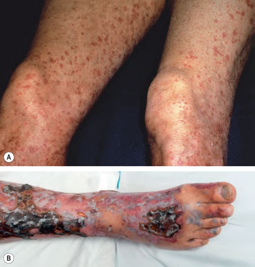
Fig. 24.15 Cryoglobulinemic vasculitis.A Palpable purpura of the lower extremities. B Purpuric papules and retiform plaques with central necrosis and ulceration in a patient with type II cryoglobulinemia secondary to hepatitis C viral infection. These cutaneous manifestations reflect a “mixed” pattern of both small and medium-sized vessel vasculitis. The patient also had an elevated rheumatoid factor, decreased C4, and an IgM monoclonal gammopathy. B, Courtesy David A. Wetter MD.
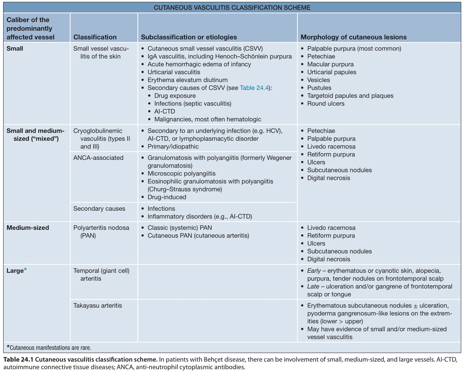
Table 24.1 Cutaneous vasculitis classification scheme. In patients with Behçet disease, there can be involvement of small, medium-sized, and large vessels. AI-CTD, autoimmune connective tissue diseases; ANCA, anti-neutrophil cytoplasmic antibodies.
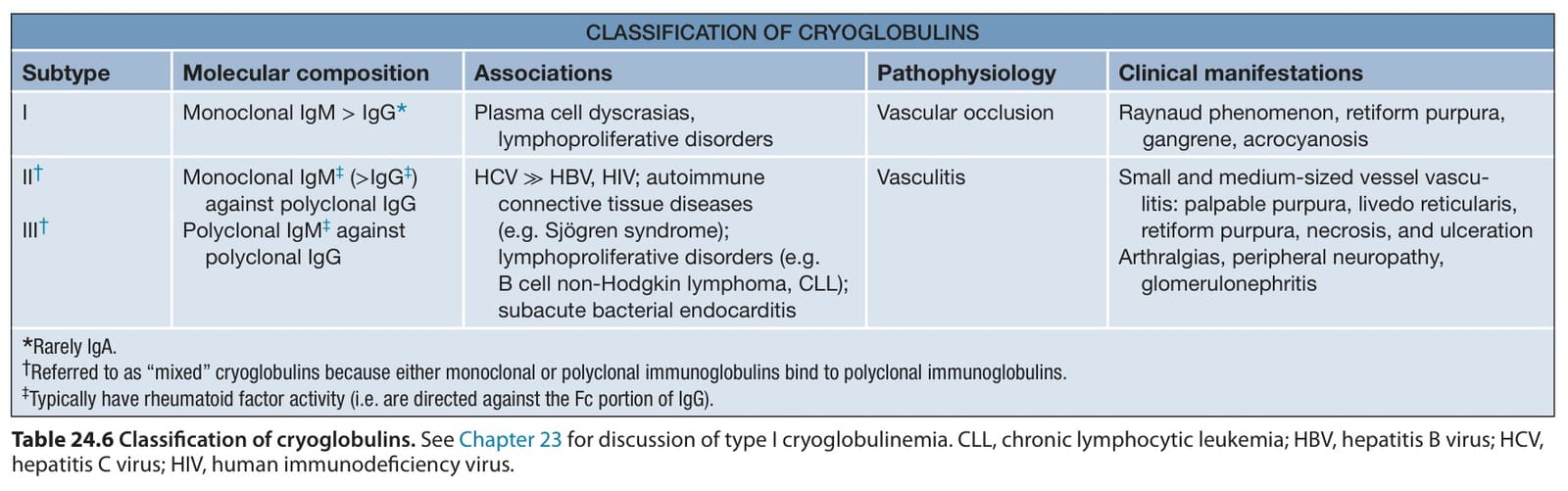
Table 24.6 Classification of cryoglobulins. See Chapter 23 for discussion of type I cryoglobulinemia. CLL, chronic lymphocytic leukemia; HBV, hepatitis B virus; HCV, hepatitis C virus; HIV, human immunodeficiency virus.

Table 24.10 Therapeutic ladder for patients with vasculitis.