ANCA-Associated Vasculitides
Introduction
The ANCA-associated vasculitides (AAVs) are characterized by vasculitis of small to medium-sized vessels, the presence of ANCAs, and an overlapping spectrum of organ involvement, with each having distinguishing clinical and laboratory features. New ACR-EULAR classification criteria for these diseases have been proposed. The three AAVs discussed here are granulomatosis with polyangiitis (GPA), microscopic polyangiitis (MPA), and eosinophilic granulomatosis with polyangiitis (EGPA) (Table 24.7).
ANCAs are predominantly IgG autoantibodies directed against components of both primary granules of neutrophils and monocyte lysosomes, and they likely have an important role in the pathogenesis of AAVs via interactions with primed neutrophils and endothelial cells. By immunofluorescent staining, there are two vasculitis-relevant patterns: (1) cytoplasmic (c)-ANCAs – directed against the antigen proteinase
3 (PR3); and (2) perinuclear (p)-ANCAs – directed against the antigen myeloperoxidase (MPO), with other antigens (e.g. lactoferrin, cathepsin G, elastase) serving as targets in the setting of nonspecific inflammation. ANCAs are useful diagnostically and for monitoring disease activity. However, these autoantibodies are also found in patients receiving certain medications (e.g. propylthiouracil, hydralazine) and who have underlying chronic infections, rheumatologic or inflammatory disorders, and malignancies (see Table 40.6).
In order to improve specificity with regard to AAVs, an indirect immunofluorescence (IIF) test should be used as a screening evaluation to detect perinuclear versus cytoplasmic staining, followed by an ELISA that specifically detects anti-PR3 and anti-MPO antibodies. However, up to 5% of serum samples are positive by ELISA only, so when there is a high index of suspicion for AAV, the ELISA should be performed even if the IIF screening test is negative. Because neither anti-PR3 ANCA nor anti-MPO ANCA is specific for a particular AAV and patients with AAV may be ANCA-negative, clinicopathologic correlation is required when evaluating a patient with suspected AAV (Fig. 24.16). Semi-quantitative analysis may allow for disease monitoring and response to treatment since the persistence of ANCAs following remission has been associated with disease relapse. Furthermore, there is growing recognition that ANCA type (anti-PR3 versus anti-MPO) is perhaps more clinically and prognostically meaningful than is disease type (GPA versus MPA), such that experts and clinical trialists increasingly refer to AAV by ANCA type and stratify clinical trial enrollment accordingly.
Epidemiology
In a population-based study of biopsy-proven small vessel vasculitis of the skin, the annual incidence of an underlying AAV was 4 cases per million, i.e. ~10% of the patients had an AAV, emphasizing the importance of considering this group of disorders. Conversely, cutaneous manifestations are a common presenting feature of AAV, occurring in a third of patients.
Variability reflects differences in cohorts studied, disease extent and severity at time of assays, diagnostic criteria, and cutoff values of various ANCA assays. Negative ANCA testing does not exclude the presence of an ANCA-associated vasculitis, as overall frequencies for ANCA positivity are 90% (generalized GPA), 60% (limited GPA), 90% (MPA), and 50% (EGPA), respectively. *MPO-ANCAs are overrepresented in Chinese patients with GPA; 10% of non-Chinese patients with GPA have MPO-ANCAs. **Examples include propylthiouracil, minocycline, hydralazine, and levamisole-tainted cocaine (see Tables 24.4 and 40.6). Note that other ANCA specificities may be detected in drug-induced vasculitis. ANCA, anti-neutrophil cytoplasmic antibody; EGPA, eosinophilic granulomatosis with polyangiitis; GPA, granulomatosis with polyangiitis; MPA, microscopic polyangiitis; MPO, myeloperoxidase; PR3, proteinase 3. Adapted from Wiik AS, Rheum Dis Clin NA. 2010;36:479–89.
Pathogenesis
The pathogenesis of AAV, depicted in Fig. 24.1B, is complex and includes genetic as well as environmental factors. Key elements include: autoreactive B-lymphocytes that produce ANCAs; abnormal T-regulatory function which allows persistent T cell activation, especially of Th17 cells; neutrophils that cause tissue damage via injury to vascular epithelium; and activation of the alternative complement pathway, namely C5a, and neutrophil extracellular traps (NETs) which drive vascular inflammation.
Natural history, clinical features, and treatment
The morbidity and mortality of AAVs are relatively high, stemming from associated end-organ damage as well as complications of immunosuppressive therapy. Delay in diagnosis, renal impairment, propensity for relapse, the presence of ANCAs, and older age are risk factors for reduced survival. The three major AAVs are discussed in the following sections, including clinicopathologic features (see Table 24.7), as well as treatment options (see Table 24.10).
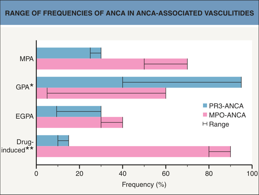
Fig. 24.16 Range of frequencies of ANCA in ANCA-associated vasculitides.
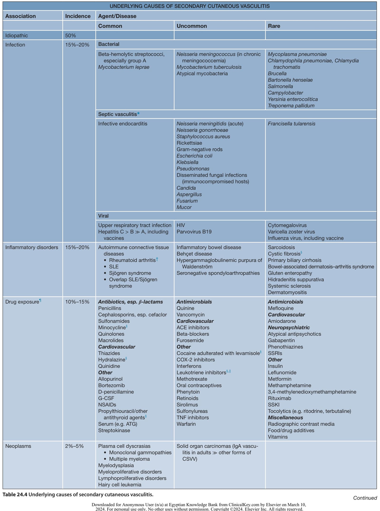
Table 24.4 Underlying causes of secondary cutaneous vasculitis. Continued
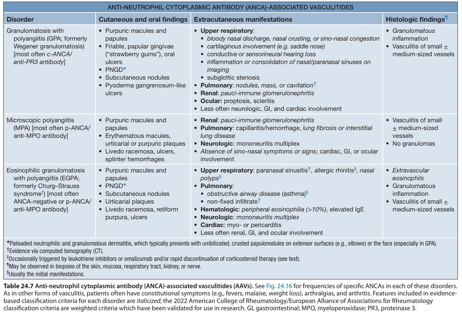
Table 24.7 Anti-neutrophil cytoplasmic antibody (ANCA)-associated vasculitides (AAVs). See Fig. 24.16 for frequencies of specific ANCAs in each of these disorders. As in other forms of vasculitis, patients often have constitutional symptoms (e.g., fevers, malaise, weight loss), arthralgias, and arthritis. Features included in evidencebased classification criteria for each disorder are italicized; the 2022 American College of Rheumatology/European Alliance of Associations for Rheumatology classification criteria are weighted criteria which have been validated for use in research. GI, gastrointestinal; MPO, myeloperoxidase; PR3, proteinase 3.

Table 24.10 Therapeutic ladder for patients with vasculitis.