Granulomatosis With Polyangiitis
Synonym: Wegener granulomatosis (historical)
Key features
Necrotizing granulomatous inflammation of the upper and lower respiratory tracts
Pauci-immune glomerulonephritis
Systemic vasculitis that can involve the skin and oral mucosa
c-ANCA and anti-PR3 antibodies most commonly detected
Introduction
Granulomatosis with polyangiitis (GPA) is classically described as the triad of granulomatous inflammation of the upper and lower respiratory tracts, systemic necrotizing vasculitis, and pauci-immune glomerulonephritis. Patients with GPA exist along a spectrum of severity, depending upon the number of organs involved and the degree of functional impairment. Those with organ- or life-threatening disease have a high mortality rate if left untreated, while patients without organ- or life-threatening GPA have primarily airway disease without constitutional symptoms or systemic vasculitis. The initial evaluation of patients with GPA should establish the extent of organ involvement, as evidence supports distinct treatment approaches based upon disease severity. However, because this disorder can evolve over time, patients initially without organ- or life-threatening disease must still be closely monitored.
Epidemiology
The incidence of GPA varies widely by geographic region and is estimated to be 0.4 to 12 cases per million per year. The disease occurs most often in White individuals, with a peak age of onset of 45–65 years.
Pathogenesis
The pathophysiology of GPA is reflected in two clinicopathologic hallmarks of the disease – granuloma formation and small to mediumsized vessel vasculitis. Activity of alpha-1 antitrypsin, a proteinase inhibitor, has been found to be markedly reduced in patients with GPA, while ANCAs, environmental factors, and genetic polymorphisms (e.g. PTPN22 620W allele) all contribute to immune dysregulation.
Although the specific pathogenic roles of PR3 or anti-PR3 antibodies remain unclear, surface expression of PR3 on apoptotic and/or activated neutrophils may stimulate dendritic cell maturation and proinflammatory cytokine release by antigen-presenting cells, thereby favoring development of Th1-mediated granuloma formation (termed “neutrophil priming”). Expression of PR3 on the cell surface of neutrophils may impede phagocytosis by macrophages, thus prolonging the opportunity for autoantibody development. In turn, ANCAs binding to PR3 on neutrophils result in vessel damage (e.g. pauci-immune or nonimmune complex-mediated vasculitis; see Fig. 24.1B).
Neutrophil priming may occur as a result of certain infections, such as Staphylococcus aureus. Of note, nasal carriage of S. aureus is associated with relapse of GPA, and treatment of nasal carriage with antibiotics improves outcomes.
Clinical features
Patients with GPA typically present with nonspecific symptoms such as fever, malaise, weight loss, arthralgias, and myalgias. In one-third of patients, mucocutaneous involvement is a presenting feature. While the most common lesions are petechiae and purpuric macules and papules that may be painful, subcutaneous nodules, nonspecific rashes, and ulcerations that can resemble pyoderma gangrenosum may also be seen (Fig. 24.17). Papulonecrotic lesions are common and usually occur on extensor surfaces, particularly the elbows. They are sometimes referred to as Churg–Strauss nodules. Cutaneous involvement may be prognostically important, as it has been associated with an increased risk of severe systemic manifestations of GPA, including glomerulonephritis and alveolar hemorrhage.
The upper or lower respiratory tracts are involved in up to 90% of patients with GPA. Nasal, sinus, tracheal, and ear involvement represent the presenting complaints in >70% of patients. Suggestive symptoms and signs include recurrent epistaxis, mucosal ulcerations, nasal septal perforation, and saddle nose deformity. Patients with pulmonary involvement typically present with dyspnea, cough, hemoptysis, or pleuritis, and chest X-rays demonstrate irregular infiltrates or nodules. Renal disease is present in only 20% of patients at presentation, but ~75% of patients eventually develop glomerulonephritis. Other organ systems commonly affected by necrotizing vasculitis in GPA include musculoskeletal (70%), ocular (30%–60%), neurologic (20%–50%), gastrointestinal (5%–10%), and cardiac (5%–40%).
Laboratory findings are consistent with an inflammatory process, including elevated acute phase reactants (ESR, C-reactive protein), anemia, and leukocytosis. The frequencies of ANCAs are depicted in Fig. 24.16 and vary depending upon disease activity and severity. Patients with GPA most frequently have circulating c-ANCAs and anti-PR3 antibodies. Those with renal involvement may have proteinuria, hematuria, or an active urine sediment with red blood cell casts, as well as progressive renal failure. Additional diagnostic testing that assists in assessing for systemic involvement and excluding alternative diagnoses is outlined in Table 24.8.
Pathology
The diagnosis of GPA should be confirmed or corroborated histologically whenever possible. In one large series of patients with GPA, skin biopsies were performed in only a minority (29%) of patients, but they were thought to be diagnostic of vasculitis in 82%. Histologic findings may be nonspecific (e.g. perivascular lymphocytic infiltrates), or they may demonstrate LCV and/or granulomatous inflammation. Biopsies of the papulonecrotic lesions (Churg–Strauss nodules) show a palisading neutrophilic dermatitis with areas of granulomatous inflammation surrounding foci of basophilic necrobiosis, i.e. palisaded neutrophilic and granulomatous dermatitis (PNGD; see Ch. 93).
Differential diagnosis
The primary differential diagnosis for GPA includes the other AAVs, given the shared and overlapping features of these diseases. Additional disorders and their distinguishing features are outlined in Table 24.9.
Treatment
Treatment of GPA consists of an initial induction regimen followed by maintenance therapy, with the goal of inducing and maintaining a long-term, relapse-free remission. The standard therapy for patients with organ- or life-threatening disease (e.g. active glomerulonephritis, pulmonary hemorrhage) is systemic corticosteroids (e.g. 1 mg/kg/day of prednisone) in conjunction with either rituximab or cyclophosphamide (see Table 24.10). There is some practice variation regarding which of the two is chosen. The combination of corticosteroids and oral cyclophosphamide results in a complete remission in up to 75% of patients and a ~90% survival rate. However, two seminal randomized trials demonstrated comparable efficacy for rituximab-based regimens. Given the more favorable side-effect profile of rituximab, many authors now prefer this agent for initial therapy. With regard to corticosteroids, a reduced-dose, 4- to 6-month tapering regimen is recommended for
most patients; this is based upon randomized trial data demonstrating similar remission rates and fewer adverse events when compared to standard regimens. Following induction of a remission, a maintenance regimen is initiated to try to prevent disease relapse. Although rituximab is generally favored for maintenance therapy, azathioprine, methotrexate, and mycophenolate mofetil may be reasonable alternatives depending on various patient-specific factors. Risk factors for relapse include PR3-ANCA positivity. The typical duration of maintenance therapy is 1 to 2 years.
Those without organ- or life-threatening disease as well as no renal dysfunction may receive weekly methotrexate (20–25 mg) as initial therapy and later as maintenance therapy, in combination with lower starting doses of corticosteroids (0.5 mg/kg/day prednisone equivalent). This regimen successfully induces a remission in most patients, but it is associated with a higher relapse rate. Rituximab is also a reasonable option for these patients.
Recent work has led to a shift away from broader, more profound immunosuppression towards more targeted agents, resulting from improved understanding of disease pathophysiology. A phase III study evaluating the safety and efficacy of avacopan (an oral C5a inhibitor) as maintenance therapy found a higher sustained remission rate in the avacopan group than in the prednisone group, with similar rates of serious adverse events and approximately one-third the mean total dose of corticosteroids.
Additional alternative treatment regimens are summarized in Table 24.10.
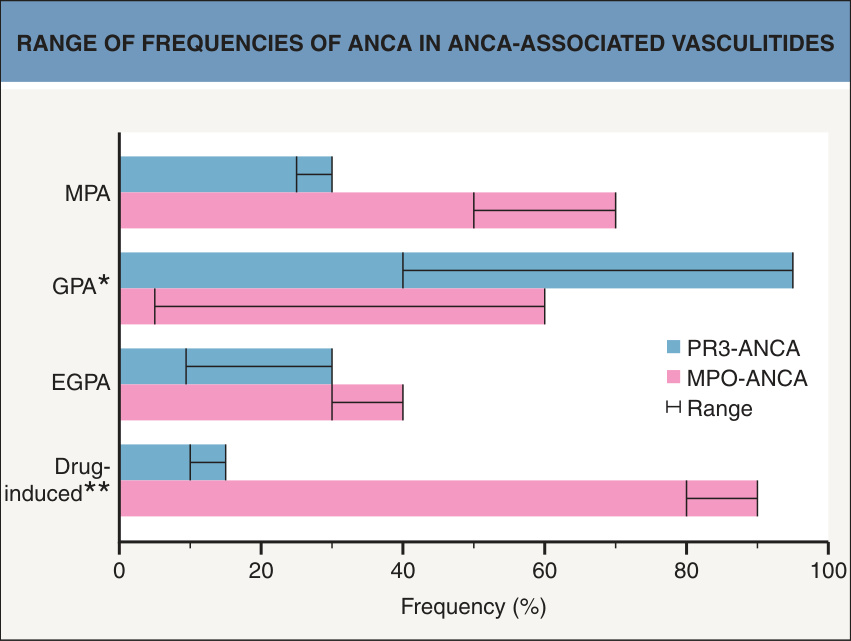
Fig. 24.16 Range of frequencies of ANCA in ANCA-associated vasculitides.
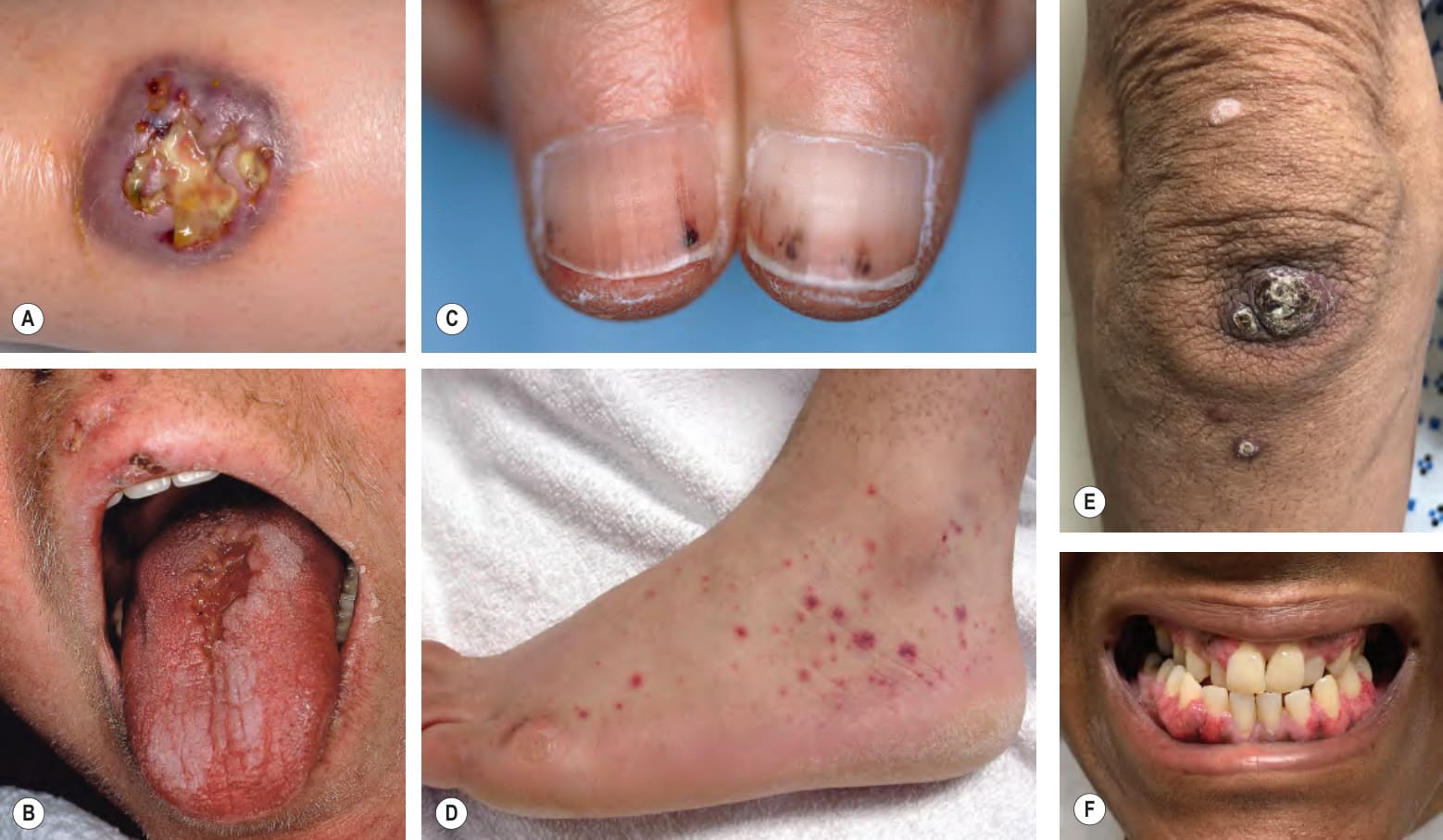
Fig. 24.17 Granulomatosis with polyangiitis.A Ulceration on the leg, which may be misdiagnosed as pyoderma gangrenosum. B Ulceration of the tongue. C Subungual digital infarcts. D Purpuric macules and papules with central crusts due to small vessel vasculitis (leukocytoclastic vasculitis). E Papules and papulonodules with central scale-crust on the elbow (also referred to as Churg–Strauss nodules). F Friable papules of the gingivae (“strawberry gums”). A, Courtesy David A. Wetter MD; E, Courtesy Jeffrey Gelhausen, MD and Nan Ring, MD.
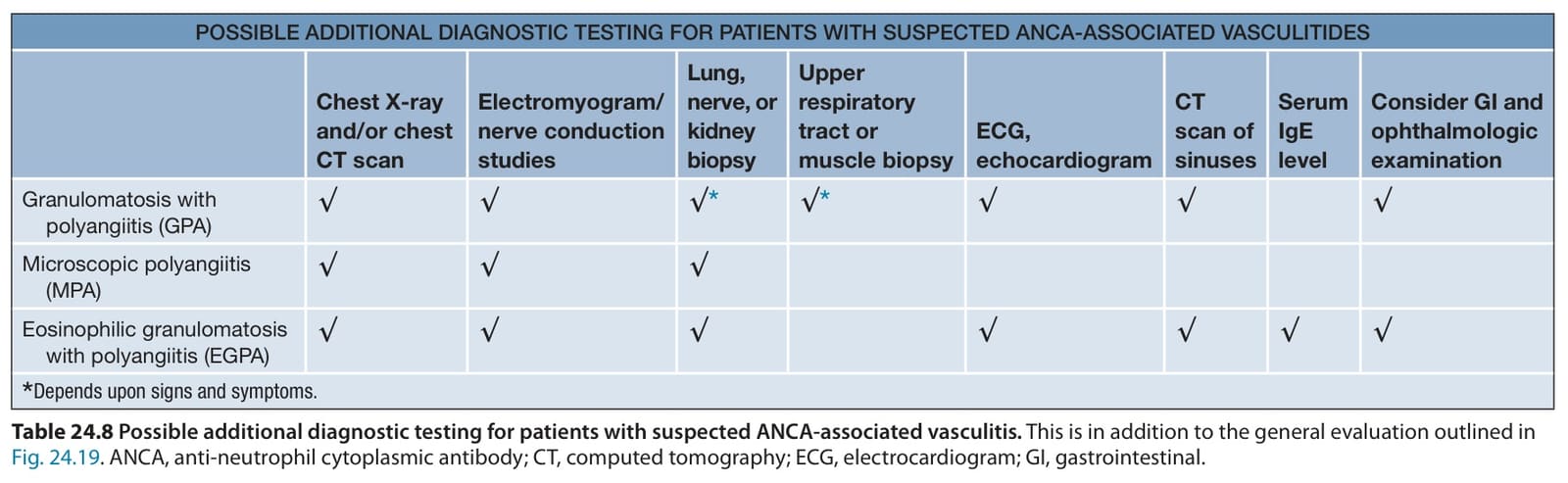
Table 24.8 Possible additional diagnostic testing for patients with suspected ANCA-associated vasculitis. This is in addition to the general evaluation outlined in Fig. 24.19. ANCA, anti-neutrophil cytoplasmic antibody; CT, computed tomography; ECG, electrocardiogram; GI, gastrointestinal.
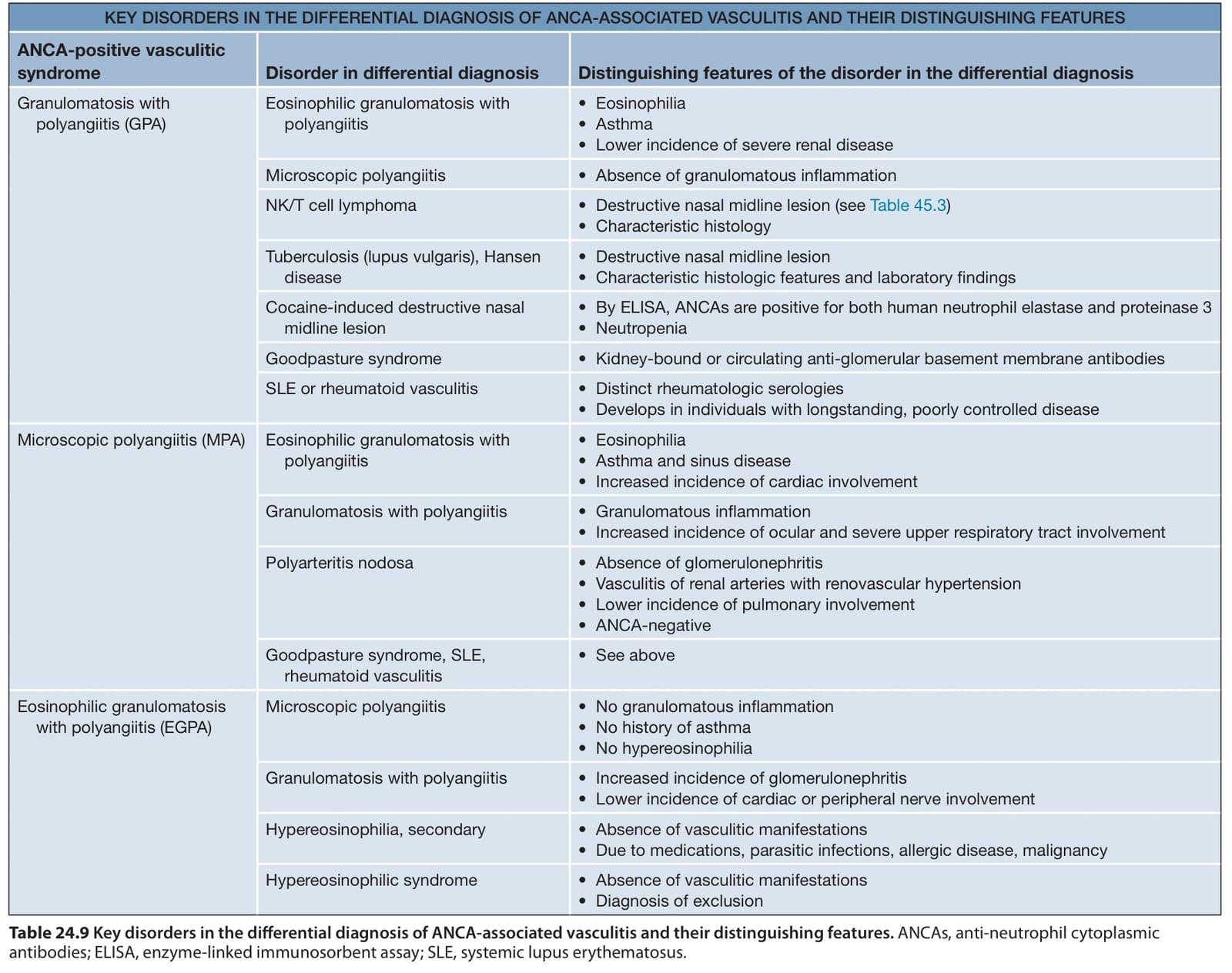
Table 24.9 Key disorders in the differential diagnosis of ANCA-associated vasculitis and their distinguishing features. ANCAs, anti-neutrophil cytoplasmic antibodies; ELISA, enzyme-linked immunosorbent assay; SLE, systemic lupus erythematosus.

Table 24.10 Therapeutic ladder for patients with vasculitis.