Microscopic Polyangiitis
Synonym: Microscopic polyarteritis
Key features
Vasculitis of capillaries, venules, and medium-sized arteries
Constitutional symptoms, crescentic necrotizing glomerulo nephritis, and alveolar hemorrhage
Purpura, livedo reticularis, and livedo racemosa
p-ANCA and anti-MPO antibodies most commonly detected
Absence of granuloma formation
Introduction
Microscopic polyangiitis (MPA) is a systemic vasculitis involving small and medium-sized blood vessels that most commonly affects the skin, lungs, and kidneys. More than 90% of patients are ANCA-positive (most commonly p-ANCA and anti-MPO antibodies), but granulomatous inflammation is usually absent. Because the clinical features of MPA and GPA overlap considerably, it may at times be difficult to differentiate between these two entities. Given these similarities, most therapeutic trials of AAV include both patients with MPA and those with GPA or stratify enrollment by ANCA type (anti-MPO or anti-PR3 antibodies).
Epidemiology
The estimated annual incidence of MPA is 0.5 to 24 cases per million. The condition occurs most commonly in East Asia. The reported mean age of onset is 57 years, with a peak incidence between the ages of 65 and 75 years.
Pathogenesis
Similar to GPA, the etiology of MPA is largely attributed to the presence of autoreactive ANCAs, which form in the presence of proinflammatory cytokines and recognize exposed neutrophilic antigens. In MPA, ANCAs bind primarily to MPO, resulting in vascular damage. The precise role of predisposing genetic, environmental, and other factors is unknown (see general Pathogenesis).
Clinical features
Most patients with MPA initially experience constitutional symptoms, such as fever, weight loss, arthralgias and myalgias, for months to years before other symptoms appear. Depending upon the series, 20%–70% of patients will have cutaneous involvement, most commonly petechiae and purpura (Fig. 24.18). Livedo reticularis and livedo racemosa are more often seen in MPA than in GPA or EGPA. Other cutaneous findings may include a nonspecific rash, painful papules or nodules, and ulcers or gangrene. Skin involvement is associated with higher rates of neurologic and musculoskeletal vasculitic manifestations but lower rates of renal and pulmonary complications.
Renal involvement occurs in nearly all patients with MPA (>90%); a pauci-immune, crescentic, necrotizing glomerulonephritis is the primary pathology, as in GPA. Pulmonary capillaritis (with dyspnea and pulmonary infiltrates) occurs in 30%–50% of patients and can result in diffuse alveolar hemorrhage (10%). Interstitial lung disease can also develop, occurring more often in MPA than in GPA. Neurologic involvement is commonly observed and it usually presents as a peripheral neuropathy or mononeuritis multiplex in up to one-third of patients. Upper respiratory tract involvement occurs as well, but it is less common and less destructive than in GPA. The progressive clinical course of MPA typically leads to renal failure and/or pulmonary hemorrhage.
Anti-MPO antibodies (55%–65% of patients) are detected more often than anti-PR3 antibodies (20%–30%) (see Fig. 24.16). Uncommonly, patients may be ANCA-negative. Additional diagnostic testing for systemic involvement is outlined in Table 24.8, and Fig. 24.19 outlines an approach to the patient with suspected vasculitis.
A Petechiae and purpuric macules on the lower extremity; some of the lesions have central hemorrhagic crusts. Histologically, leukocytoclastic vasculitis was seen. B Petechiae and multiple purpuric macules with central necrosis on the plantar surface. A, Courtesy David A. Wetter, MD; B, Courtesy Cara Whitney Hannon, MD and Robert Swerlick, MD.
Pathology
The characteristic histologic findings include a segmental necrotizing vasculitis of small blood vessels (capillaries, venules, and arterioles) and, less often, a vasculitis of small and/or medium-sized arteries. Granulomatous inflammation is absent.
Differential diagnosis
MPA frequently presents with petechiae and purpura and constitutional symptoms. If no additional internal organ involvement is evident at initial presentation, it may be difficult to distinguish MPA from CSVV or other types of vasculitis involving small blood vessels. The differential diagnosis includes the entities listed in Table 24.9.
Treatment
The treatment approach for MPA is the same as that for GPA, consisting of both an induction phase and a maintenance phase. As stated previously, because these two conditions are related, with overlapping features and outcomes, recent clinical trials have included patients with both diseases. To induce remission in patients with organ- or lifethreatening disease such as active glomerulonephritis or pulmonary hemorrhage, corticosteroids (e.g. initial prednisone dose of 1 mg/kg/ day) plus either rituximab (usually preferred) or cyclophosphamide are recommended (see Table 24.10). A reduced-dose, tapering regimen of corticosteroids should be employed, if possible. Maintenance therapy with rituximab should be initiated after remission has been achieved; alternatives include azathioprine, methotrexate, or mycophenolate mofetil. Those without organ- or life-threatening disease may be treated with corticosteroids and weekly methotrexate or rituximab. The role of plasma exchange in the initial management of GPA and MPA is controversial.
Patients with MPA have a lower risk of relapse compared to those with GPA. In both disorders, the presence of anti-PR3 antibodies and persistent or rising ANCA titers are associated with an increased risk of relapse after remission.
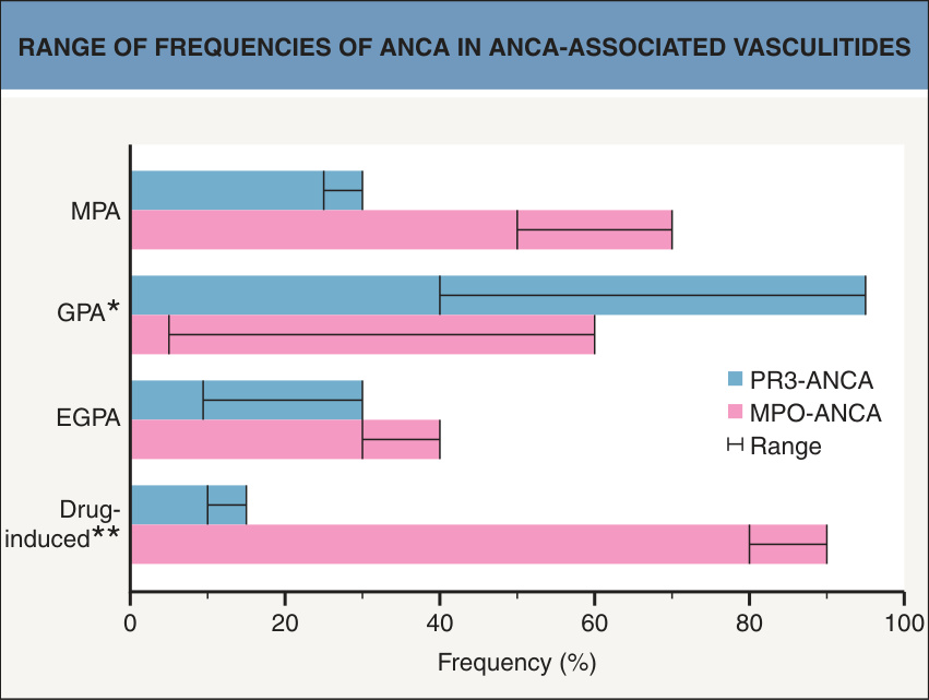
Fig. 24.16 Range of frequencies of ANCA in ANCA-associated vasculitides.
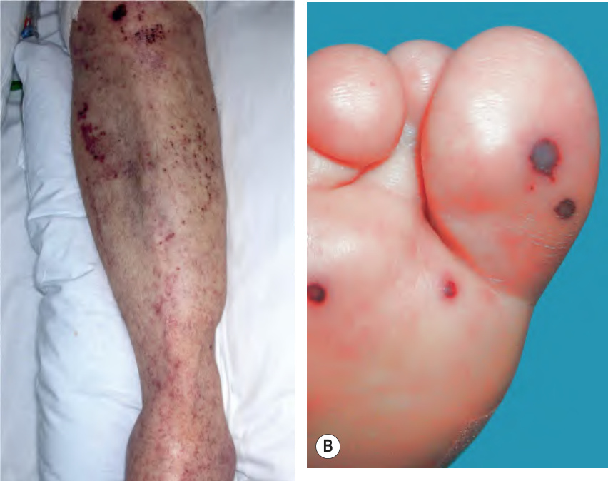
Fig. 24.18 Microscopic polyangiitis.
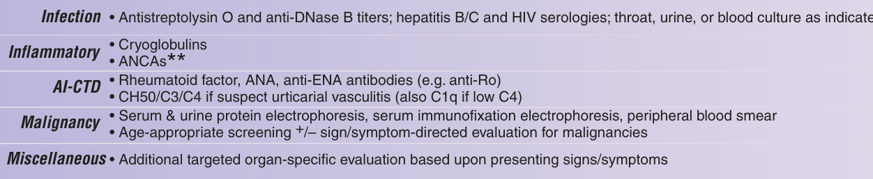
Fig. 24.19 Approach to the patient with suspected cutaneous vasculitis. AI-CTD, autoimmune connective tissue disease; ANA, anti-nuclear antibody; ANCA, anti-neutrophil cytoplasmic antibody; BMZ, basement membrane zone; BUN, blood urea nitrogen; CBC, complete blood count; CT, computerized tomography; DIF, direct immunofluorescence; ELISA, enzyme-linked immunosorbent assay; ENA, extractable nuclear antigen; ESR, erythrocyte sedimentation rate; H&E, hematoxylin and eosin; HIV, human immunodeficiency virus; Ig, immunoglobulin; IgAV, IgA vasculitis (Henoch– Schönlein purpura); SLE, systemic lupus erythematosus. Adapted from Goeser MR, et al. Am J Clin Dermatol 2014;15:299–306.
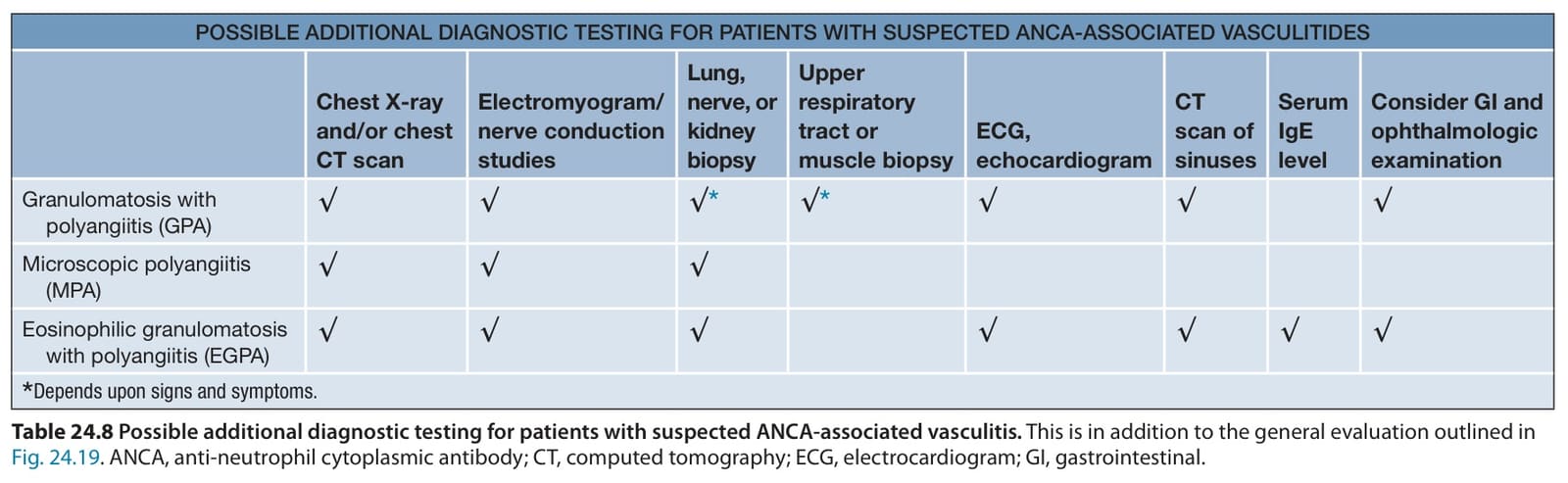
Table 24.8 Possible additional diagnostic testing for patients with suspected ANCA-associated vasculitis. This is in addition to the general evaluation outlined in Fig. 24.19. ANCA, anti-neutrophil cytoplasmic antibody; CT, computed tomography; ECG, electrocardiogram; GI, gastrointestinal.
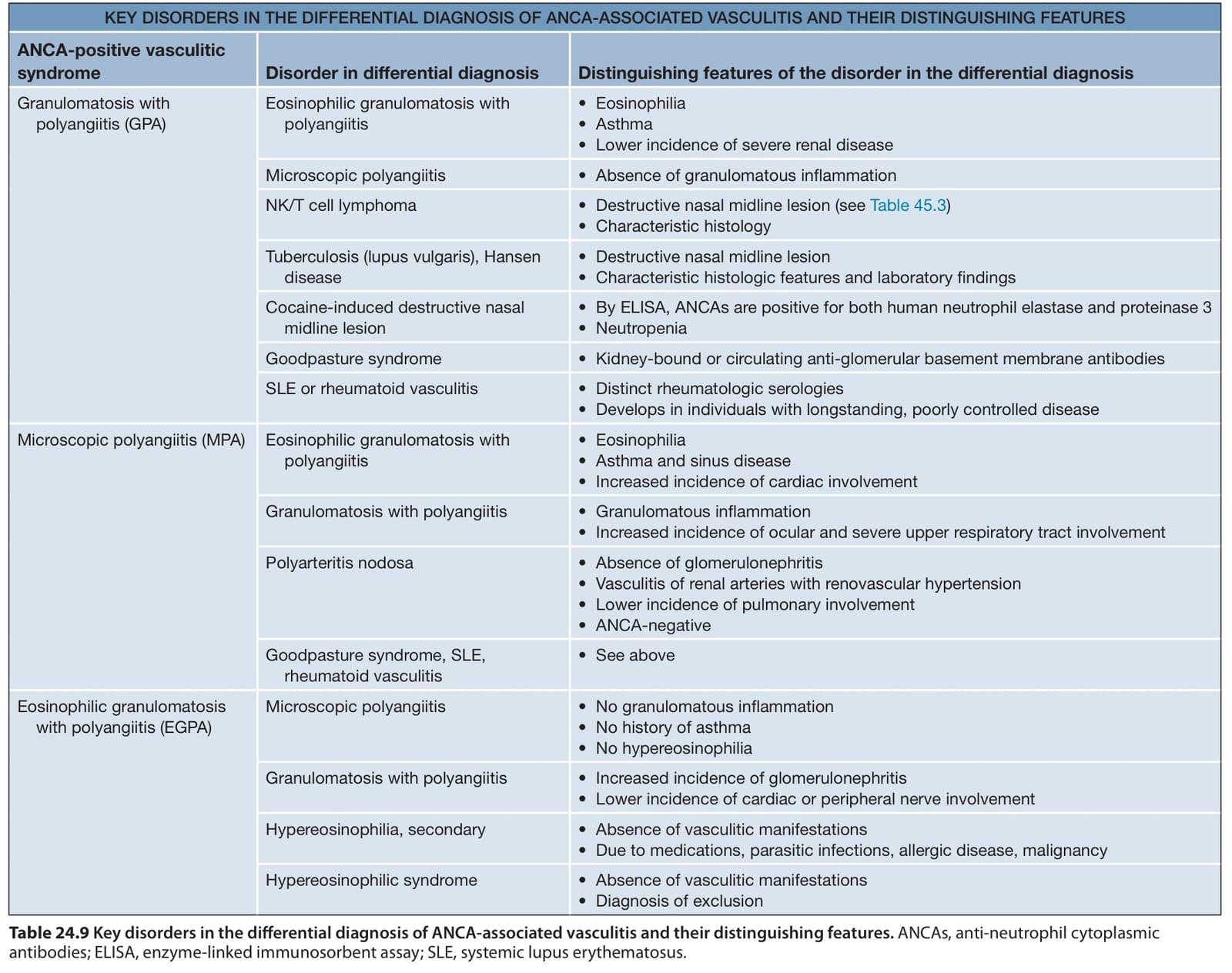
Table 24.9 Key disorders in the differential diagnosis of ANCA-associated vasculitis and their distinguishing features. ANCAs, anti-neutrophil cytoplasmic antibodies; ELISA, enzyme-linked immunosorbent assay; SLE, systemic lupus erythematosus.

Table 24.10 Therapeutic ladder for patients with vasculitis.