Eosinophilic Granulomatosis With Polyangiitis
Synonyms: Churg–Strauss syndrome Churg–Strauss vasculitis Allergic angiitis and granulomatosis
Key features
Prodromal asthma and allergic rhinitis are followed by eosinophilic and vasculitic phases
Peripheral eosinophilia and ANCA-positivity in half of patients
(usually p-ANCA and anti-MPO antibodies)
Cutaneous manifestations in ~50% of patients
Most commonly pulmonary, neurologic, renal, and cardiac involvement
Histologic features consist of eosinophils, extravascular granu- lomas, and vasculitis
Introduction
Eosinophilic granulomatosis with polyangiitis (EGPA), also referred to as Churg–Strauss syndrome, is characterized by vascular and extravascular granulomas, eosinophil-rich pulmonary infiltrates, and necrotizing vasculitis that involves small to medium-sized vessels within multiple organ systems. It is distinguished by an association with asthma and eosinophilia.
Epidemiology
Although the true incidence of EGPA is unknown due to uncertainties surrounding its diagnosis, it is thought to be the least common of the ANCA-associated vasculitides. EGPA has an estimated incidence ranging from 0.5 to 2.7 cases per million per year. The mean age at diagnosis is 40 years, and it has no sex predominance. The incidence of EGPA in patients with asthma ranges from 35 to 65 per million per year.
Pathogenesis
The onset of symptoms has been associated with various triggering factors, including vaccination, desensitization therapy, leukotriene inhibitors, omalizumab, and rapid discontinuation of corticosteroids. However, in some patients, this may more accurately represent an “unmasking” of the disease. T lymphocytes, eosinophils, and ANCAs all play a role in disease pathogenesis. Tissue infiltration and degranulation of eosinophils can lead to tissue injury, while T cells, both Th1 and Th2, are postulated to contribute to granuloma formation and allergic features, respectively. ANCA-dependent activation of neutrophils results in vasculitis (see Fig. 24.1B).
Two subsets of EGPA have been identified in which several risk genes (e.g. HLA-DQ) are associated with ANCA-positivity, while variants in GPA33 and IL5 are associated with ANCA-negative EGPA. The former group of patients is more likely to develop glomerulonephritis and neuropathy while the latter group more often has cardiomyopathy and pulmonary infiltrates.
Clinical features
The clinical presentation can be divided into three successive phases: (1) prodromal phase – symptoms of allergic rhinitis, nasal polyps, and asthma, which may persist for years; (2) eosinophilic phase – peripheral eosinophilia and eosinophilic infiltration of internal organs, especially the lungs and gastrointestinal tract; and (3) vasculitic phase – systemic necrotizing vasculitis with granulomatous inflammation, which can occur several years to decades after the initial symptoms. Asthma, frequently severe, affects almost all patients and often precedes the onset of additional systemic manifestations by a decade or more. Otitis media, allergic rhinitis, nasal polyposis, and sinusitis are also exceedingly common.
Cutaneous findings occur in 40%–75% of patients, typically during the third phase of the disease, but are the presenting sign in ~15% of patients. Although petechiae and purpura are most often seen, a variety of nonspecific, non-vasculitic manifestations are also frequently observed. The latter include “rash”, pruritus, and urticaria. Because these “allergic” skin findings occur more frequently in EGPA than in other types of ANCA-associated vasculitides, recognizing them as potentially consistent with EGPA in the right clinical setting is important. Subcutaneous nodules, livedo racemosa, retiform purpura, and papulonecrotic lesions, particularly on the extensor surfaces (i.e. Churg–Strauss nodules, PNGD), may also be seen (Fig. 24.20). In addition to the respiratory tract, patients with EGPA often have neurologic and cardiac involvement, presenting as mononeuritis multiplex and cardiomyopathy, pericarditis, or an arrhythmia. Peripheral neuropathy affecting more than one site (mononeuritis multiplex) is seen in up to 75% of patients and may result in pain, numbness, or weakness. Cardiac manifestations occur in up to half of patients, developing more often in those with higher peripheral blood eosinophil counts, and are the leading cause of death. Necrotizing glomerulonephritis and pulmonary capillaritis resulting in diffuse alveolar hemorrhage occur less commonly than in the other AAVs. Musculoskeletal, gastrointestinal, and ocular involvement can also occur.
There are no specific laboratory findings, but peripheral eosinophilia (≥1500 cells/microL or ≥10% of the total leukocyte count) should prompt suspicion in the right clinical context. In addition, patients frequently have elevated serum IgE levels. ANCAs are present in roughly half of those with EGPA, with 75% of these antibodies p-ANCA and anti-MPO (see Fig. 24.16). ANCA positivity has been associated with a higher risk of vasculitic features, including peripheral neuropathy, glomerulonephritis, and histologic findings of vasculitis, as well as higher rates of disease relapse. On the other hand, cardiac involvement is more commonly observed in ANCA-negative patients.
Pathology
The histopathologic hallmarks are infiltrates of eosinophils, formation of extravascular granulomas, and necrotizing vasculitis of small to medium-sized vessels; both arteries and veins are affected. Biopsy specimens obtained from papulonecrotic lesions demonstrate a palisading dermatitis with eosinophil infiltration, granuloma formation, and eosinophilic necrobiosis.
Differential diagnosis
EGPA is suspected on the basis of its typical clinical findings of asthma, rhinosinusitis, and peripheral eosinophilia in addition to manifestations of vasculitis (e.g. cutaneous, neurologic, renal), biopsy findings of vasculitis, and/or ANCA positivity (if present). Due to the tendency
Purpuric macules and papules on the buttocks due to small vessel vasculitis (leukocytoclastic vasculitis). B Purpuric dermal plaques on the palm that histologically demonstrated vasculitis of a small muscular artery. C Crusted, firm papules on the elbow (Churg–Strauss nodules; palisaded neutrophilic and granulomatous dermatitis). D A few scattered purpuric papules, some of which have central pinpoint crusts, on the ankles. A, B, Courtesy Lindy Fox, MD and Kanade Shinkai, MD; C, Courtesy Kalman Watsky, MD.
for this disease to evolve over time or to lack one or more of these features, diagnosis can be challenging and delayed. As with GPA and MPA, revised EULAR/ACR diagnostic criteria have been published.
The differential diagnosis of EGPA includes other vasculitides (see Table 24.9), other causes of secondary hypereosinophilia (e.g. parasitic infections), and hypereosinophilic syndrome (HES; see Ch. 26), as well as chronic eosinophilic pneumonia and allergic bronchopulmonary aspergillosis.
Treatment
Systemic corticosteroids (usually 0.5–1 mg/kg/day prednisone equivalent) are the mainstay of therapy, and >90% of patients respond to treatment with corticosteroids alone. However, relapse is common, and patients with more severe internal organ involvement (e.g. cardiac, renal, gastrointestinal, CNS) generally require additional immunosuppressive therapy. In such patients, either cyclophosphamide or rituximab is typically added to the corticosteroid. The five-factors score (FFS), which is based on the presence or absence of various clinical factors associated with worse prognosis, can be used for risk stratification. Guidelines recommend cyclophosphamide in those who have active cardiac involvement and in those who are ANCA-negative and have severe neurologic or gastrointestinal manifestations. Rituximab may be preferred in those with: ANCA positivity, active glomerulonephritis, prior cyclophosphamide treatment, or risk of gonadal toxicity. For patients with active, non-severe EGPA, corticosteroids combined with the anti-IL-5 monoclonal antibody mepolizumab are recommended. Alternative options for patients without organ-threatening disease include methotrexate or azathioprine plus corticosteroids.
Once remission has been achieved, corticosteroids should be tapered over ~12–18 months. However, many patients will need to continue low-dose corticosteroids (e.g. ≤10 mg/day prednisone) long-term. In addition, cyclophosphamide or rituximab can be replaced by less toxic immunosuppressive drugs such as azathioprine, methotrexate, or mycophenolate mofetil. The data supporting azathioprine and other second- and third-line agents (e.g. methotrexate, leflunomide) for treatment of relapsed or refractory EGPA are limited (see Table 24.10). Of note, mepolizumab is FDA-approved for the treatment of EGPA based on a randomized trial in which increased rates of remission and reduced corticosteroid use were observed in patients with relapsing or refractory non-severe disease. Still, only about half of patients who received mepolizumab achieved remission. Clinical trials of other anti-IL-5 drugs are ongoing.
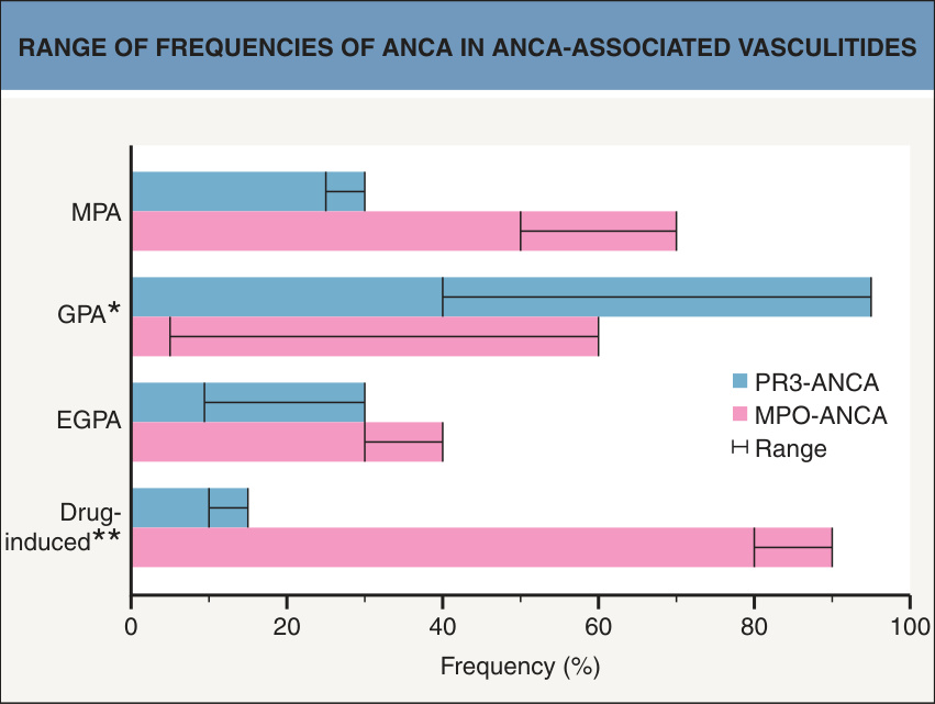
Fig. 24.16 Range of frequencies of ANCA in ANCA-associated vasculitides.
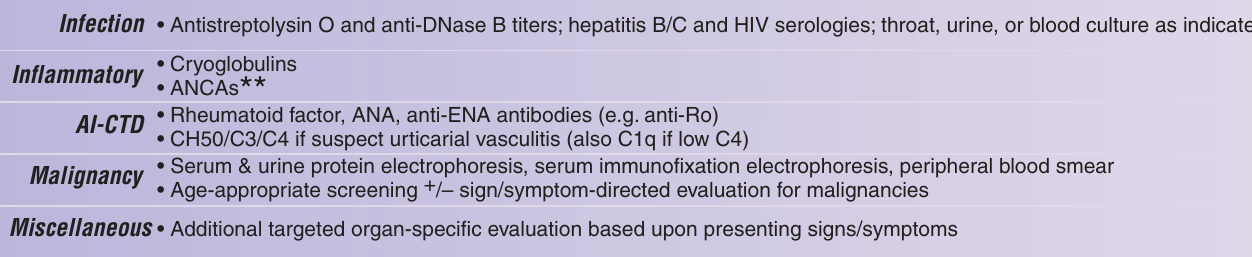
Fig. 24.19 Approach to the patient with suspected cutaneous vasculitis. AI-CTD, autoimmune connective tissue disease; ANA, anti-nuclear antibody; ANCA, anti-neutrophil cytoplasmic antibody; BMZ, basement membrane zone; BUN, blood urea nitrogen; CBC, complete blood count; CT, computerized tomography; DIF, direct immunofluorescence; ELISA, enzyme-linked immunosorbent assay; ENA, extractable nuclear antigen; ESR, erythrocyte sedimentation rate; H&E, hematoxylin and eosin; HIV, human immunodeficiency virus; Ig, immunoglobulin; IgAV, IgA vasculitis (Henoch– Schönlein purpura); SLE, systemic lupus erythematosus. Adapted from Goeser MR, et al. Am J Clin Dermatol 2014;15:299–306.
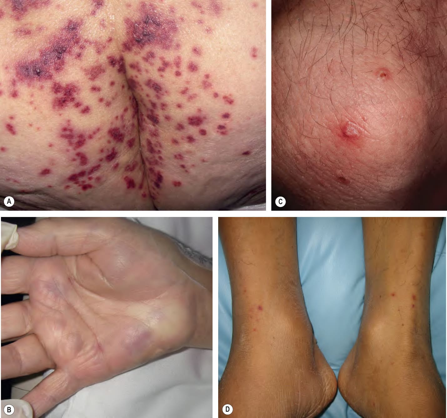
Fig. 24.20 Eosinophilic granulomatosis with polyangiitis (Churg– Strauss syndrome).A
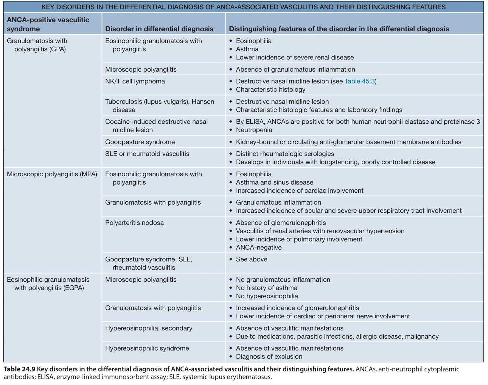
Table 24.9 Key disorders in the differential diagnosis of ANCA-associated vasculitis and their distinguishing features. ANCAs, anti-neutrophil cytoplasmic antibodies; ELISA, enzyme-linked immunosorbent assay; SLE, systemic lupus erythematosus.

Table 24.10 Therapeutic ladder for patients with vasculitis.