TEMPORAL ARTERITIS (GIANT CELL ARTERITIS)
Rarely, cutaneous ulcers, primarily of the frontolateral scalp and tongue, can develop due to large vessel vasculitis (Fig. 24.23; see Table 24.1). Patients may have a history of headaches, jaw claudication, tenderness along the course of the temporal artery, and visual disturbances. They may also experience the muscle weakness associated with polymyalgia rheumatica. Cranial duplex ultrasound of the temporal artery is a non-invasive, but operator-dependent, option for initial diagnosis. In some centers, it has replaced temporal artery biopsy as the preferred initial diagnostic procedure. High-resolution MRI of cranial arteries is an alternative imaging modality with good sensitivity and specificity. Plasma interleukin (IL)-6 levels may represent a marker of disease activity, and tocilizumab, an anti-IL-6 receptor antibody, is approved for the treatment of large vessel vasculitis. High-dose corticosteroids are also employed as first-line therapy. In a phase 2 trial, mavrilimumab, an IgG4 monoclonal antibody which blocks GM-CSF signaling, demonstrated efficacy in maintaining clinical remission123a.
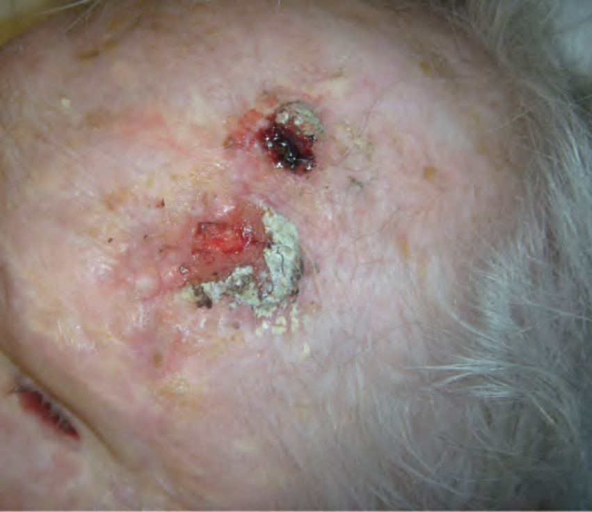
Fig. 24.23 Temporal arteritis. Two ulcerations of the scalp in association with alopecia and scarring in an elderly patient. Courtesy Steve Feldman, MD.
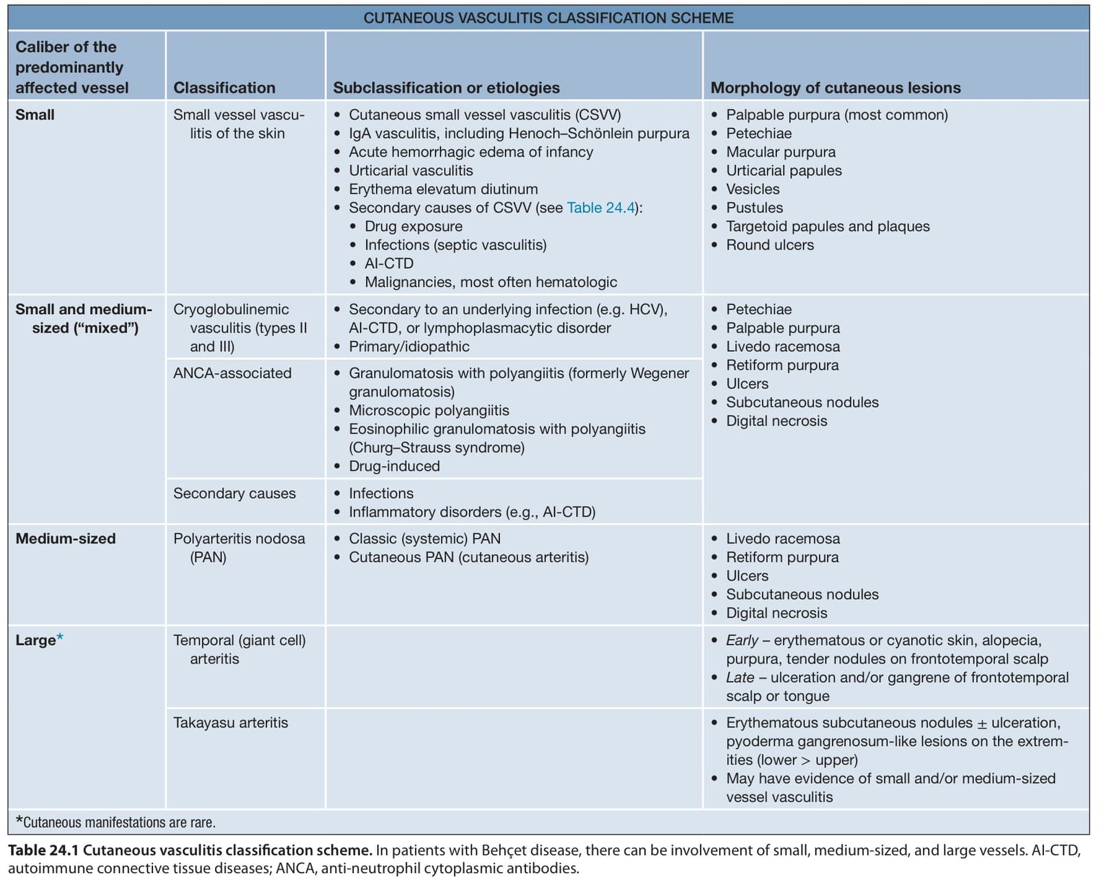
Table 24.1 Cutaneous vasculitis classification scheme. In patients with Behçet disease, there can be involvement of small, medium-sized, and large vessels. AI-CTD, autoimmune connective tissue diseases; ANCA, anti-neutrophil cytoplasmic antibodies.