SWEET SYNDROME
Synonym: Acute febrile neutrophilic dermatosis
Key features
Constitutional signs and symptoms such as fever and malaise Clinically, erythematous plaques that are occasionally bullous Histologically, dense perivascular neutrophilic infiltrate, edema and, infrequently, bullae; leukocytoclasia with minimal to no evidence of vasculitis Associated conditions include infections, malignancies (especially acute myelogenous leukemia), inflammatory bowel disease, autoimmune disorders (e. g. systemic lupus erythematosus [SLE]), drugs, and pregnancy
History
The prototype of the neutrophilic dermatoses is Sweet syndrome. In 1964, Sweet described eight middle-aged women who had an acute onset of fever and erythematous plaques associated with a nonspecific infection of the respiratory tract or gastrointestinal tract. Histologically, the skin lesions were characterized by a neutrophilic infiltrate. He named this constellation of findings “acute febrile neutrophilic dermatosis”. In 1968, Whittle and colleagues reported a similar case and named it “Sweet’s syndrome”.
Epidemiology
Sweet syndrome is an uncommon disease, with a worldwide distribution and no obvious racial predilection, although the disorder appears to be more frequent in Japan. The average age of onset is 30–60 years, but infants, children, and the elderly may be affected; there is a female predominance of 4 to 1. At least half of patients have an identifiable associated disorder or trigger: 15%–30% have an internal malignancy (hematologic ≫ solid organ), ~25% a preceding infection, and ~10% exposure to a potentially causative drug (Table 25.2). While the epidemiologic statistics in part reflect associated underlying disorders including internal malignancies, inflammatory bowel disease, and autoimmune connective tissue diseases such as SLE, they do not completely explain the female predominance.
Pathogenesis
The pathogenesis of Sweet syndrome is poorly understood, but the association with underlying diseases suggests a hypersensitivity reaction. Local and systemic dysregulation of cytokine secretion, including inflammasome activation, has been observed. Implicated cytokines include IL-1, tumor necrosis factor (TNF), G-CSF,
granulocyte–macrophage CSF (GM-CSF), and interferon-γ. A familial form of Sweet syndrome due to heterozygous mutations in MEFV (e.g. S242R) has been described. Of note, this gain-of-function mutation in MEFV differs from those seen in familial Mediterranean fever. The term pyrin-associated autoinflammatory disease has been applied to these patients. As in PG and a murine model of neutrophilic dermatoses, the possibility that splice variants in PTPN6 play a role in Sweet syndrome has been raised.
In hematologic malignancy-associated Sweet syndrome, the neutrophils in the cutaneous infiltrate and the malignant myeloid clone appear to share a clonal progenitor. This was observed when the skin lesions were present at the time of diagnosis as well as when they developed during treatment. Thus, the inciting factors for malignancy-associated and familial Sweet syndrome likely differ from those of “classic” or drug-induced disease. However, downstream effectors presumably overlap with an end result of aberrant activation of neutrophils.
Clinical Features
The initial cutaneous lesions are tender, non-pruritic, erythematous plaques or papules, which may enlarge or coalesce to form plaques with an uneven mammillated surface (Fig. 25.2 & 25.3). Because of the pronounced associated edema, the lesions may have a pseudovesi cular or pseudopustular appearance; however, some patients actually develop vesiculation, bullae, or pustules within their plaques. Occasionally, the plaques have a central yellowish discoloration creating a targetoid appearance. A facial erysipelas-like appearance has also been described as has a giant cellulitis-like variant. A relapsing localized variant can develop within areas of lymphedema or at sites of orthopedic implants. The vesiculobullous variant, which is most frequently associated with acute myelogenous leukemia, can progress to ulceration resembling superficial pyoderma gangrenosum (PG). In this form, there may be a single lesion or multiple, asymmetrically distributed lesions.
The cutaneous eruption of Sweet syndrome favors the head, neck, and upper extremities (including the dorsal aspect of the hands), but can occur anywhere. In true malignancy-associated cases, the lesions tend to have a more widespread distribution. Papulonodules involving the lower legs may resemble erythema nodosum and although erythema nodosum has been reported in patients with Sweet syndrome, such nodules more likely represent a neutrophilic panniculitis. When isolated, the latter is often referred to as subcutaneous Sweet syndrome. A subset of patients, often with systemic involvement, can develop lesions that mimic necrotizing fasciitis, referred to as necrotizing neutrophilic dermatosis; such lesions are also seen in patients with PG.
As with PG, specific lesions may be initiated by wounding injuries such as needle sticks, thermal burns, and surgical incisions. This phenomenon is known as pathergy. Oral lesions are uncommon, except in patients with hematologic disorders; initially they appear pseudopustular, and later ulcerate and appear as aphthae. The cutaneous eruption of Sweet syndrome usually resolves spontaneously within 5–12 weeks but recurs in up to 30% of patients.
An upper respiratory tract infection or flu-like illness frequently pre-cedes the development of the syndrome. Fever occurs in 40%–80% of patients and can be intermittent. Extracutaneous involvement is also frequently seen (Fig. 25.4; Table 25.3).
Associated Diseases
These are summarized in Table 25.2 and should guide the history, including all medications, physical examination, and focused laboratory evaluation.
Pathology
The histopathologic features are contingent upon the type of cutaneous lesion sampled. The characteristic histologic presentation is a dense, diffuse dermal nodular and perivascular neutrophilic infiltrate without evidence of vasculitis (Fig. 25.5), although occasionally leukocytoclastic vasculitis (LCV) can be observed. Leukocytoclasia with endothelial swelling, but without the fibrinoid necrosis that fulfills
the criteria for LCV, is the usual finding. Occasionally, the dermal infiltrate may extend into the subcutis, creating a septal or, less frequently, a lobular panniculitis. Isolated neutrophilic panniculitis has been described.
In general, epidermal changes are not significant. However, neutrophils occasionally invade the epidermis, producing subcorneal pustules, and they may also infiltrate the adnexa. In the setting of significant edema, there can be epidermal spongiosis and sometimes reticular degeneration as well as intraepidermal and subepidermal vesiculation.
Three histologic variants of Sweet syndrome, histiocytoid, lymphocytic and eosinophilic, have been increasingly recognized. The histiocytoid variant is characterized by a dermal, and sometimes sub-cutaneous, infiltrate composed of histiocyte-like immature myeloid cells. These cells have myeloperoxidase activity (Fig. 25.6) and must be distinguished from those of leukemia cutis (bearing in mind that cells related to the malignant clone have been identified in some cases of histiocytoid Sweet syndrome associated with myelogenous leukemia). The lymphocytic variant is often associated with, and sometimes precedes, underlying myelodysplasia. More recently, an eosinophilic variant
has been described. In a retrospective review, patients with the histiocytoid variant or subcutaneous involvement were more likely to have malignancy-associated disease.
Diagnosis and Differential Diagnosis
Although there are no specific diagnostic findings in this disease, a peripheral leukocytosis with neutrophilia and an elevated ESR and C-reactive protein (CRP) are frequently seen. In patients with an associated hematologic malignancy or myelodysplasia, there may be a high or low white cell count, lymphocytosis or lymphopenia, and thrombocytosis or thrombocytopenia. Although elevated serum levels of anti-neutrophil cytoplasmic antibody (ANCA) have been reported, in particular pANCA or an atypical ANCA, there is no strong evidence that it is a serologic marker for this disease.
In 1986, Su and Liu proposed two major and four minor criteria for the diagnosis of Sweet syndrome, which have been widely accepted and are shown with minor revisions in Table 25.4. The differential diagnosis of the syndrome depends on the nature and age of the lesions and on the associated disorder (Table 25.5; see Fig. 25.1).
Patients with neutrophilic dermatosis of the dorsal hands develop tender, erythematous to violaceous plaques that may become bullous or ulcerative (Fig. 25.7). There is a spectrum of associated histologic features, from pustular vasculitis to those of PG or Sweet syndrome. Hence, some authors consider this to be a variant of Sweet syndrome.
In patients with hematologic malignancies, especially acute myelogenous leukemia, several neutrophilic dermatoses, e.g. Sweet syndrome, atypical (bullous) PG, and neutrophilic eccrine hidradenitis, can occur alone or in combination. The approach to such a patient is presented in Table 25.6.
In patients with rheumatoid arthritis, the possibility of rheumatoid neutrophilic dermatitis, including the uncommon annular variant, needs to be considered. As noted in Table 25.5, Sweet syndrome can also mimic soft tissue infections, including cellulitis and necrotizing fasciitis. This distinction can prove challenging, especially in immunocompromised hosts.
Treatment
When limited to the skin, Sweet syndrome is a benign condition which, if left untreated, may persist for weeks or months. Cutaneous lesions then involute spontaneously, rarely leaving scars. However, recurrences develop in ~30% of patients (with or without treatment) and occur even more often in those with hematologic disorders (~50%). Although the initial clinical presentation often suggests sepsis, antibiotics, in general, are ineffective. However, when the disease is associated with a recognized infection, treatment of the underlying infection may result in improvement.
The most effective therapy for Sweet syndrome is oral prednisone (0.5– 1.0 mg/kg/day) for 2–6 weeks. There is prompt relief of not only the cutaneous, but also the extracutaneous, manifestations. In some patients, prolonged low-dose prednisone for an additional 2–3 months may be necessary to suppress recurrences. When the lesions are few and localized, topical superpotent or intralesional corticosteroids may prove helpful.
The major alternative drugs are potassium iodide (900 mg/day; see Table 100.6), dapsone (100–200 mg/day), and colchicine (1–1.5 mg/day or 1.2–1.8 mg/day, depending upon dosage of commercially available tablets). They can also be used as corticosteroid-sparing agents. Nonsteroidal anti-inflammatory drugs (e.g. indomethacin, naproxen, sulindac), cyclosporine, thalidomide, lenalidomide, anakinra, methotrexate, and rituximab have also been reported to lead to improvement of Sweet syndrome. Evidence for any of these therapeutic regimens is based on case reports or small case series. Of note, while TNF inhibitors have been used to treat refractory Sweet syndrome, they can also be a cause of drug-induced Sweet syndrome.
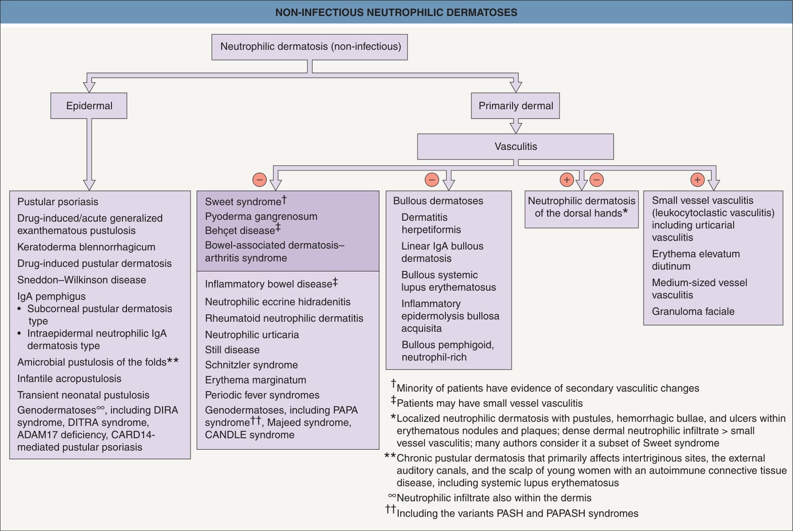
Fig. 25.1 Non-infectious neutrophilic dermatoses. Entities in the darker box are discussed in this chapter. CANDLE, chronic atypical neutrophilic dermatosis with lipodystrophy and elevated temperature; DIRA, deficiency of the interleukin-1 receptor antagonist; DITRA, deficiency of the interleukin-36 receptor antagonist; PAPA, pyogenic arthritis, pyoderma gangrenosum, and acne; PAPASH, pyogenic arthritis, pyoderma gangrenosum, acne, and suppurative hidradenitis; PASH, pyoderma gangrenosum, acne, and suppurative hidradenitis.
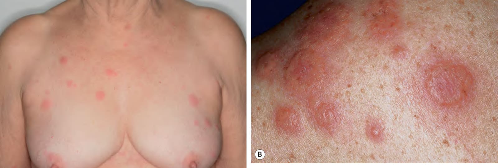
Fig. 25.2 Sweet syndrome.A Scattered edematous pink papules and plaques on the chest. B The edema can be quite marked as seen in these lesions on the upper back. B, Courtesy Kalman Watsky, MD.
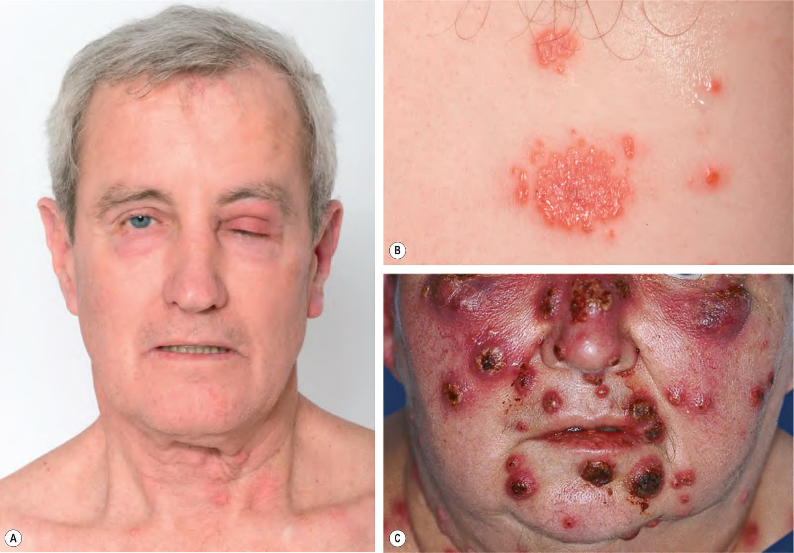
Fig. 25.3 Sweet syndrome.A The periocular lesion demonstrates how some lesions can mimic cellulitis (pseudocellulitis). This patient also had neutrophilic esophageal ulcerations and subsequently developed colon cancer. B The plaques can have a pseudomammillated appearance due to the associated edema. C Hemorrhagic crusts may develop within the papules and plaques and in patients with acute leukemia and/or neutropenia, infectious etiologies are often the initial clinical diagnosis. C, Courtesy Kalman Watsky, MD.
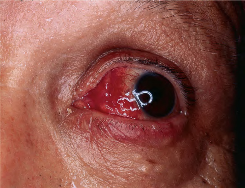
Fig. 25.4 Sweet syndrome – ocular involvement. Obvious erythema and hemorrhage involving the sclera and conjunctiva. Courtesy Kalman Watsky, MD.
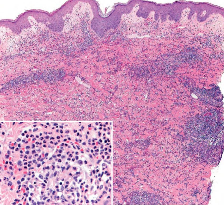
Fig. 25.5 Sweet syndrome – histopathologic features. Mild edema in the papillary dermis and a rather dense dermal infiltrate composed predominantly of neutrophils (inset). Courtesy Lorenzo Cerroni, MD.
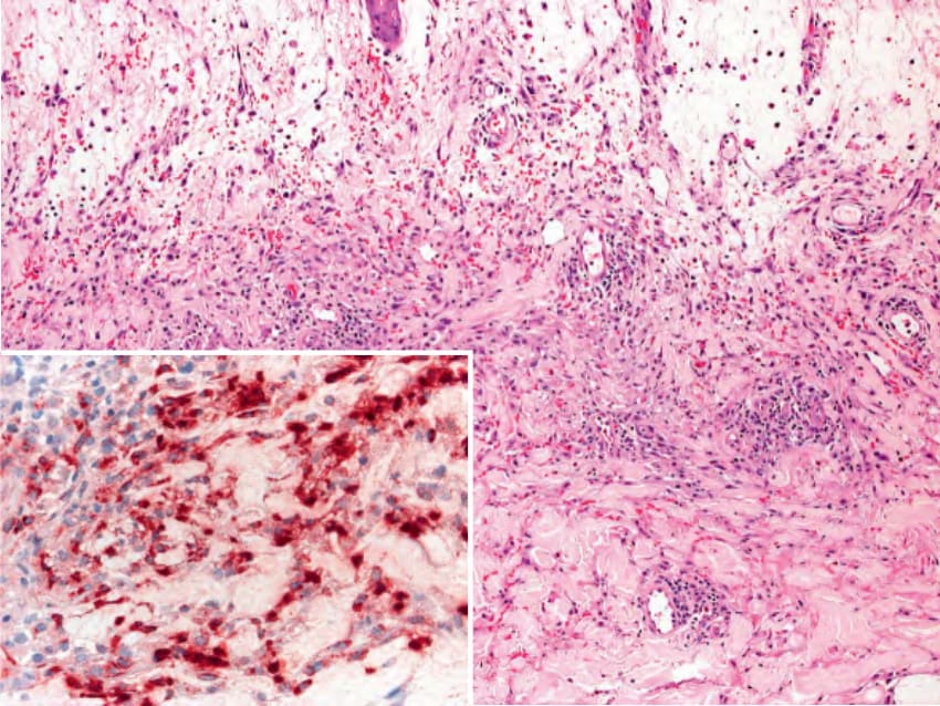
Fig. 25.6 Histiocytoid Sweet syndrome – histopathologic features. There is edema in the papillary dermis as well as a dermal infiltrate composed of histiocytoid cells admixed with neutrophils, a few eosinophils, and hemorrhage. The histiocytoid cells represent immature myeloid cells and therefore have stained positively for myeloperoxidase (inset). Courtesy Lorenzo Cerroni, MD.
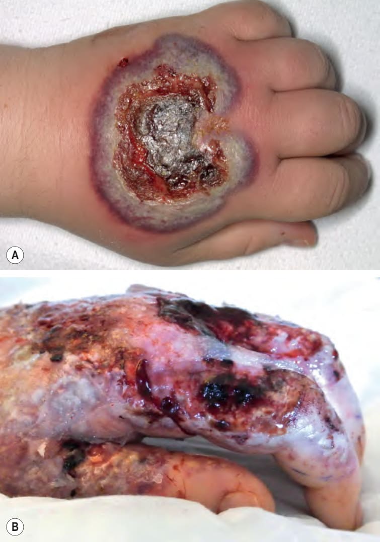
Fig. 25.7 Neutrophilic dermatosis of the dorsal hands.A Clinically and histologically, there is overlap with Sweet syndrome and bullous pyoderma gangrenosum. B More extensive necrotizing disease can be confused with an infectious process such as necrotizing cellulitis or necrotizing fasciitis.
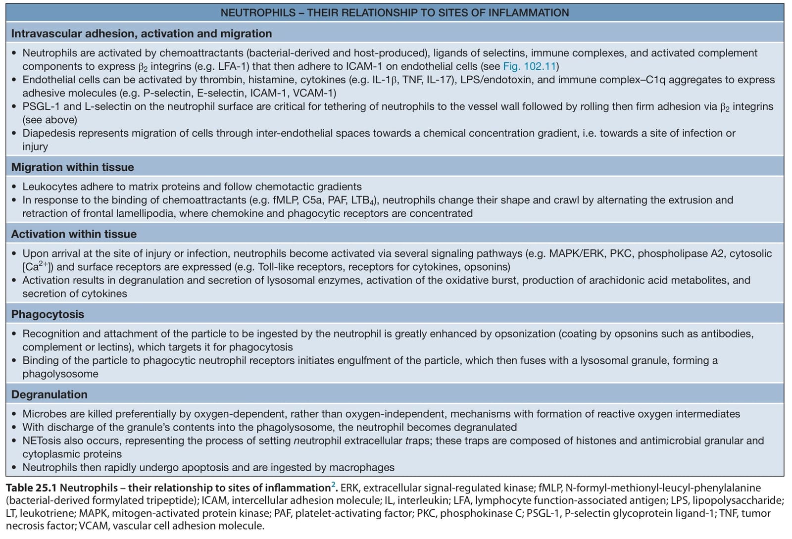
Table 25.1 Neutrophils – their relationship to sites of inflammation. ERK, extracellular signal-regulated kinase; fMLP, N-formyl-methionyl-leucyl-phenylalanine (bacterial-derived formylated tripeptide); ICAM, intercellular adhesion molecule; IL, interleukin; LFA, lymphocyte function-associated antigen; LPS, lipopolysaccharide; LT, leukotriene; MAPK, mitogen-activated protein kinase; PAF, platelet-activating factor; PKC, phosphokinase C; PSGL-1, P-selectin glycoprotein ligand-1; TNF, tumor necrosis factor; VCAM, vascular cell adhesion molecule.
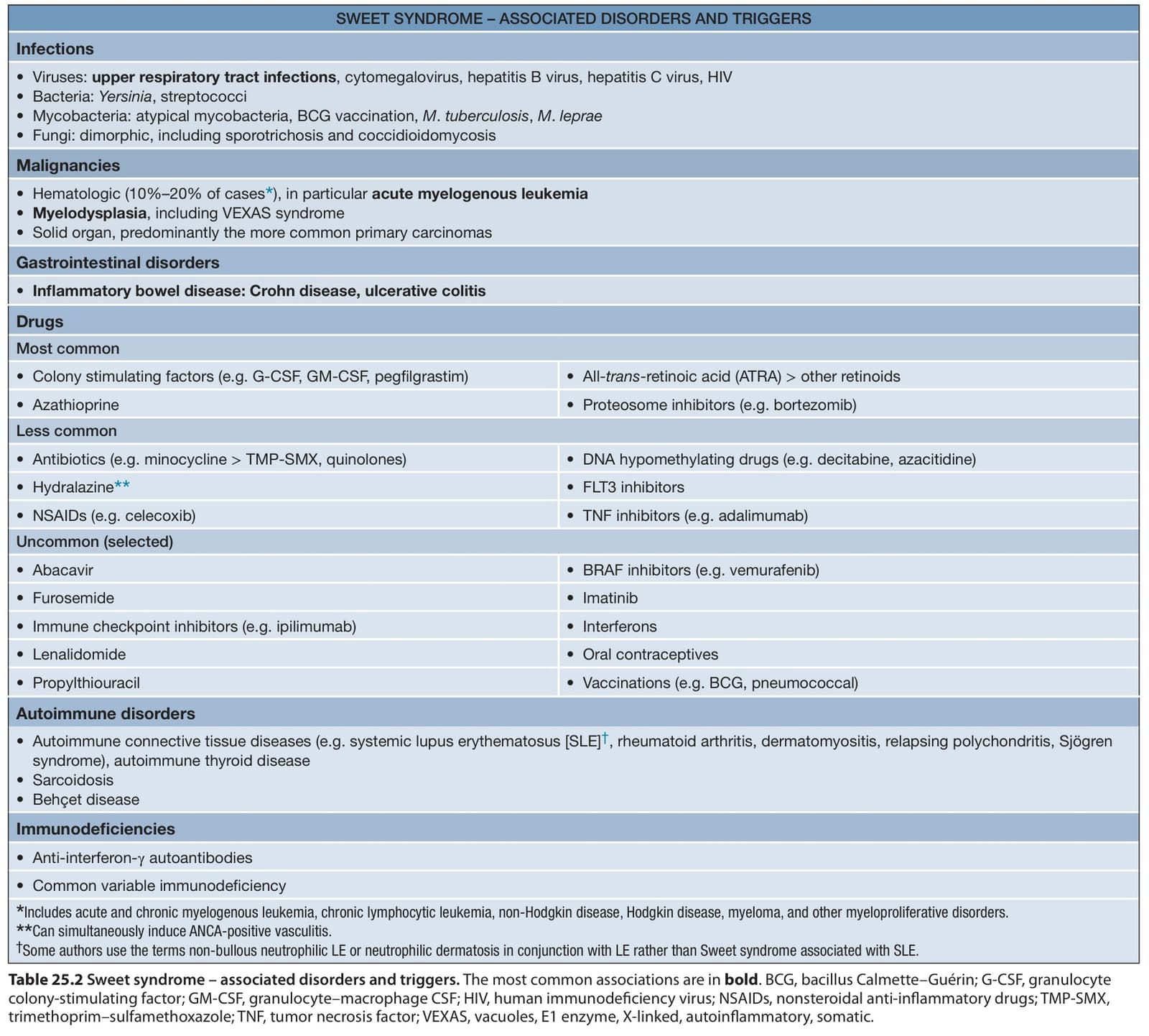
Table 25.2 Sweet syndrome – associated disorders and triggers. The most common associations are in bold. BCG, bacillus Calmette–Guérin; G-CSF, granulocyte colony-stimulating factor; GM-CSF, granulocyte–macrophage CSF; HIV, human immunodeficiency virus; NSAIDs, nonsteroidal anti-inflammatory drugs; TMP-SMX, trimethoprim–sulfamethoxazole; TNF, tumor necrosis factor; VEXAS, vacuoles, E1 enzyme, X-linked, autoinflammatory, somatic.
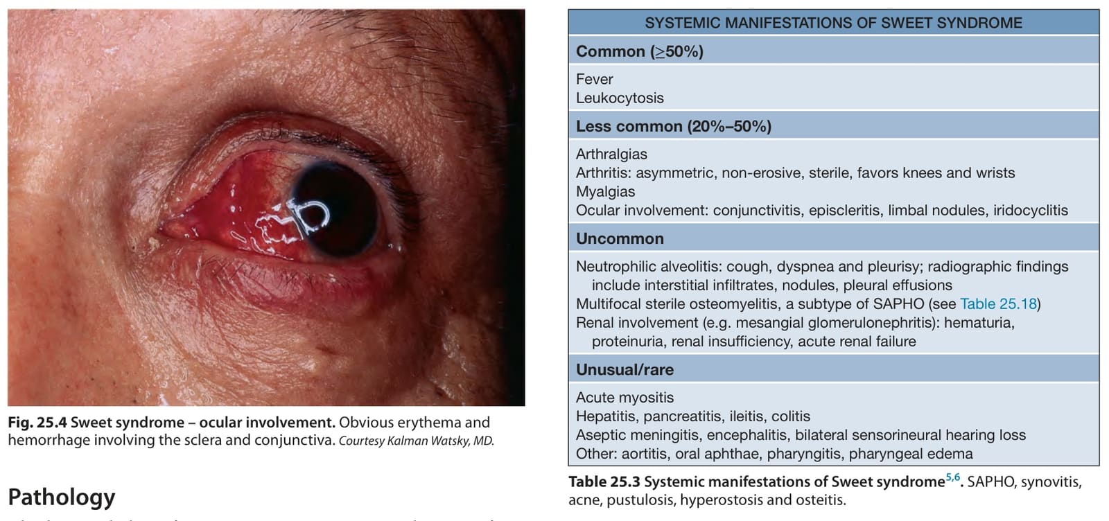
Table 25.3 Systemic manifestations of Sweet syndrome. SAPHO, synovitis, acne, pustulosis, hyperostosis and osteitis.
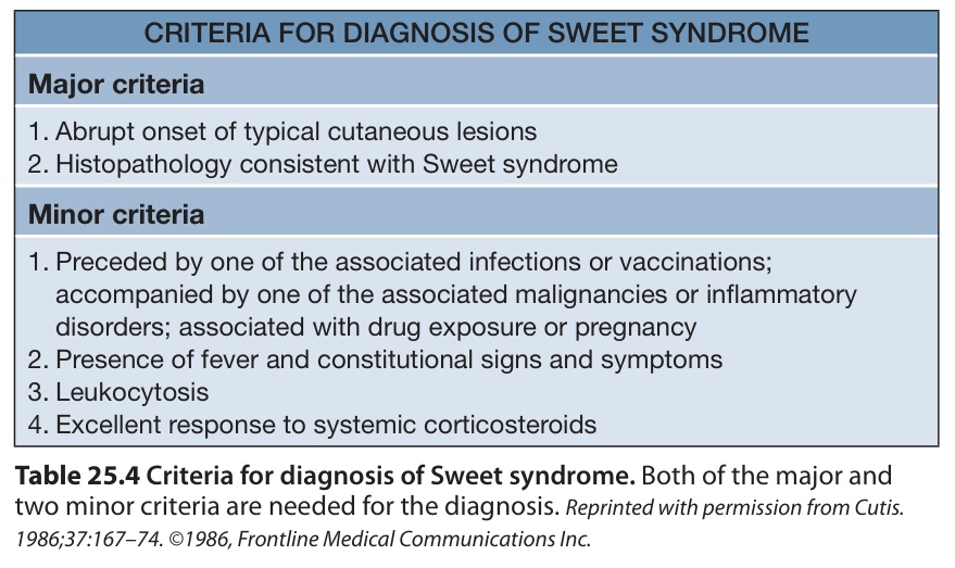
Table 25.4 Criteria for diagnosis of Sweet syndrome. Both of the major and two minor criteria are needed for the diagnosis. Reprinted with permission from Cutis. 1986;37:167–74. ©1986, Frontline Medical Communications Inc.
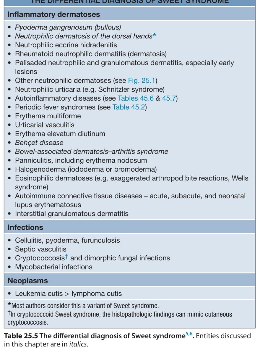
Table 25.5 The differential diagnosis of Sweet syndrome. Entities discussed in this chapter are in italics
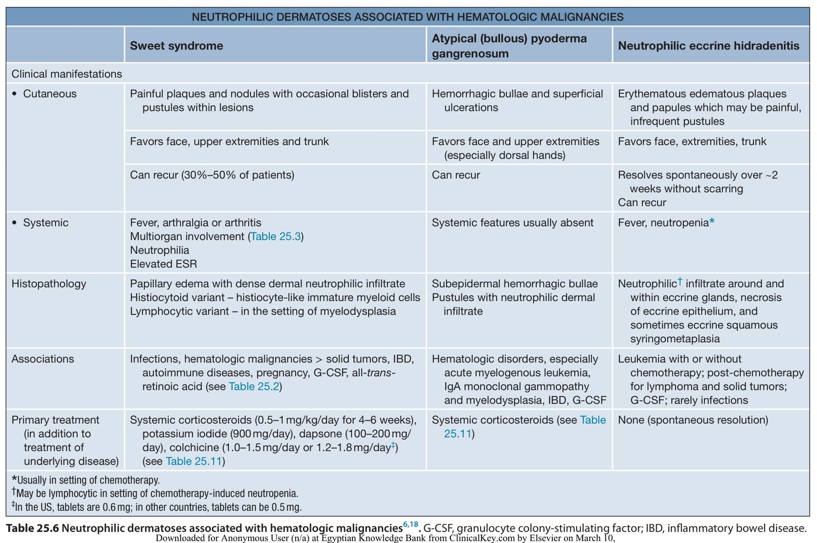
Table 25.6 Neutrophilic dermatoses associated with hematologic malignancies. G-CSF, granulocyte colony-stimulating factor; IBD, inflammatory bowel disease.