PYODERMA GANGRENOSUM
Key features
Four major clinical forms: ulcerative, bullous, pustular, and superficial granulomatous; additional variants include mucosal, peristomal, postsurgical, and necrotizing neutrophilic dermatosis Initial lesion is often a pustule on an erythematous or violaceous base, an erythematous nodule, or a bulla Characteristic lesion is an expanding ulcer with a necrotic undermined border; the base may be purulent or vegetative Histologically, a sterile dermal or adnexal abscess (without a vasculopathy) is seen in active, untreated, expanding lesions Associated illnesses include inflammatory bowel disease, arthritis, monoclonal gammopathies, and other hematologic disorders
Introduction
Pyoderma gangrenosum (PG) is an uncommon, chronic, recurrent, cutaneous ulcerative disease with a distinctive morphologic presentation. The laboratory and histopathologic findings can vary and therefore the diagnosis requires clinicopathologic correlation. This neutrophilic dermatosis is frequently associated with systemic disease.
History
In 1908, Brocq described a series of patients with skin ulcers he called “geometric phagedism”. Two decades later, Brunsting, Goeckerman, and O’Leary coined the term “pyoderma gangrenosum” and advanced the theory that it had an infectious etiology (streptococci and staphylococci).
Epidemiology
PG is a global disease. It can occur at any age but most commonly afflicts women between 20 and 50 years of age. Nearly half of the patients have an underlying systemic disease, most commonly inflammatory bowel disease, arthritis, or a hematologic disorder (e.g. IgA monoclonal gammopathy, acute myelogenous leukemia, myelodysplasia). Approximately 4% of cases of PG occur in infants and children. PG is also a cutaneous manifestation of several monogenic autoinflammatory diseases (see Tables 45.6 and 45.7).
Pathogenesis
Although the disease is idiopathic in 25%–50% of patients, an underlying immunologic abnormality is currently favored, given its frequent association with systemic diseases that have a suspected autoimmune pathogenesis. The association of PG with several autoinflammatory diseases points to aberrant activation of the innate immune system. In particular, enhanced activity of the IL-1 pathway is thought to play a role, as it does in several autoinflammatory disorders (see Fig. 4.2). In lesional skin biopsies, overexpression of IL-1α, IL-1β, and the IL-1 receptor as well as IL-8, IL-17, and TNF has been reported. Elevated serum levels of IL-1β have also been observed, and clinical improvement of PG following administration of the anti-IL-1β antibody canakinumab provides additional support for this proposed pathogenesis.
The adaptive immune system may also play a role as cytokines involved in the Th1/Th17 pathways (e.g. IL-23) and the JAK/STAT pathway have been implicated in the pathogenesis of PG. Pathergy is seen in 20%–30% of patients with PG and not only does this phenomenon lead to new cutaneous lesions but it can also aggravate them. Overexpression of IL-8 and IL-36 may contribute to this reaction to minor trauma.
Additional insights into the pathogenesis of PG should come from a better understanding of the monogenic primary immunodeficiencies and autoinflammatory disorders associated with PG (see below). As noted previously, based upon findings from a murine model of neutrophilic dermatoses, splice variants in PTPN6 may play a role in PG as well as Sweet syndrome.
Clinical Features
Although the classic morphologic clinical presentation of PG is an ulcer, there are several variants (bullous, pustular, and superficial granulomatous/vegetative) which differ by their clinical presentation, location, and associated diseases (Table 25.7; Fig. 25.8 & 25.9). Cutaneous lesions are painful and most frequently occur on the lower extremities in patients with typical (classic) PG, especially the pretibial area (Fig. 25.10A,B,D), but they can occur anywhere (Fig. 25.10C,E), including
A Pyostomatitis vegetans in a patient with ulcerative colitis. B Vegetative form following trauma to the skin. Courtesy Samuel L. Moschella, MD.
on mucous membranes and peristomal sites (Fig. 25.11A). A vulvovaginal variant has been observed in patients who have received rituximab.
The lesions of PG usually begin as a tender papulopustule (Fig. 25.12) with surrounding erythematous or violaceous induration, an erythematous nodule, or a bulla on a violaceous base; the papulopustule may be follicular. All these lesions then undergo necrosis leading to a central shallow or deep ulcer; loss of tissue can expose underlying tendons or muscles. When fully developed, the ulcer has a purulent base with an irregular, undermined and overhanging, gunmetal-colored border which extends centrifugally (see Fig. 25.10A,B). This border may have surrounding erythema. Re-epithelialization occurs from the margins and the ulcers heal with atrophic cribriform pigmented scars (Fig. 25.10F).
In PG, the number of ulcers can vary from one to over a dozen, and sometimes they coalesce (Fig. 25.11B). Although the ulcers are classically described as rapidly expanding, some are less inflammatory and expand more slowly. The latter may require less aggressive therapeutic interventions.
In patients with hematologic disorders or in drug-induced cases, the clinical course is often characterized by an acute onset of hemorrhagic or purulent bullous lesions, with a more widespread distribution that can include the upper extremities, particularly the dorsal hands, as well as the face. These lesions rapidly necrose and are often associated with fever and signs of toxicity. In those patients with associated inflammatory diseases, such as inflammatory bowel disease or arthritis, the more characteristic presentation is a chronic, slowly enlarging ulcer with excessive granulation tissue in its base and sometimes with evidence of spontaneous regression.
Clinical variants have been described based on the clinical presentation (see Table 25.7) or the location of the lesion (e.g. peristomal, mucosal including the genitalia) or the age of the afflicted. While the
A Several ulcers surround an ileostomy in a patient who had a total proctocolectomy performed because of refractory chronic ulcerative colitis. B Multiple ulcerations of the breasts following breast reduction. Because the original diagnosis postoperatively was soft tissue infection, multiple debridements had been performed and systemic antibiotics administered.
clinical appearance of PG in children is similar to that in adults, the lesions more frequently involve the head and anogenital region. As with Sweet syndrome, extracutaneous sterile neutrophilic infiltrates have been reported in the bones, lung, liver, pancreas, spleen, kidneys, and CNS of patients with PG.
Associated Diseases
Between 50% and 70% of patients with PG have an antecedent, coincident, or subsequent associated disease or condition. The most common associations are inflammatory bowel disease (ulcerative colitis or Crohn disease [20%–30%]), arthritis (seronegative arthritis, spondylitis of inflammatory bowel disease or rheumatoid arthritis [20%]), and hematologic disease (in particular, acute > chronic myelogenous leukemia, myelodysplasia, hairy cell leukemia, and monoclonal gammopathy [15%–25%]). The monoclonal gammopathy is often composed of IgA.
Individuals with the autoinflammatory PAPA (pyogenic [sterile] arthritis, PG and acne) syndrome have been shown to have mutations in PSTPIP1, which encodes a CD2-binding protein, presumably leading to abnormal inflammatory responses (Fig. 25.13). PG-like lesions are also seen in patients with PASH and PAPASH syndromes (see Table 45.6), as well as in primary immunodeficiencies, e.g. leukocyte adhesion deficiency-1, common variable immunodeficiency due to NFKB1 mutations (see Table 60.1).
Other neutrophilic dermatoses that have been reported in association with PG are subcorneal pustular dermatosis, Behçet disease, and Sweet syndrome.
Individuals with this monogenic autoinflammatory disorder have pyogenic sterile arthritis, pyoderma gangrenosum and acne. Courtesy Maria Chanco Turner, MD.
Pathology
The histopathology of PG can be nonspecific, especially when the disease is partially treated or minimally inflamed, and therefore be non-diagnostic. In early lesions, there is a neutrophilic vascular reaction, which can be folliculocentric. Neutrophilic infiltrates, often with leukocytoclasia, are seen in active, untreated, expanding lesions (Fig. 25.14). In fully developed ulcers, there is marked tissue necrosis with surrounding mononuclear cell infiltrates. In most patients with typical PG,
chronic ulcers have fibrosing inflammation at the edge of the ulcer bed. Although biopsy specimens of PG may show nonspecific changes, histopathologic examination, often accompanied by tissue culture, assists in excluding other entities that can mimic PG.
Differential Diagnosis
Since there are no specific or diagnostic laboratory tests or histopathologic features and some of the associated conditions may not have declared themselves, the physician often needs to exclude other causes of cutaneous ulceration and search for a treatable associated disease. PG can be misdiagnosed: of 157 consecutive patients seen at one tertiary care medical center with a referral diagnosis of resistant ulcers due to PG, 15 (~10%) were found not to have PG. To assist the clinician, three validated diagnostic schemes have been proposed (Table 25.8); all emphasize that no single criterion should be used in isolation. The evaluation of a patient with presumed PG is summarized in Table 25.9.
The differential diagnosis clearly depends on the evolutionary stage of the process – the initial inflammatory process characterized by erythematous papules, pustules, plaques, and nodules versus the later characteristic vegetative or ulcerative lesions (Table 25.10). Failure to establish the correct diagnosis can have serious consequences, including continued aggressive debridement and even amputation.
Treatment
There is neither specific nor uniformly effective therapy for PG. The nature and intensity of the therapeutic approach depend on the number, size and depth of the lesions, the rate of expansion and appearance of new lesions, the associated disorder, the medical status of the patient, and the risk and patient tolerance of prolonged therapy. Prior to instituting treatment, evaluation for an associated disease is recommended and this assessment may be guided by the age of the patient.
The therapeutic goals are to reduce the inflammatory process of the wound, thereby promoting healing and reducing pain, and to control the contributing underlying disease (especially leukemias and inflammatory bowel disease), while producing the least adverse side effects. The standard medical treatment of PG is local or combined local and systemic corticosteroid therapy, with or without adjunctive systemic therapy; based upon an evidence-based review, cyclosporine is also considered first-line therapy.
When delivered in adequate dosage, systemic corticosteroids have generally been the most predictable and effective medication. Unfortunately, the more resistant lesions require more protracted therapy (>3 months) at a higher than desirable dosage, thus inviting adverse side effects. Such patients should be closely monitored and should receive supplemental calcium (1500 mg/day), vitamin D (800 IU/day) and, in most instances, bisphosphonates (see Ch. 125). Additional therapies, e.g. classic steroid-sparing agents, JAK, TNF and IL-12/23 inhibitors, are outlined in Table 25.11. Once the inflammation is under control, gentle debridement of necrotic debris may be performed, but a test area, as with intralesional corticosteroids, allows for assessment of pathergy.
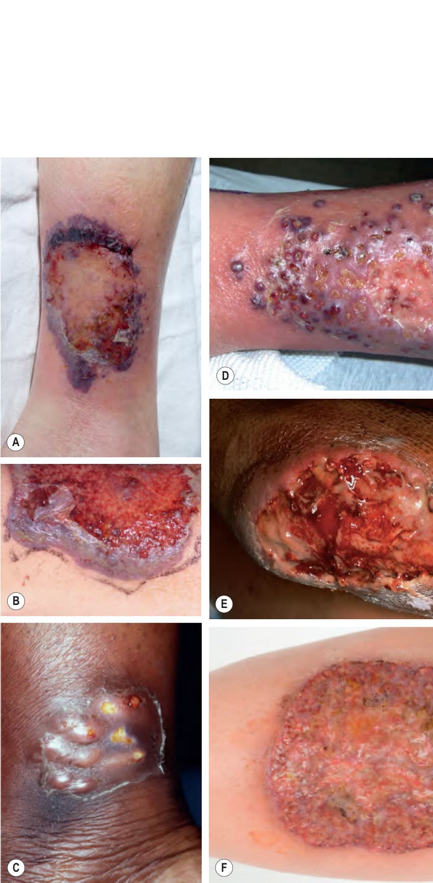
Fig. 25.8 Bullous pyoderma gangrenosum. This patient had ulcerative colitis.
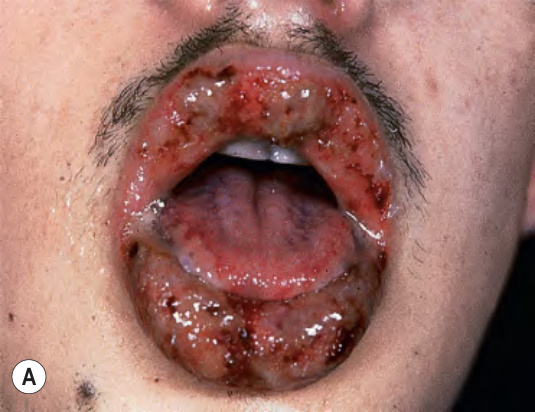
Fig. 25.9 Variants of pyoderma gangrenosum.
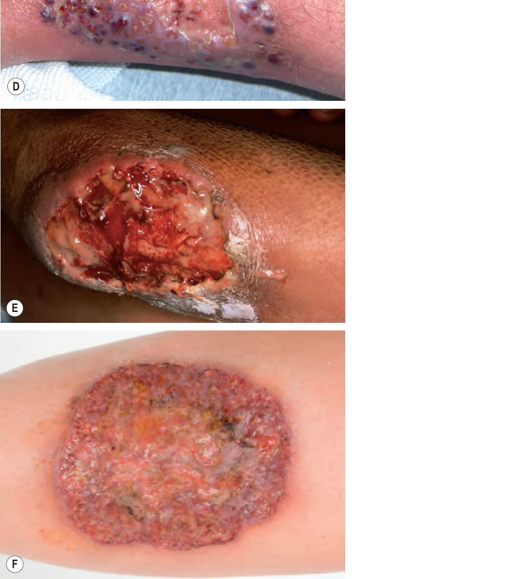
Fig. 25.10 Pyoderma gangrenosum (PG) – clinical presentations.A, B Typical (classic) form in which the ulcer has an undermined and overhanging violet– gray edge as well as a surrounding violaceous border. C Grouped sterile pustular nodules. D Central ulceration surrounded by inflammatory papules and pustules. E Deeper ulcer with vegetative and pustular base. F Centrally, there is healing with cribriform (sievelike) scarring.
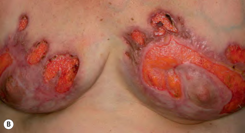
Fig. 25.11 Peristomal and postsurgical pyoderma gangrenosum (PG).
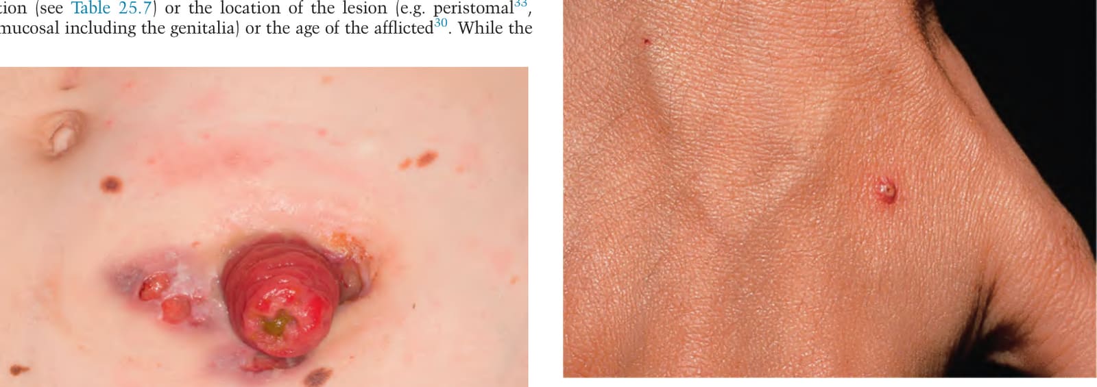
Fig. 25.12 The earliest clinical lesion in pyoderma gangrenosum is a pustule with an inflammatory base. This patient had Crohn disease.
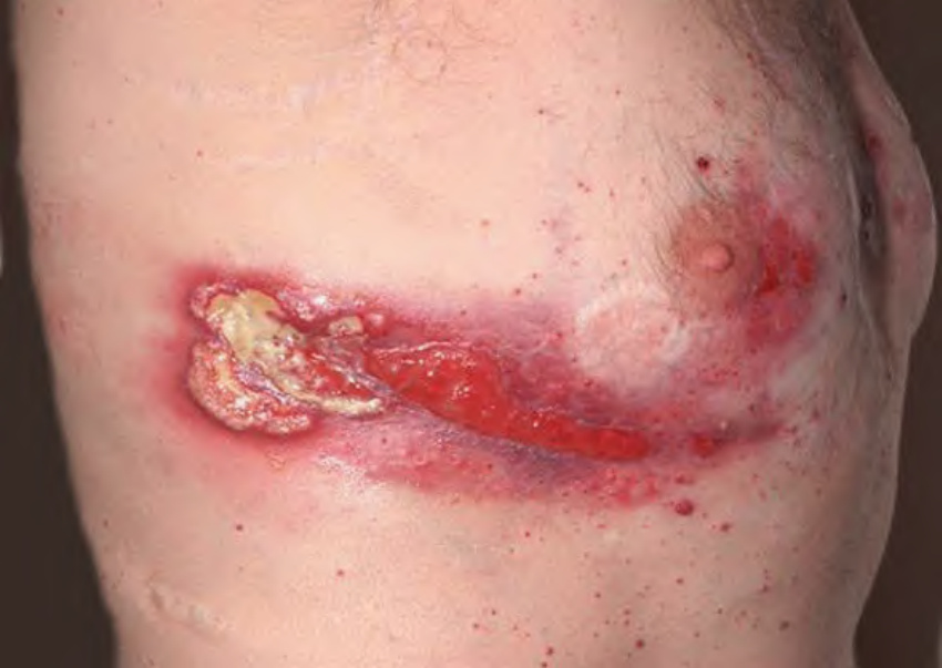
Fig. 25.13 Pyoderma gangrenosum in a patient with PAPA syndrome.
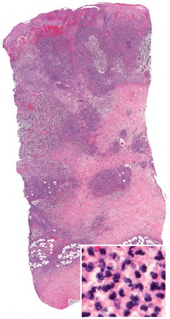
Fig. 25.14 Pyoderma gangrenosum. In expanding untreated lesions, a diffuse infiltrate of neutrophils (inset) is present at the edge of an ulceration. Courtesy Lorenzo Cerroni, MD.
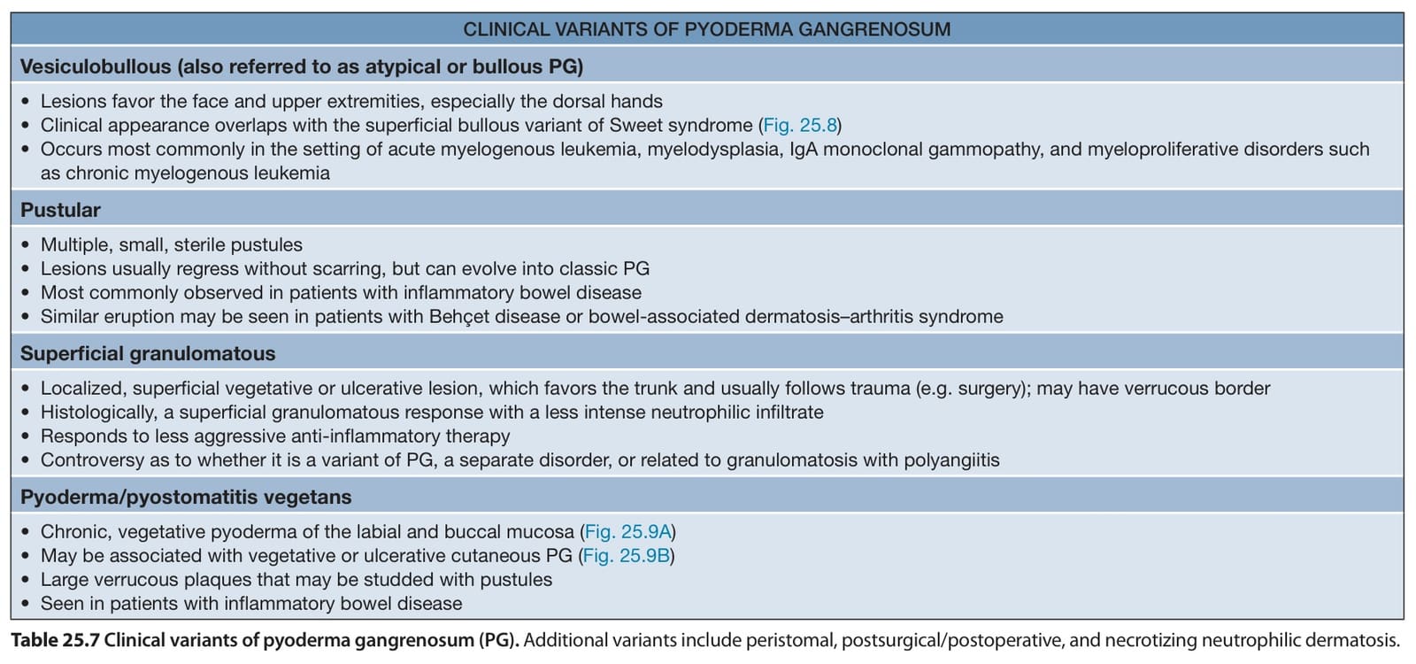
Table 25.7 Clinical variants of pyoderma gangrenosum (PG). Additional variants include peristomal, postsurgical/postoperative, and necrotizing neutrophilic dermatosis.
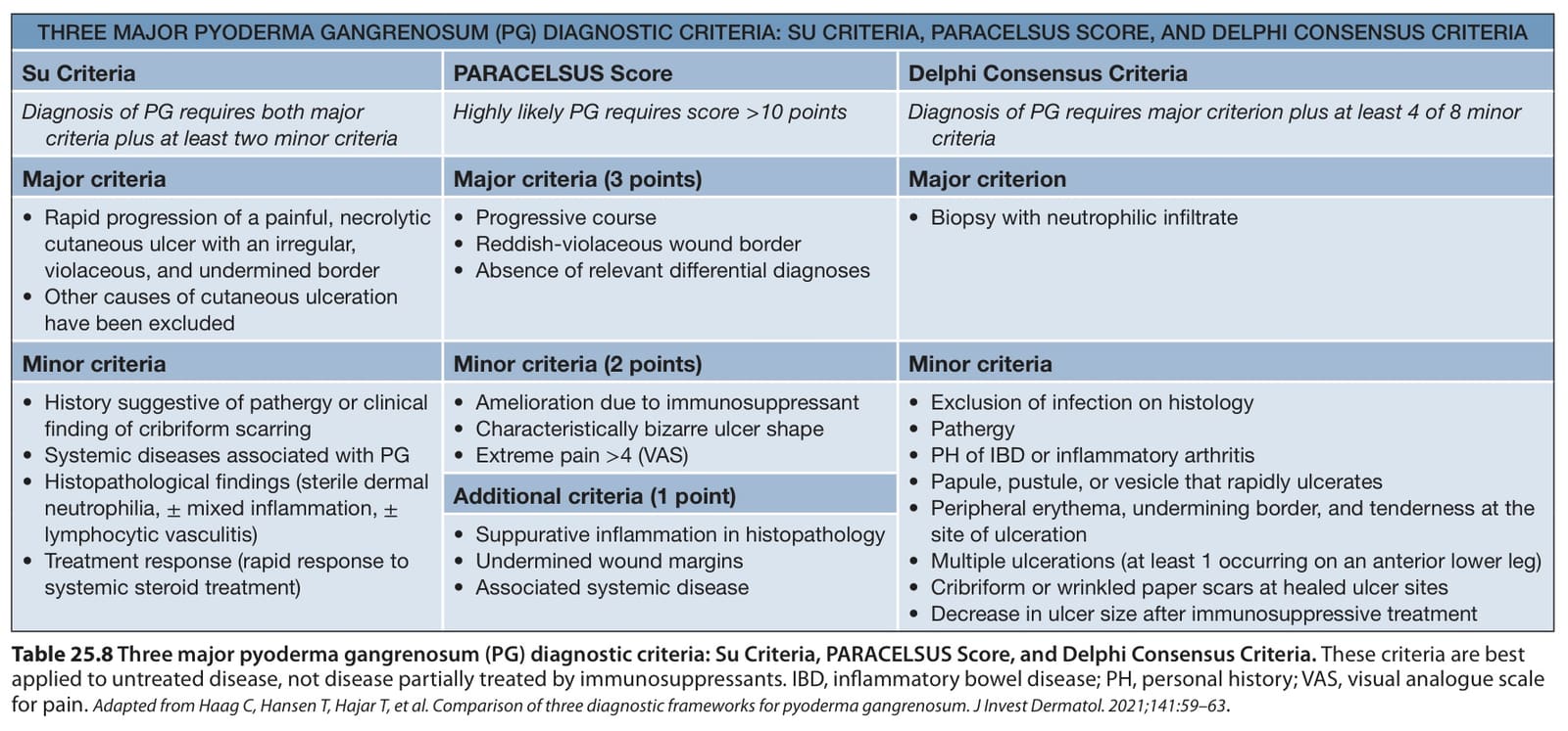
Table 25.8 Three major pyoderma gangrenosum (PG) diagnostic criteria: Su Criteria, PARACELSUS Score, and Delphi Consensus Criteria. These criteria are best applied to untreated disease, not disease partially treated by immunosuppressants. IBD, inflammatory bowel disease; PH, personal history; VAS, visual analogue scale for pain. Adapted from Haag C, Hansen T, Hajar T, et al. Comparison of three diagnostic frameworks for pyoderma gangrenosum. J Invest Dermatol. 2021;141:59–63
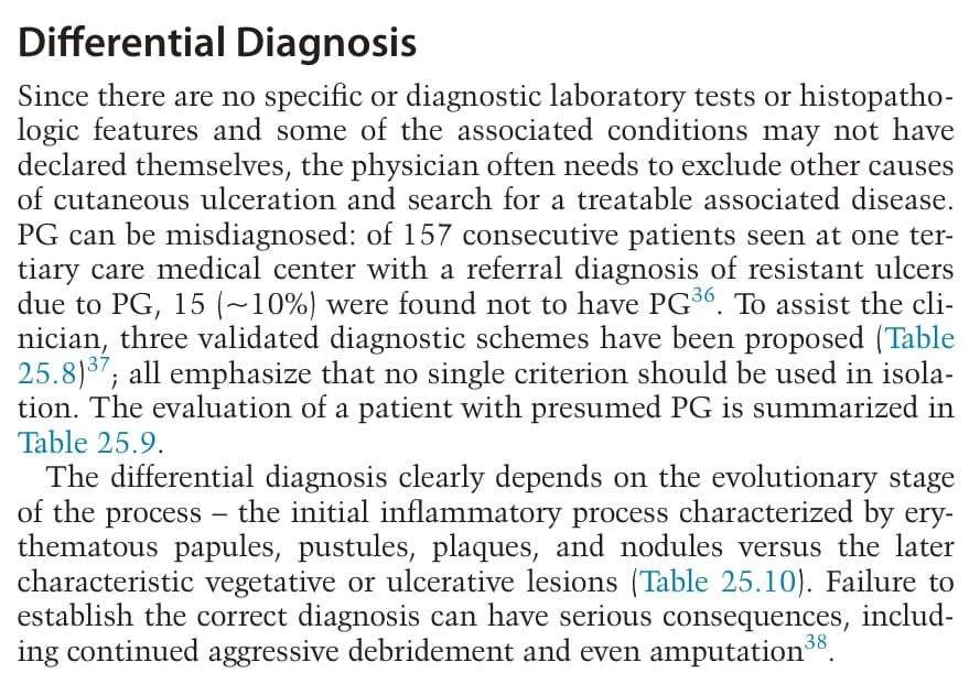
Table 25.9 Evaluation of a patient with presumed pyoderma gangrenosum. ANCA, anti-neutrophil cytoplasmic antibodies.
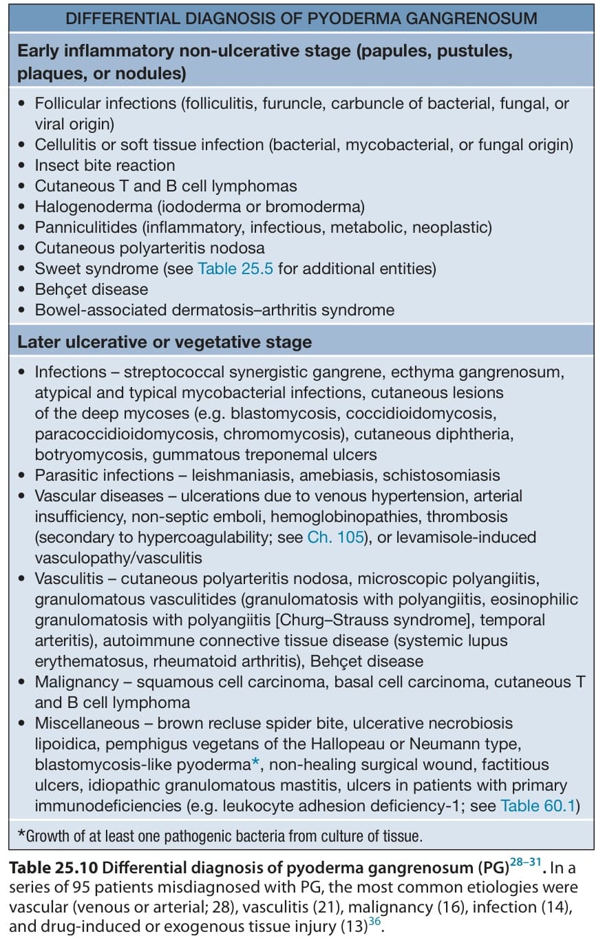
Table 25.10 Differential diagnosis of pyoderma gangrenosum (PG). In a series of 95 patients misdiagnosed with PG, the most common etiologies were vascular (venous or arterial; 28), vasculitis (21), malignancy (16), infection (14), and drug-induced or exogenous tissue injury (13)
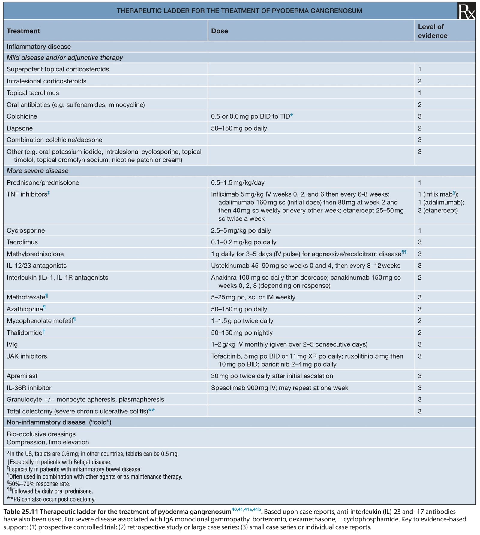
Table 25.11 Therapeutic ladder for the treatment of pyoderma gangrenosum40,41,41a,41b. Based upon case reports, anti-interleukin (IL)-23 and -17 antibodies have also been used. For severe disease associated with IgA monoclonal gammopathy, bortezomib, dexamethasone, ± cyclophosphamide. Key to evidence-based support: (1) prospective controlled trial; (2) retrospective study or large case series; (3) small case series or individual case reports.