BEHÇET DISEASE
Key features
A multisystem, polysymptomatic disease Diagnosis is based on International Study Group criteria of recurrent oral ulceration, recurrent genital ulceration, ocular abnormalities (e. g. uveitis, retinal vasculitis), and cutaneous lesions Cutaneous findings range from sterile papulopustules and palpable purpura to erythema nodosum-like lesions Histologically, a neutrophilic angiocentric infiltrate with leukocytoclastic (early) or lymphocytic (late) vasculitis is the characteristic finding
Introduction
Behçet disease is a multisystem, polysymptomatic disease with unpredictable exacerbations and remissions. All organs of the body can be affected concomitantly or consecutively. As a result, all subspecialties can be involved in the care of these patients.
History
In the fifth century BCE, Hippocrates first described the symptomatology of this disease, and in 1936, Behçet reported a patient with eye disease and orogenital ulcers.
Epidemiology
Most patients are from regions along the ancient Silk Route, which stretched from the Korean peninsula and Japan to the Mediterranean Sea. Turkey has the highest prevalence, at 80 per 100 000. In Japan, it is 10 per 100 000; in the UK, 0.64 per 100 000; and in the US, 0.12 per 100 000. In Japan and Korea, the incidence of Behçet disease is greater in women, but in Middle Eastern countries it is seen more frequently in men. The peak incidence is between 20 and 35 years of age. The familial form usually comprises 2%–5% of cases, except in the Middle East, where it represents 10%–15%.
Pathogenesis
Epidemiologic evidence suggests that genetic and environmental factors contribute to the development of this disease. More than 80% of Asian patients have the HLA-B51 allele, but it is present in only about 15% of White individuals from Western countries; thus, the allele appears to be an important risk factor for those who live along the Silk Route.
Earlier theories of pathogenesis entertained the possibility of an infectious etiology (viral or bacterial), but subsequent evidence failed to substantiate this hypothesis. Among the infectious agents investigated were HSV, hepatitis C virus, parvovirus B19, and streptococci. These infections may potentially trigger an immunoregulatory defect in a genetically predisposed individual.
The pathomechanisms of Behçet disease involve vascular injuries and autoimmune responses. Circulating immune complexes and neutrophils appear to be responsible for the mucocutaneous lesions that are characterized histologically by a neutrophilic vascular reaction or even leukocytoclastic vasculitis. The neutrophils of Behçet disease produce an increased amount of superoxides and an excess of lysosomal enzymes, and they have enhanced chemotaxis, all of which lead to tissue injury. The elevated circulating levels of TNF, IL-1β, and IL-8 may result in activation of neutrophils and augmentation of cellular interactions between neutrophils and endothelial cells. Clonal expansion of autoreactive T cells that recognize a peptide derived from heat shock protein 60 has also been described. More recent genome-wide association studies found that single nucleotide polymorphisms in IL-10 and IL-23R regions were associated with the disease.
Clinical Features and Differential Diagnosis
Mucocutaneous involvement
Aphthous stomatitis, the major criterion, is frequently the first symptom of Behçet disease (65%–70% of patients), and it is almost always present during the course of the disease. The aphthosis may precede the other manifestations by many years, but a dramatic increase in severity often occurs in concert with the onset of other features. Oral ulcers begin as an erythematous papule, which develops a yellowish pseudomembrane and then forms a painful usually non-scarring ulcer (Fig. 25.15A,B); it heals over a period of weeks. The ulcers are indistinguishable from those that occur with complex aphthosis or inflammatory bowel disease and they must be differentiated from other causes of oral ulcers, e.g. pemphigus vulgaris, Marshall syndrome. The latter patients also have periodic fevers, pharyngitis, and cervical adenitis.
The genital aphthae involve primarily the scrotum and penis in men and the vulva in women. Compared to oral lesions, the anogenital aphthae tend to be larger with irregular margins, but can vary in size (Fig. 25.15C); they are more painful. Distinction of genital lesions from lesions of HSV requires PCR, viral culture, or direct fluorescent antibody stain. Both immunobullous diseases (e.g. mucous membrane pemphigoid) and erosive lichen planus can also lead to oral and genital ulcerations.
Among the primary cutaneous lesions are acral and facial sterile vesiculopustules as well as pustular and/or purpuric papules. Although the papulopustular lesions are usually not folliculocentric, they have nonetheless been described as acneiform, leading to some confusion (see Pathology below). The panniculitic erythema nodosum-like lesions favor women and appear on their legs and buttocks, and less often on the face and neck; they have to be differentiated from both superficial thrombophlebitis (which occurs in 30% of patients with Behçet disease) and erythema nodosum (which can develop in patients with Behçet disease). Pathergy also occurs (Fig. 25.15D), as in PG. Patients with features of both Behçet disease and relapsing polychondritis have been described and reported as MAGIC (mouth and genital ulcers with inflamed cartilage) syndrome (see Ch. 45).
Patients with A20 haploinsufficiency, also referred to as familial Behçet-like autoinflammatory syndrome, can also have oral and genital ulcers as well as Sweet-like and erythema nodosum-like lesions and pustular eruptions (palmoplantar, folliculitis, pseudofolliculitis). This disorder is due to TNFAIP3 mutations.
Systemic involvement
Systemic manifestations are summarized in Table 25.12 and distinctions between Behçet disease with bowel involvement and inflammatory bowel disease are outlined in Table 25.13.
Pathology
The histopathology of the cutaneous lesions is nonspecific. Vasculitic changes, which can involve all sizes of blood vessels within the
Oral aphthosis involving the tongue and lip; the ulcerations can be deep. These ulcers may be diagnosed as infectious (e. g. due to herpes simplex virus) or neoplastic rather than inflammatory. C Aphthae of the vulva and inguinal region. D Pathergy – a papulopustule appeared at the site of insertion of an intravenous catheter.
dermis and subcutis, are probably secondary to the intense inflammation observed. Both leukocytoclastic and lymphocytic vasculitis may be seen, and the affected vessels are sometimes occluded by fibrin thrombi.
The histologic features of the erythema nodosum-like lesions can vary from a neutrophilic lobular panniculitis to a septal and lobular panniculitis with a mixed inflammatory infiltrate, fat necrosis, and evidence of vasculitis.
Diagnosis
Since Behçet disease has no diagnostic test and is characterized by a complex constellation of signs and symptoms, various sets of clinical criteria have been created to assist in the diagnosis. Among the reported diagnostic criteria are those of the Japanese (1974), O’Duffy (1974), Zhang (1980), James (1986), and the International Study Group (ISG; 1990). The latter are presented in Table 25.14. The development of the ISG criteria was based on the discriminating performance of various criteria-grouped combination sets, and the ISG criteria exhibited the highest sensitivity, specificity, and relative value. Controversy exists, however, about the failure of these criteria to require exclusion of inflammatory bowel disease.
Treatment
The management of the disease is difficult because of its variable course and the lack of sufficient double-blind studies. The therapeutic approach is a symptomatic one and is dictated by the nature of the mucocutaneous and visceral involvement. Because of the frequent vital organ involvement, e.g. the eye, and the tendency for the disease to recur, early and aggressive treatment is vital. CNS and large artery and vein involvement respond less favorably to therapy. There are multiple drugs available, both topical and systemic, that can be used as monotherapy or as combination therapy (Table 25.15). Evidence for many of the therapies is based on case-series data.
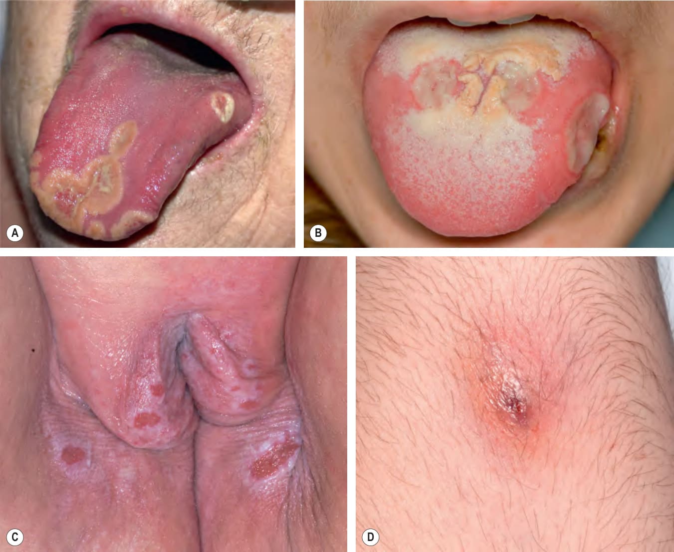
Fig. 25.15 Behçet disease – mucocutaneous lesions.A, B
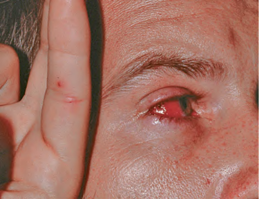
Fig. 25.16 Behçet disease – systemic involvement. Iritis and cutaneous pustular vasculitis. Courtesy Samuel L. Moschella, MD.
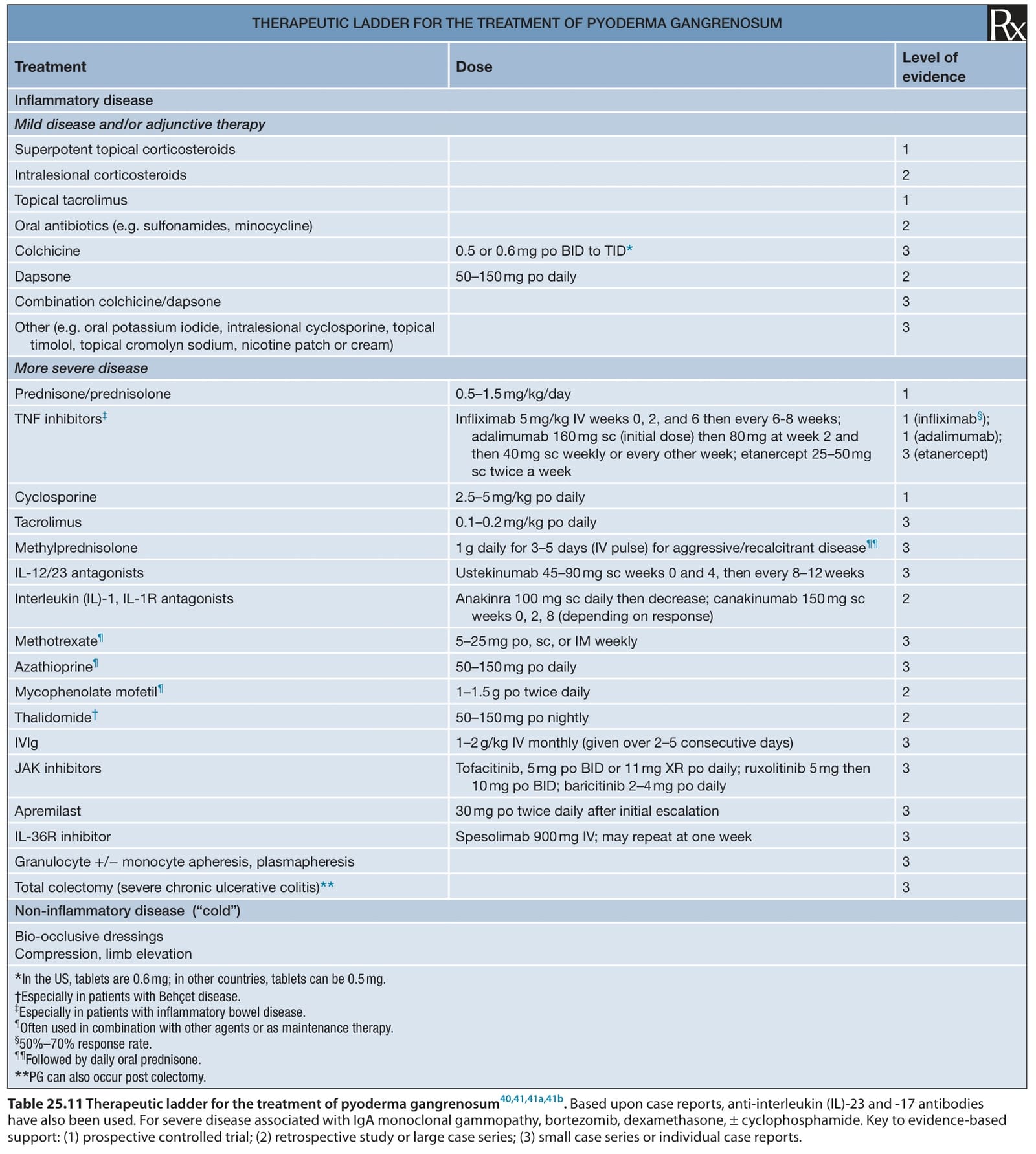
Table 25.11 Therapeutic ladder for the treatment of pyoderma gangrenosum40,41,41a,41b. Based upon case reports, anti-interleukin (IL)-23 and -17 antibodies have also been used. For severe disease associated with IgA monoclonal gammopathy, bortezomib, dexamethasone, ± cyclophosphamide. Key to evidence-based support: (1) prospective controlled trial; (2) retrospective study or large case series; (3) small case series or individual case reports.
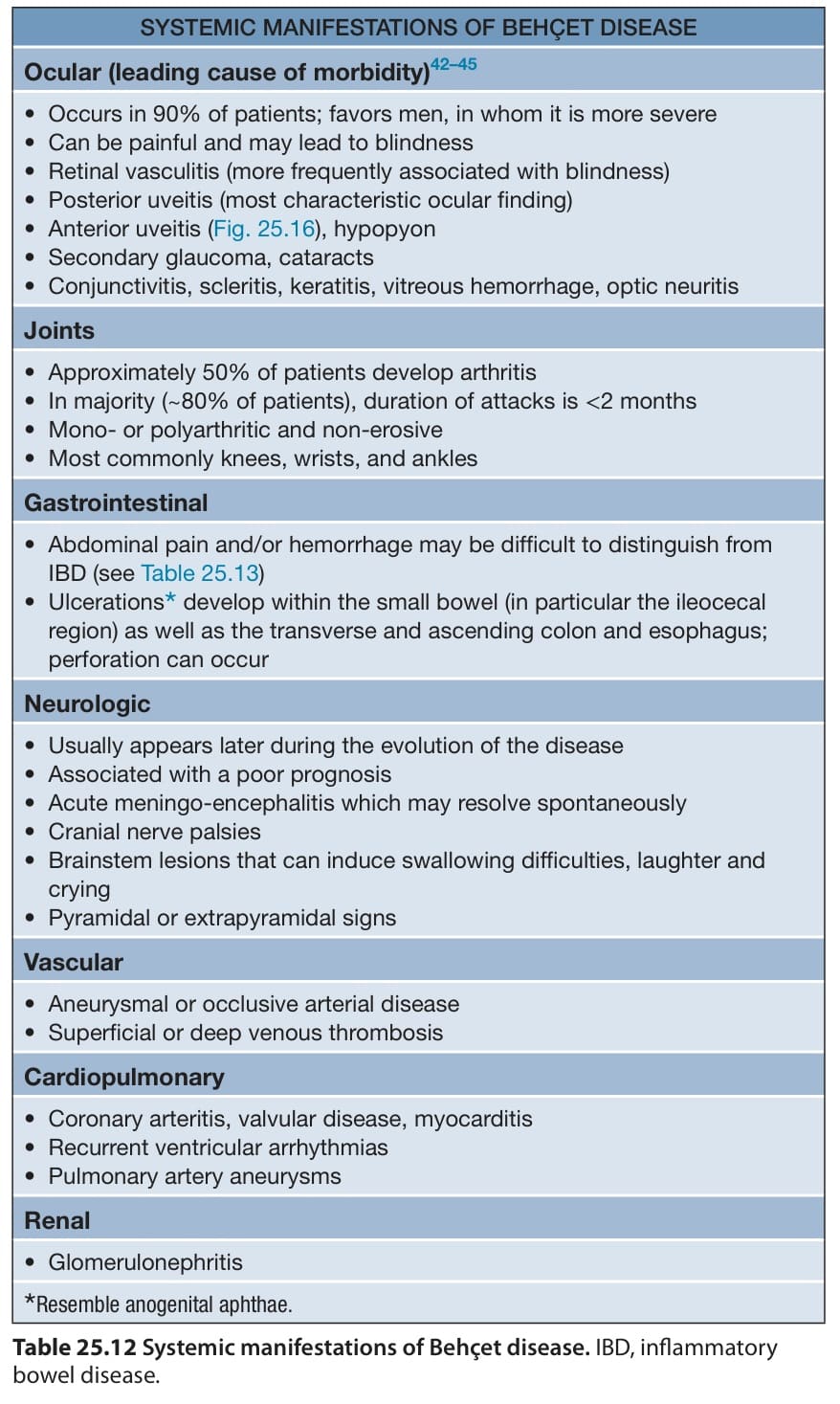
Table 25.12 Systemic manifestations of Behçet disease. IBD, inflammatory bowel disease.
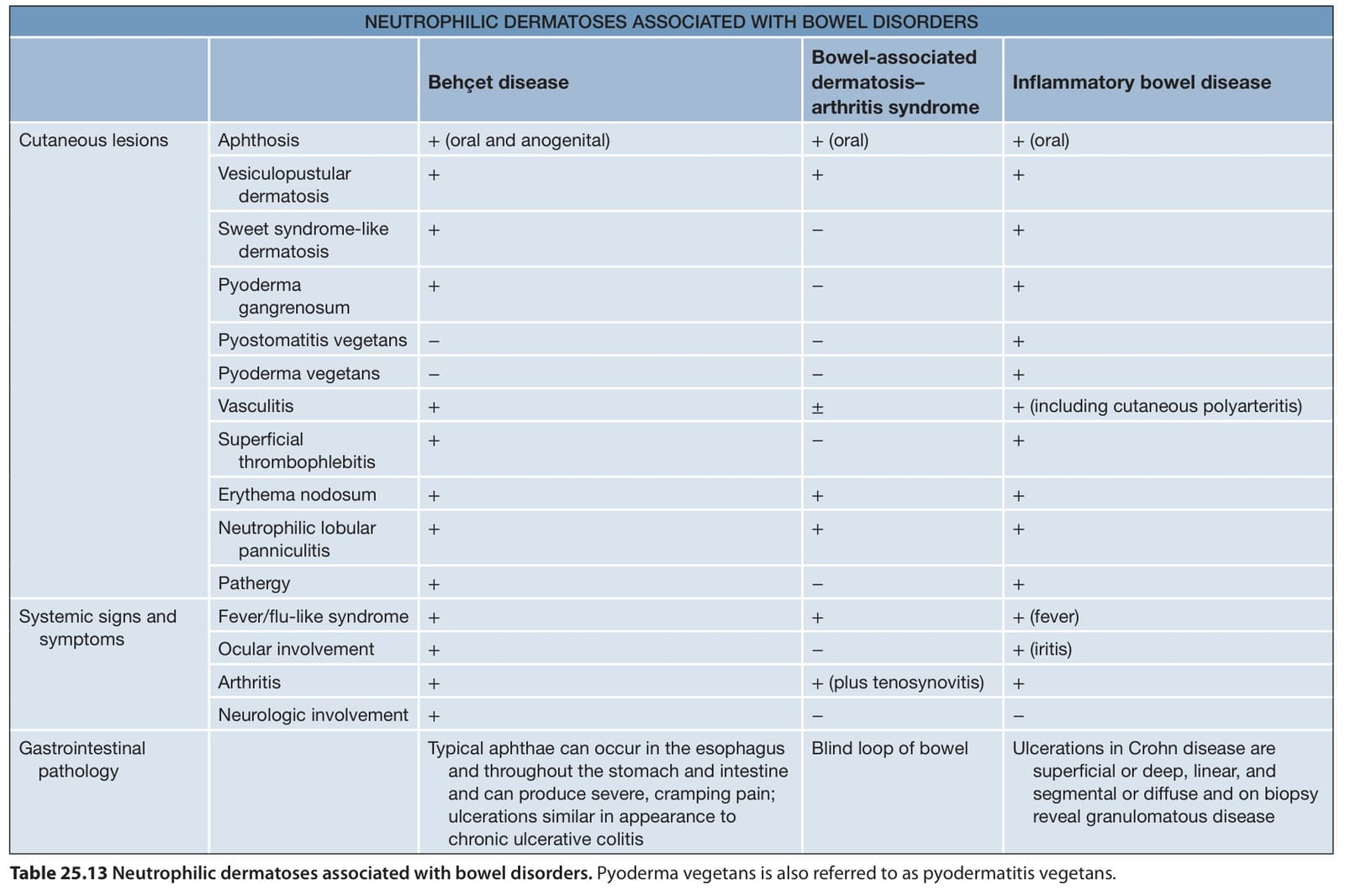
Table 25.13 Neutrophilic dermatoses associated with bowel disorders. Pyoderma vegetans is also referred to as pyodermatitis vegetans.
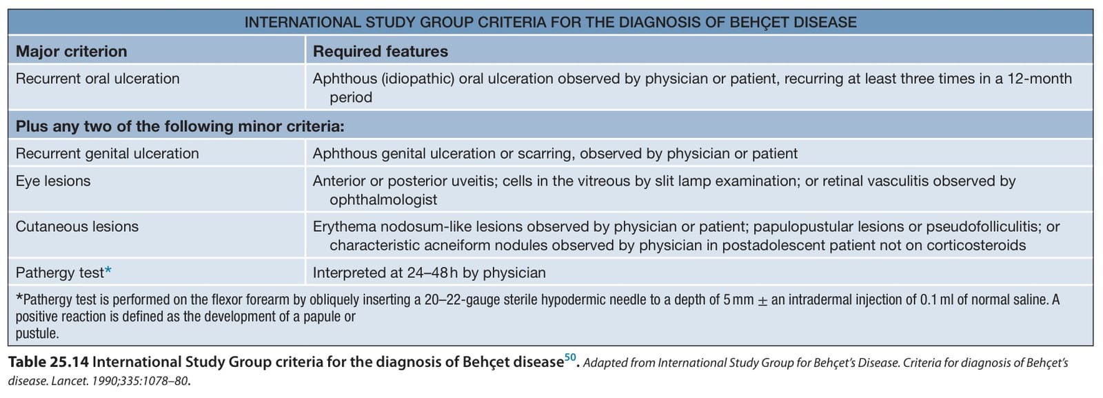
Table 25.14 International Study Group criteria for the diagnosis of Behçet disease. Adapted from International Study Group for Behçet’s Disease. Criteria for diagnosis of Behçet’s disease. Lancet. 1990;335:1078–80
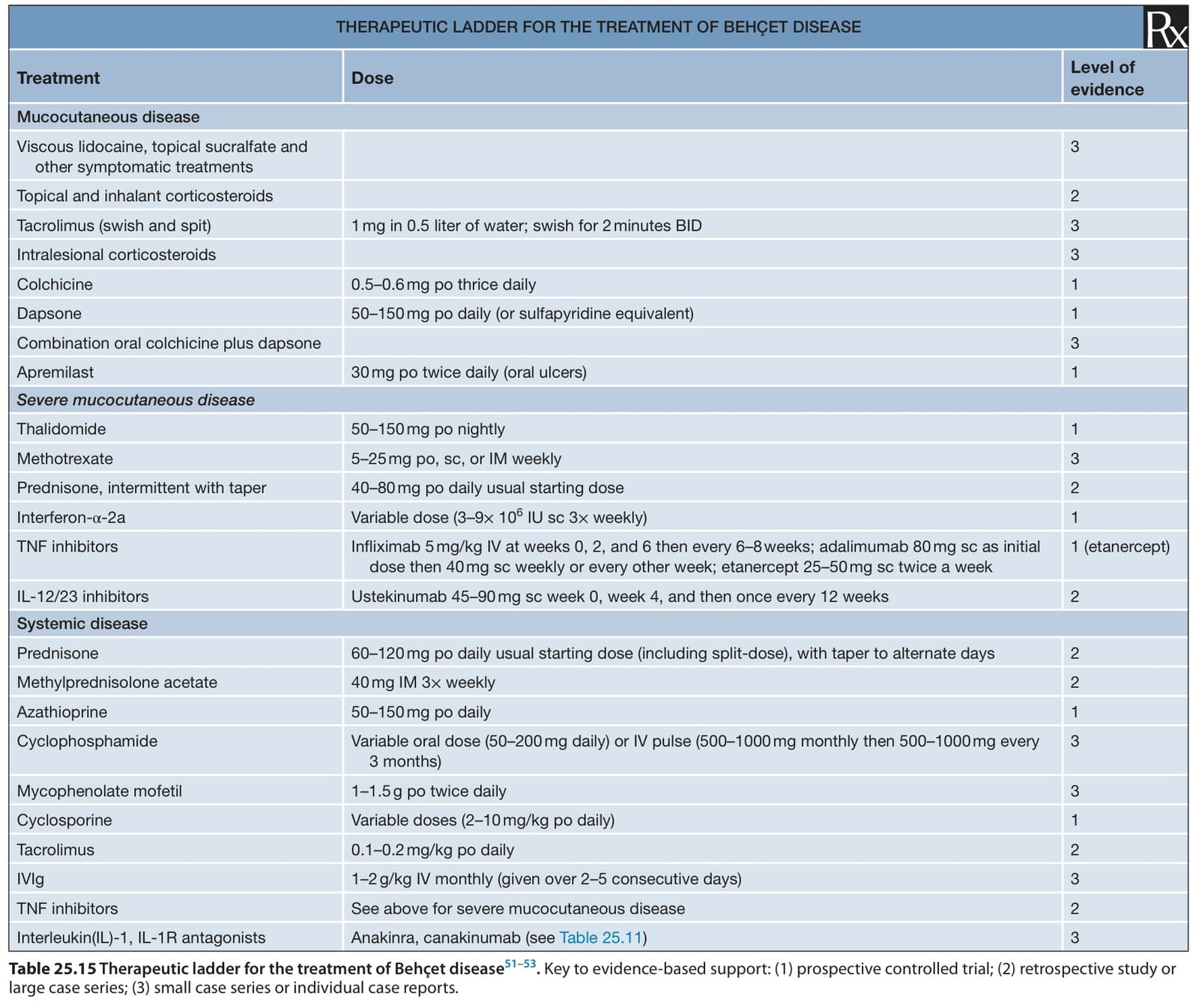
Table 25.15 Therapeutic ladder for the treatment of Behçet disease. Key to evidence-based support: (1) prospective controlled trial; (2) retrospective study or large case series; (3) small case series or individual case reports.